
Remember me
Thermoluminescent dosimeters (TLDs) serve as compact and user-friendly tools for various applications, including personal radiation dosimetry and radiation therapy. This study explores the potential of utilizing TLD-100 personal dosimetry, conventionally applied in PET/CT (positron emission tomography/computed tomography) settings, in the PET/MRI (magnetic resonance imaging) environment. The integration of MRI into conventional radiotherapy and PET systems necessitates ionizing radiation dosimetry in the presence of static magnetic fields. In this study, TLD-100 dosimeters were exposed on the surface of a water-filled cylindrical phantom containing PET-radioisotope and positioned on the patient table of a 3 T PET/MRI, where the magnetic field strength is around 0.2 T, aiming to replicate real-world scenarios experienced by personnel in PET/MRI environments. Results indicate that the modified MR-safe TLD-100 personal dosimeters exhibit no significant impact from the static magnetic field of the 3 T PET/MRI, supporting their suitability for personal dosimetry in PET/MRI settings. This study addresses a notable gap in existing literature on the effect of MRI static magnetic field on TLDs.
Thermoluminescent dosimeters (TLDs) are compact and user-friendly solution for diverse applications, encompassing personal radiation dosimetry, environmental monitoring, and dosimetry within radiation therapy. They can be used to measure a wide range of radiation types, including x-rays, gamma rays, and beta particles. TLDs are created by adding impurities to a perfect crystalline insulator, thereby introducing intermediary energy levels between the valence and conduction bands. Upon exposure to ionizing radiation, electrons and holes are captured by electron- and hole-traps, respectively, remaining trapped unless additional stimuli like heat are applied. Subsequent heating of irradiated TLDs in a calibrated 'reader' leads to the release of trapped electrons, recombination with trapped holes, and the creation of defects in an excited state, which finally relax by emitting light. The quantity of emitted light from each TLD serves as an indicator of the absorbed radiation dose during irradiation of that TLD [1].
One of the applications of TLD is in personal dosimetry. Healthcare professionals employed in radiology, nuclear medicine, and radiation therapy departments encounter ionizing radiation, with the extent of exposure contingent on various factors including the individual's role within the respective department, workload, effectiveness of shielding measures, and individual practices at the workplace. To monitor radiation exposure levels, personal dosimeters are employed, providing a means to measure the radiation dose over a specific time period, typically on a monthly basis. Regular monitoring ensures that the received dose by personnel remains below the levels set by national and international regulations.
On other hand, magnetic resonance imaging (MRI) is a medical imaging technique that utilizes a strong static magnetic field and radiofrequency pulses to generate high contrast images of soft tissues. Unlike computed tomography (CT), radiation therapy, and positron emission tomography (PET), MRI does not utilize ionizing radiation. Consequently, traditional practices of dosimetry for ionizing radiation and personal dosimetry such as TLD in MRI environments have not been necessitated, underlining the non-ionizing nature of MRI technology. However, recent advancements in radiation therapy and positron emission tomography (PET) have involved the integration of MRI into conventional radiotherapy and PET systems, which necessitates dosimetry of ionizing radiation in presence of the strong magnetic field of MRI.
An instance where ionizing radiation dosimetry in the presence of a magnetic field becomes relevant is in the context of MRI-guided radiotherapy (MRIgRT). This technique integrates a conventional linear accelerator (linac) with an MRI scanner, which allows real-time imaging during radiation treatment without adding extra radiation dose from the imaging system. This approach incorporates advanced optimization techniques to tailor treatment delivery to tumor in the moving anatomical structures [2]. The static magnetic field of MRI may affect dosimetry results in MRIgRT. The constant magnetic field of the MRI scanner alters the direction of motion of charged particles, due to the Lorentz force, and consequently affects the detector response and the dose to medium. Several studies have investigated the influence of the magnetic field on different detectors, such as ionization chambers, Gafchromic film, alanine, Fricke dosimeters, and Presage [2–4]. However, there is a scarcity of studies examining the impact of magnetic fields on the response of thermoluminescent dosimeters.
Another scenario demanding dosimetry in the presence of a magnetic field is observed in PET/MRI, an advanced technology that harnesses the benefits of both MRI and high-sensitivity functional PET imaging. The combination of these modalities elevates diagnostic capabilities in neurological and oncological imaging and facilitates the monitoring of tumor response to therapy, delivering comprehensive anatomical and functional insights. Patients undergoing PET/MRI receive a lower radiation dose compared to those undergoing PET/CT. This is attributed to the fact that, unlike CT, MRI does not subject patients to any ionizing radiation dose apart from the dose from the PET tracer. On the other hand, personnel working with PET/MR may experience elevated levels of radiation dose compared to their counterparts in PET/CT. This is due to the necessity of coil positioning for PET/MRI scans, especially in whole-body scans, which may result in extended exposure periods for workers in comparison to PET/CT scans. In a study by Itikawa et al, no significant difference was found for whole-body dose of personal between PET/MR and PET/CT, despite the prolonged time of patient positioning in PET/MRI compared to PET/CT. They encourage further investigations on this subjects, especially on dynamic studies [5]. However, the TLD material was not mentioned and the possible effect of magnetic field of PET/MRI on TLD measurements was not included in that study.
The aim is to explore the suitability of TLD-100 personal dosimetry, traditionally employed for monitoring personal dose in PET/CT settings, for application in the PET/MRI environment.
Personal dosimeters are designed to measure the dose within soft tissue at specified depths, such as Hp(10) and Hp(0.07), which represent the personal dose equivalent at a depth of 10 mm and 0.07 mm, respectively [6].
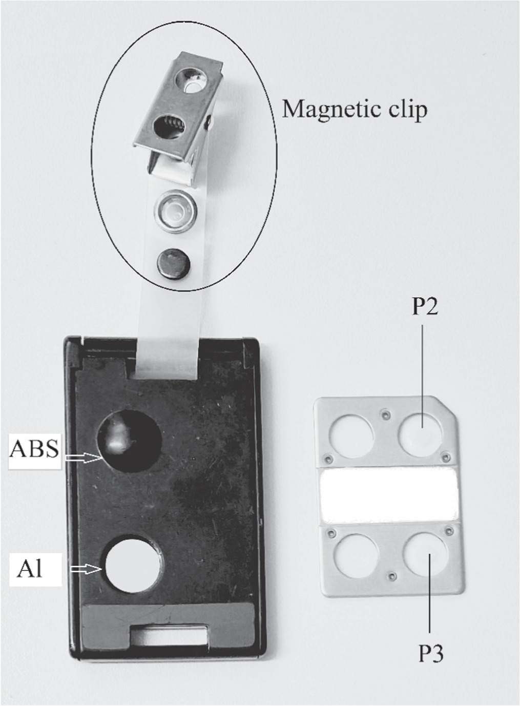
The Sahlgrenska University Hospital's personal dosimetry department has approval from the Swedish Radiation Safety Authority, responsible for the National Metrology Laboratory for ionizing radiation. The approval encompasses Hp(10) and Hp(0.07) personal dosimetry, utilizing TLD-100, the Thermo Scientific Harshaw Model 6600 Plus reader, and the dosimeter holder model 8814. In this system, TLD-100 elements (chips) are mounted between two Teflon (PTFE) sheets on an aluminum substrate, TLD Card, which is placed in a holder. The TLD material is LiF:Mg Ti and the holder is of tissue equivalent ABS material and protects the card from environmental damage and retains the filtration media. TLD Card can have places for four chips, but only two of them (positions 2 and 3) are used in this application. Total filtration in position 2 is 1000 mg cm−2 (combined ABS and PTFE) for measurement of Hp(10), while the total filtration in position 3 is 17 mg cm−2 (combined 0.06 mm aluminized Mylar and PTFE) for measurement of Hp(0.07). The TLD-Card and the card holder are shown in figure 1. The calibration and quality control of the personal dosimetry system is done regularly according to the corresponding standards and N–80 reference radiation quality is used [7, 8].
Figure 1. The card holder with ABS and Al filtration elements (left) and the TLD-card (right) used in personal dosimetry.
Download figure:
Standard image High-resolution imageThe dosimeters underwent assessment for MR-safety, revealing that the only magnetic component was the band containing a metal clip designed for dosimeter attachment to clothing (figure 1). For this study, the dosimeters were utilized without these bands. Additionally, a decision was made to substitute the bands with plastic counterparts for dosimeters designated for personnel working in the 3 T Signa PET/MRI, GE Healthcare, room.
Twenty-one personnel dosimeters were acquired and were divided into three groups: 9 were designated for measurements in the absence of a magnetic field (Room 1), another 9 for measurements in the presence of the magnetic field of the PET/MRI (Room 2), and the remaining 3 for background measurements in both Room 1 and Room 2, as well as the space between these two rooms.
A water-filled cylindrical phantom with dimensions 20 cm × 18 cm (D × H) was infused with 50 MBq of 18F-FDG. Subsequently, nine TLD-100 staff dosimeters were affixed to the surface of the phantom (figure 2), which was then placed in a room isolated from the PET/MRI room and devoid of any other radiation sources. The phantom remained in this room for 24 h. Background radiation levels in the room were measured on a separate day, also over a 24 h period. The entire process was replicated within the PET/MRI room, with the phantom positioned on the cradle where the patient's head is usually placed, and the magnetic field is about 0.2 T. The irradiated dosimeters, along with those used for background measurements, were subsequently submitted to the personal dosimetry department for dose reading.
Figure 2. The water-filled cylindrical 20 cm × 18 cm (D × H) phantom infused with 50 MBq of 18F-FDG and nine TLD-100 staff dosimeters affixed to its surface. In the setup, the side of the dosimeters typically affixed to personnel clothing faces outward on the cylinder, while the filter side is attached to the surface of the cylinder. Note that the band containing a metal clip designed for personal dosimeter attachment to clothing has been removed from dosimeters to make them MR-safe.
Download figure:
Standard image High-resolution imageThe personal dosimetry department reported the measurement results as Hp(10) and Hp(0.07), consistent with the usual reporting format for dosimeters used by staff in clinical practice. These measurement reports were utilized to evaluate the recorded dose disparities between dosimeters exposed in the presence of the magnetic field (on the PET/MRI cradle) and those exposed in its absence. The initial radioactivity of the phantom at the start of dosimeters irradiation was 46.9 ± 5% MBq and 46.3 ± 5% MBq in the presence and absence of the magnetic field, respectively. To account for this radioactivity difference, the registered dose values of dosimeters in the absence of a magnetic field were adjusted by multiplying them by the ratio 46.9/46.3. The statistical significance was evaluated using two-sided t-test with p-value<0.05.
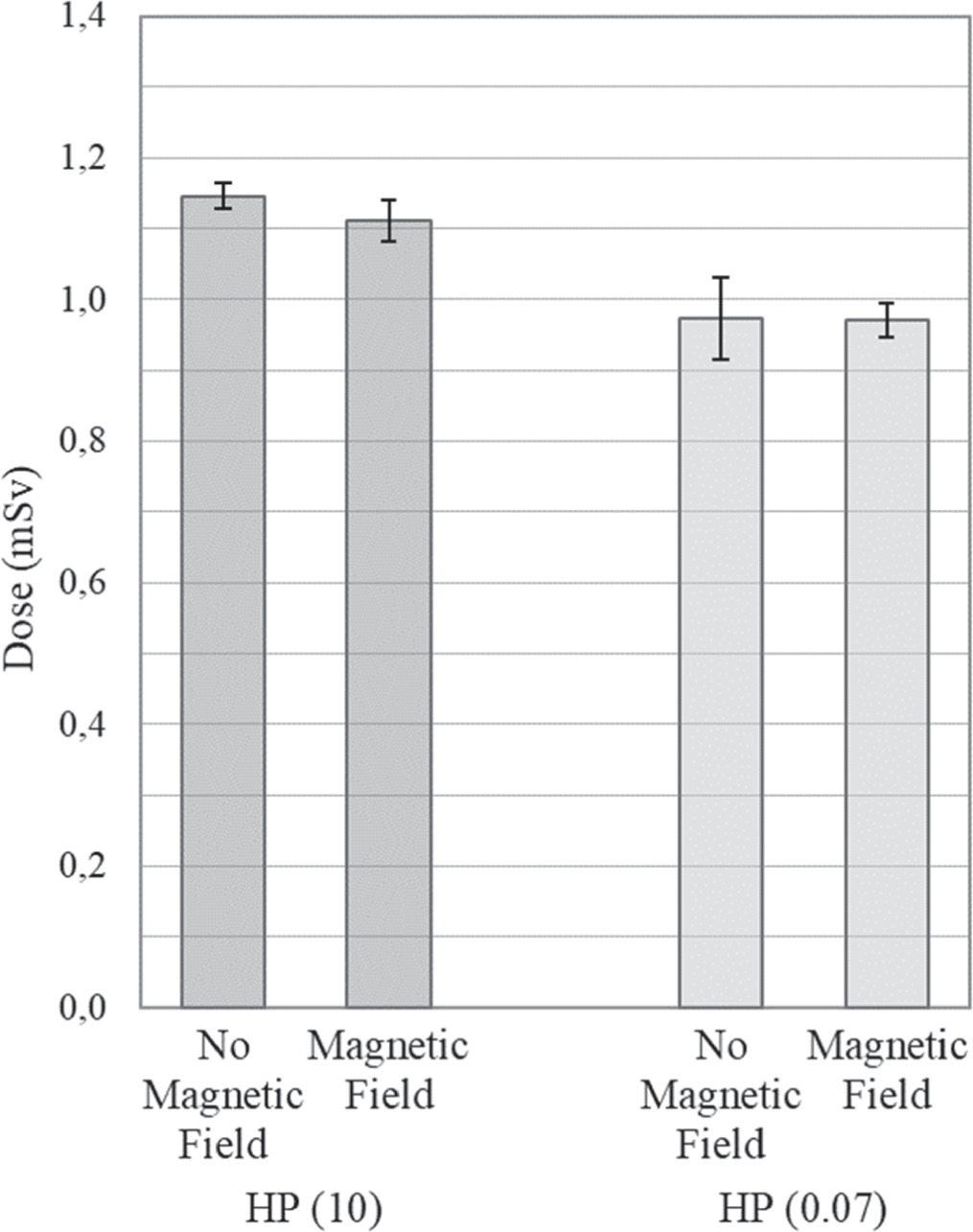
The measurement outcomes are presented in figure 3. The mean Hp(10) dose amounted to 1.15 mSv and 1.11 mSv for dosimeters irradiated in the absence and presence of a magnetic field, respectively. The standard deviation was 2.4% and 4.0% for dosimeters irradiated in the absence and presence of a magnetic field, respectively. The difference between two groups was not statistically significant (p>0.05). The mean Hp(0.07) dose amounted to 0.97 mSv regardless of presence or absence of a magnetic field.
Figure 3. TLD measurement results in the absence and presence of the magnetic field of a 3 T PET/MRI close to the bore where the patient's head is usually positioned, and the strength of the magnetic field is about 0.2 T. Background subtraction applied to all readings. The mean deep dose equivalent Hp(10) under magnetic field conditions showed a 3% reduction compared to the mean dose without the magnetic field, but the difference was not statistically significant (p>0.05). The mean skin dose equivalent Hp(0.07) was the same for irradiation of dosimeter in absence or presence of the magnetic field. Error bars show the 95% confidence interval.
Download figure:
Standard image High-resolution imageIt's important to mention that in this study, we utilize the Hp(10) and Hp(0.07) designations as provided by our personal dosimetry department. However, it's worth noting that these measurements do not equate or fully conform to the standard definition of Hp(X), as certain elements such as back-scattered dose from the wearer's body are absent in this particular setup. Nonetheless, our objective was to compare dosimetry results in the presence and absence of a magnetic field and not the absolute dose values, hence we use the designations reported by our personal dosimetry department.
There is a scarcity of publications specifically addressing the influence of magnetic fields on TLD measurements. Existing studies on this subject exhibit variations in TLD material, radiation type or energy, strength of magnetic field, and dose levels. Additionally, certain studies either omit information on the TLD material used or employ doses at the scale of radiation therapy, exceeding typical occupational doses for healthcare professionals [9–13].
Steinmann A et al investigated the TLD performance with a prescribed dose of 3 Gy to the maximum depth deposition under the presence of 1.5 T, 0.35 T, and 0 T magnetic field strengths in MRgRT systems. The effect of magnetic field on TLD was limited and not more than 0.6% in presence of 1.5 T [12].
Je J et al conducted a study examining the impact of a magnetic field on the glow curve of TLD-100. The TLDs were irradiated with a 100 kVp x-ray beam, and a subset was placed within the magnetic field generated by a neodymium magnet, with an intensity comparable to that of a 3 T MRI environment where personnel are involved in patient positioning. The magnetic field led to an approximately 8% reduction in the measured dose, specifically 2642 μGy compared to 2859 μGy, along with a 48% decrease in the glow peak at 145 °C, corresponding to the thermoluminescent peak of the low trap. Notably, the glow peak at 230 °C, representing the thermoluminescent peak of the deep trap, showed no significant impact [9]. The observed 8% decrease in dose in that study, in contrast to the more substantial 48% decrease in the glow peak at 145 °C, is likely attributed to the shorter height of the peak compared to the peak at 230 °C. The 145 °C peak contributes less to the measured dose compared to the 230 °C peak, which remained unaffected by the magnetic field.
Tchistiakova E et al confirmed that the influence of a magnetic field (within the 0.2 T line of a 3 T scanner) on Optically Stimulated Luminescence Dosimeters (OSLD) sensitivity is within acceptable radiation protection limits. They concluded that OSLD personal dosimeters are suitable for MRI-guided Radiotherapy (MRIgRT), providing a technical solution for clinical centers seeking MR-compatible radiation dosimeters. The modified OSLD dosimeter used in the MRI suite demonstrated negligible impact from the magnetic field, ensuring accurate readings. The study employed a low dose (∼1 mSv), more representative of typical radiation protection dosimetry [13]. Our result for TLD personal dosimetry is in line with their result for OSLD personal dosimetry.
One concern about the staff working with PET/MRI compared to those working with PET/CT is the longer time they need to spend close to patient. Soret M et al reported that, for an equal injected activity to patient, the radiation dose to technologists working with PET/MR was double that of PET/CT. The difference was associated with extended contact with injected patients during patient positioning with the PET/MR and MR coil placement, especially in whole-body studies [11]. However, electronic dosimeters were used in that study and the possible effect of the magnetic field of MRI on dosimeters was not included in the study.
Another interesting aspect of the possible effect of static magnetic field on TLDs is when the dosimeters are exposed to magnetic field before or after irradiation. In a study conducted by Mathis M et al, dosimeters underwent exposure to a 1.5 T magnetic field either concurrently or sequentially, both before and after irradiation. Their findings indicated that sequential exposure to the magnetic field, both pre- and post-irradiation, does not induce discernible alterations in the dosimetric characteristics of TLDs. In instances of simultaneous exposure to both the magnetic field and radiation, TLDs exhibited no significant impact from the magnetic field within the experimental uncertainty of 6%. This uncertainty is ascribed to various factors, including machine output, experiment setup, beam profile, dosimeter variance, and other pertinent considerations. It is crucial to emphasize that the dose of 200 cGy (2000 mSv) significantly exceeds the typical dose range in occupational dosimetry. While the specific type of TLD material employed in the publication was not mentioned, our own results within the dose ranges typical of occupational dosimetry align with their observations within therapeutic dose ranges [10].
The current study distinguishes itself through a unique methodology, exposing dosimeters to radiation on the surface of a water-filled cylindrical phantom containing PET-radioisotope. The phantom is positioned on the PET/MR cradle near the bore, where the strength of the static magnetic field is around 0.2 T, emulating real-world scenarios encountered by technologists in personal dosimetry in PET/MRI imaging. In this context, technologists actively engage in patient positioning, securing, and managing MR coils. The chosen TLD material is TLD-100 (LiF:Mg Ti), consistent with the standard dosimetry material employed in our hospital. Moreover, the dose level used in this study, i.e., 1 mSv, is relevant to occupational dosimetry for staff working with PET cameras [14]. Notably, there is no prior publication known to us that explores similar conditions.
The measurement results obtained from TLD-100 (LiF:Mg, Ti) dosimeters show no significant impact from the static magnetic field of the 3 T PET/MRI at the patient cradle, where the strength of magnetic field is around 0.2 T. The modified MR-safe dosimeters prove suitable for applications in personal dosimetry within a PET/MRI environment.
The author would like to thank Dieudonne Daba and Isabelle Nilsson from personal dosimetry and Linnéa Andersson, Evin Papalini, and Jesper Brovall from MR at Sahlgrenska university hospital, Sweden and Tobias Rosholm.
All data that support the findings of this study are included within the article (and any supplementary files).
This study was supported by grants 2022:420 and 2021:374 from King Gustav V Jubilee Clinic Cancer Research Foundation (Göteborg, Sweden).
Comments (0)