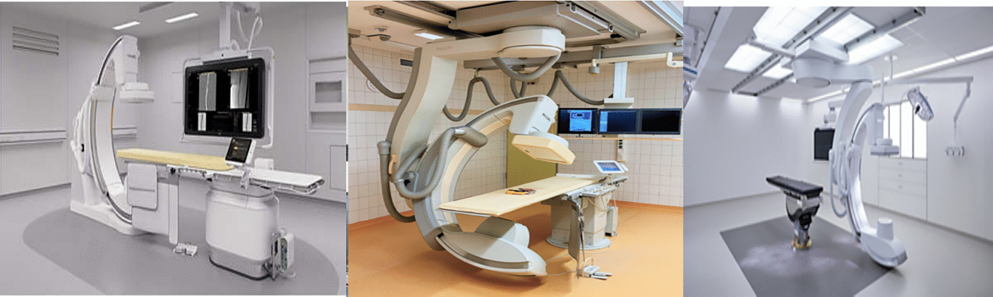
Exposure outside the area of the primary beam occurs from internal scatter. We demonstrated that using Pb Shielding in CT was statistically non-significant in minimising internal scatter. There was however a 2.0% reduction recorded in data with Pb Shielding, which was attributed to the absorption of scattered radiation from the head and gantry of the CT unit. Similarly, a study assessing the application of Pb Shielding for chest CT examination observed a reduction in the dose outside of the scan field, although the reduction was minimal [17]. Breast skin dose reductions were reported when Pb Shielding was employed for CT examinations of the brain, abdomen-pelvis, liver dynamic, lumbar spine, and neck. The reduction in breast skin dose with the use of Pb Shielding, due to its proximity to the exposed area and the absorption of scattered radiation from the head and gantry of the CT unit is widely recommended and reported in literature [19, 20].
This study demonstrated that internal scatter levels measured with Pb and without Pb Shielding was higher closest to the exposed area and decreased distant from it. Chung et al. [18] confirmed an inverse square law relation during CT when entrance surface doses were investigated. The entrance surface doses closest to the primary radiated area were greater than those in distant areas. The geometry of radiation is particularly important in determining the scatter exposure of an organ. It is essential to emphasise that internal scatter is the primary contributor to the radiation dose to radiosensitive tissues. The exposure to CT radiation from the primary beam is influenced by scatter radiation from the head and gantry, spreading to other secondary areas. The intensity of external scatter decreases as the distance from its source increases. Additionally, the larger the volume of tissue, the more internal scatter occurs. Most of the exposure outside the primary beam area comes from internal scatter. It is essential to note that external shielding does not reduce internal scatter. Internal scatter is a significant source of radiation exposure for sensitive tissues, particularly those deep within the body. Therefore, shielding is less effective when the scatter source is within a patient’s body.
The levels of internal scatter for the CT abdomen differ when Pb Shielding is applied, compared to when Pb shielding is absent. However, the abdomen contains a peritoneal cavity consisting of soft tissue organs, with only the spine providing a bony structure. Hsieh et al. [21] showed that the effective doses of kidney-ureter-bladder, intravenous urography and abdominal CT were 0.22, 1.51, and 8.21–9.27 mSv respectively where the CT yielded the highest effective dose. The chest, on the other hand, is filled with air due to the presence of lungs and has a thinner bony structure from the ribs. Therefore, there is minimal attenuation, and only a low radiation-absorbed dose is received. The brain has a small area of view and thinner cranial bones, requiring less dose for penetration.
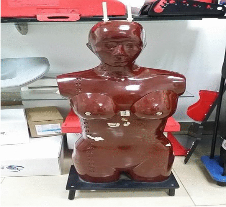
A limitation of our study was the use anthropomorphic phantom to study internal scatter, as it does not fully replicate the radiation scatter that can occur in actual human bodies with organic tissues. The phantom also simulates an average human size, but skinny and obese patients may yield different radiation dose estimates. Another limitation is the use of only 0.35 mm equivalent Pb and no other thicknesses. However, the authors believe that the internal scatter reduction and no difference with Pb and without Pb Shielding will still be seen with actual human tissue and other Pb equivalent thicknesses, hence the use of the RANDO phantom and 0.35 mm equivalent Pb was acceptable.


Comments (0)