
Remember me
Polymer bags are one of the most popular methods for collecting gaseous samples prior to offline analysis [1]. Sampling bags have been widely used in air quality assessment [2], environmental and agricultural research [3], exhaled breath analysis [4–7], and other areas [8–12]. Sampling bags are a relatively cheap alternative to other offline sampling methods, such as thermal desorption tubes or solid-phase microextraction fibers. Considering their universal use across many disciplines, in-depth research on sampling bags is necessary for accurate and repeatable analysis of offline gaseous samples.
Secondary electrospray ionization (SESI) is a soft ionization method employed in direct-infusion mass spectrometry, particularly beneficial in the context of breath analysis [13, 14], and also other fields where ambient ionization can be applied [15–17]. It exhibits very low limits of detection, down to the parts-per-trillion range, and enables the identification of even low-volatility species [17]. When combined with high-resolution mass spectrometry, it offers very high sensitivity and selectivity [18], and owing to the possibility of acquiring tandem mass spectrometry (MS) spectra, it enables reliable compound identification.
Proton transfer reaction MS (PTR-MS) is a well-established analytical method in volatile organic compound (VOC) research, employing hydronium (H3O+) reagent ions for the ionization of gaseous analytes. It has been applied to many fields where analysis of VOCs is used, e.g., analysis of breath [19–21], food science [22], and environmental research [23].
In this work, we present a comparison of storing gaseous samples in various polymer bag materials using SESI-MS at time points up to 48 h. We propose and examine a new material for the preparation of sampling bag materials, ethylene vinyl copolymer (EVOH), a barrier-like polymer mix commonly used to store aromas and chemicals [24]. We present the application of a simple, custom-made offline system for controlled sample introduction into the mass spectrometer. We demonstrate the effect of ultraviolet light on all polymer materials, simulating the process of accelerated aging. We also compare the most prevalent impurities observed in the bags, their initial levels, and their release rate after undergoing a cleaning procedure. We show an evaluation of the storage capabilities of five different bag materials using SESI-MS and compare our findings with another well-established VOC analysis technique, PTR-MS.
Although ample evidence on the stability of VOCs in different bag materials is available [4, 6, 25], until now most works made use of standard mixtures of VOCs introduced into sampling bags or examined only a single material in a specific context [4, 6, 7, 25, 26]. In this work, we use real exhaled breath as our sample matrix, stored and examined in five different materials, which considering the breath's complex composition, can largely influence the recovery of VOCs. Figure S18 (supporting information) illustrates the differences in composite spectra and m/z feature profiles between a gas standard sample, a sampling bag filled with breath, and a direct online exhaled breath sample. We also investigate the influence of ambient light on the humid samples stored in EVOH, Kynar, Nalophan, Teflon, and Tedlar. We examine the effect of sample humidity on both analytical methods. Finally, we make recommendations for offline sampling using polymer bags, specifically focusing on light exposure, an area previously under-emphasized in sampling bag usage.
2.1. Sampling bag preparationExhaled breath samples were collected into sampling bags made of five different materials: Nalophan (PET, MediSense, Netherlands, measured film thickness 20 μm), Tedlar (PVF, Sigma-Aldrich, Switzerland, measured film thickness 60 μm, polypropylene push/pull lock valve), Teflon (PTFE, Scentroid, Canada, measured film thickness 50 μm, PTFE lock valve), EVOH (ethylene vinyl alcohol copolymer, GreenPak, China, film thickness 110 μm) and Kynar (PVDF, MediSense, Netherlands, measured film thickness 80 μm, PTFE lock valve). The nominal volume of every bag was 5 l. Both EVOH and Nalophan were purchased as film rolls, and the sampling bags were produced in-house using a previously described method [6]. A PFA plug valve (Swagelok, Switzerland) was fit to ensure proper opening and closing of the bags. A visualization of all the bags can be found in the supporting information (figure S1).
2.2. Preconditioning of sampling bagsTo minimize plasticizer impurities emitted from the bags, all sampling bags underwent a cleaning procedure before and after use, as recommended previously [4]. The procedure involved flushing the bags with inert nitrogen gas, filling them to approximately 90% capacity (manufacturer's recommendation), and then fully deflating them using a membrane air pump (KNF, Switzerland). For each bag, this procedure was repeated five times (figure S2). The relative concentration levels of impurities recorded before and after the procedure can be found in the supporting information (figures S3–S7). During bag handling, it was crucial to ensure as little mechanical stress on the bag as possible, including during filling, as any scratches on the bag materials were prone to tears and film damage.
2.3. Sample collectionSamples were collected into 5-L bags of the respective materials from a single participant, who was asked to abstain from consuming food and brushing teeth prior to the sample collection. Exhaled breath was collected by breathing into the bag with multiple exhalations until the bag reached approximately 90% of its filling volume. The samples were subsequently analyzed at eight different time points after collection: 0 (immediately after exhaling into the bag), 30 min, 1 h, 2 h, 4 h, 8 h, 24 h, and 48 h. These eight time points constituted one technical replicate, which was then repeated for experiments conducted using both the SESI and PTR setups, recorded on separate days. In total, two replicates were measured using SESI-HRMS, and three replicates were measured using PTR-MS.
2.4. Offline system for sample introductionThis study utilized an offline system (figure 1) developed for the collection and analysis of breath samples stored in bags. The system consists of a polypropylene storage box, into which the sampling bag is first inserted and connected, either directly with an attached fitting or via the PFA plug valve (Swagelok, Switzerland). The box is then closed and pressurized using a diaphragm air pump (KNF, Switzerland). Once the desired overpressure (around 5 mbar) is reached, the sample is released by opening the outer PFA plug valve. The system was kept at ambient temperature (25 °C) during the time of measurement.
Figure 1. Top: offline system used for introducing breath samples from bags into the mass spectrometer. Bottom: schematic drawing of all components of the offline system.
Download figure:
Standard image High-resolution image 2.5. Accelerated aging by long-term exposure to ultraviolet lightFor the irradiation of sampling bags, a commercial laboratory UV lamp was used (Vilber, France) with a center wavelength of 365 nm, and irradiance of 2.3 mW cm−2.
2.6. Data acquisition by SESI-HRMSData was acquired using a commercial SESI source (Fossil Ion Tech, Spain) coupled to a Q Exactive Plus Orbitrap high-resolution mass spectrometer (ThermoFisher Scientific, Germany). During measurements, a stable vacuum pressure of 8 × 10⁻¹¹ mbar was maintained. The SESI sampling line and the ionization chamber were heated at 130 °C and 90 °C, respectively. For each measurement, both positive and negative ionization modes were recorded. The electrospray was generated using a 0.1% aqueous (LC-MS grade water, ThermoFisher Scientific) formic acid solution (99.5% formic acid, VWR Chemicals, Switzerland), which passed through a nano-electrospray capillary (outer diameter = 365 μm, inner diameter = 20 μm, Fossil Ion Tech) under a positive pressure of 800 mbar. Sheath gas was kept at 15 a.u., and auxiliary gas at 2 a.u. The electrospray voltage was set to ±3.5 kV, depending on the polarity mode. The MS inlet capillary was heated to 250 °C. Automatic gain control in the Orbitrap C-trap was set to 106 and the maximum injection time to 500 ms. The mass resolution was set to 140 000. The S-lens RF level was set to 50 and the voltage of the central electrode was 5 kV. The C-trap used a drive voltage of 1 kV. For each time point, the signal was acquired for a duration of 30 s in both polarity modes. After 15 full MS scans covering the mass range from 50 Da to 500 Da (approximately 8 s of acquisition), which constituted the background signal, the outer PFA plug valve was opened, and the resulting ion current was recorded for the rest of the 30 s window. For assigning compounds to m/z values in the data, tandem mass spectra were acquired and analyzed with SIRIUS [27–33]. The settings for molecular formula assignment and chemical classification were left as default. Only [M + H]+ or [M−H]− adducts were chosen, and compounds were referenced from the Human Metabolome Database (HMDB [34–38]).
2.7. Data acquisition by PTR-MSPTR-MS data was acquired using a PTR-ToF-MS-1000 (Ionicon, Austria) mass spectrometer connected to the offline sample introduction system. The key operating principles of the PTR-MS are described in detail elsewhere [39]. Briefly, H3O+ primary ions were generated in a hollow cathode and supplied by water vapor from a reservoir of pure water (Milli-Q A10, Merck, Switzerland). The ion source was operated at an ion source current of 3.5 mA, an ion source voltage of 145.0 V, a source-out voltage of 78.6 V, and a source valve opening of 55%. The drift tube was operated at a voltage of 600 V, a pressure of 2.3 mbar, and a constant temperature of 60 °C. The protonated VOCs were then analyzed using a time-of-flight mass analyzer. After data acquisition, mass calibration was performed at m/z 21.02 and 203.94 for every sample. Analyte concentrations were determined by measuring the counts per second at the m/z values listed in table S2. All analyte concentrations were compared to calibration curves measured routinely using a mixing set-up [40] with certified gas standards (PanGas, Switzerland).
2.8. Data processingAll acquired scans for SESI-HRMS were saved in the ThermoFisher Scientific. RAW file format. They were subsequently converted into the mzML open file format using MSConvert (ProteoWizard package) [41]. mzML files were then parsed using custom-made Python (v3.7.4) scripts, making use of the Pyteomics library [42, 43]. For PTR-MS, the scans were saved in the HDF5 open file format, and then parsed and processed using a custom Python script. The entirety of the codebase used for the data analysis is included in a curated data archive alongside the raw data.
The processing consisted of resampling all recorded individual MS scans onto a uniformly spaced m/z axis from 50 to 500 Da, with a step size of 10−5 Da. All scans were then averaged over all the measurements in the same polarity mode, resulting in composite mass spectra, for both positive and negative ion modes. Potential metabolic features were then determined by detecting peaks in the composite mass spectra that had an intensity above a threshold of 1000 cps. The peaks were then traced over time in every measurement and their interpolated intensities were integrated within the respective peak's m/z range. The time traces of each peak were tested for positive correlation with the total ion current (TIC) using Pearson's r correlation coefficient. This step was omitted for PTR-MS data, as no TIC was extracted from the HDF5 files. The mean intensity of all accepted peaks (features) was then calculated and stored in an n × m intensity matrix, where n represents the number of measurements and m is the number of features. Within the intensity matrix, further feature filtering criteria were applied. If a feature did not appear in at least 80% of the given set of measurements, it was excluded from further analysis.
2.9. Postprocessing and statistical analysisThe intensity matrices obtained from the preprocessing step were utilized for the analysis and differentiation of bag materials. To assess the compound storage capabilities, known human breath markers (table S2) were traced. Their mean intensities at each time point were normalized to the first time point (time point '0', immediately after acquisition) and averaged across all technical replicates (figure 2). Concentration levels of emitted impurities were determined by analyzing the composite spectra (figure 6) of bags filled with medical air and comparing their levels up to 48 h after the conditioning procedure (supporting information, figures S8–S12). To visualize the differentiation between bag materials across all measurements, multiclass linear discriminant analysis (LDA) was performed. The samples were grouped into five categorical variable classes (one for each material) with equal probability for each class and labeled accordingly. The feature number was limited to the features found in all bag materials in at least 80% of all measurements. Subsequently, the LDA model was fitted, and the data were transformed by projecting into the two most feature discriminative directions (figure S13). All other statistical analyses and graphing were performed using GraphPad Prism software, version 10.1.0.
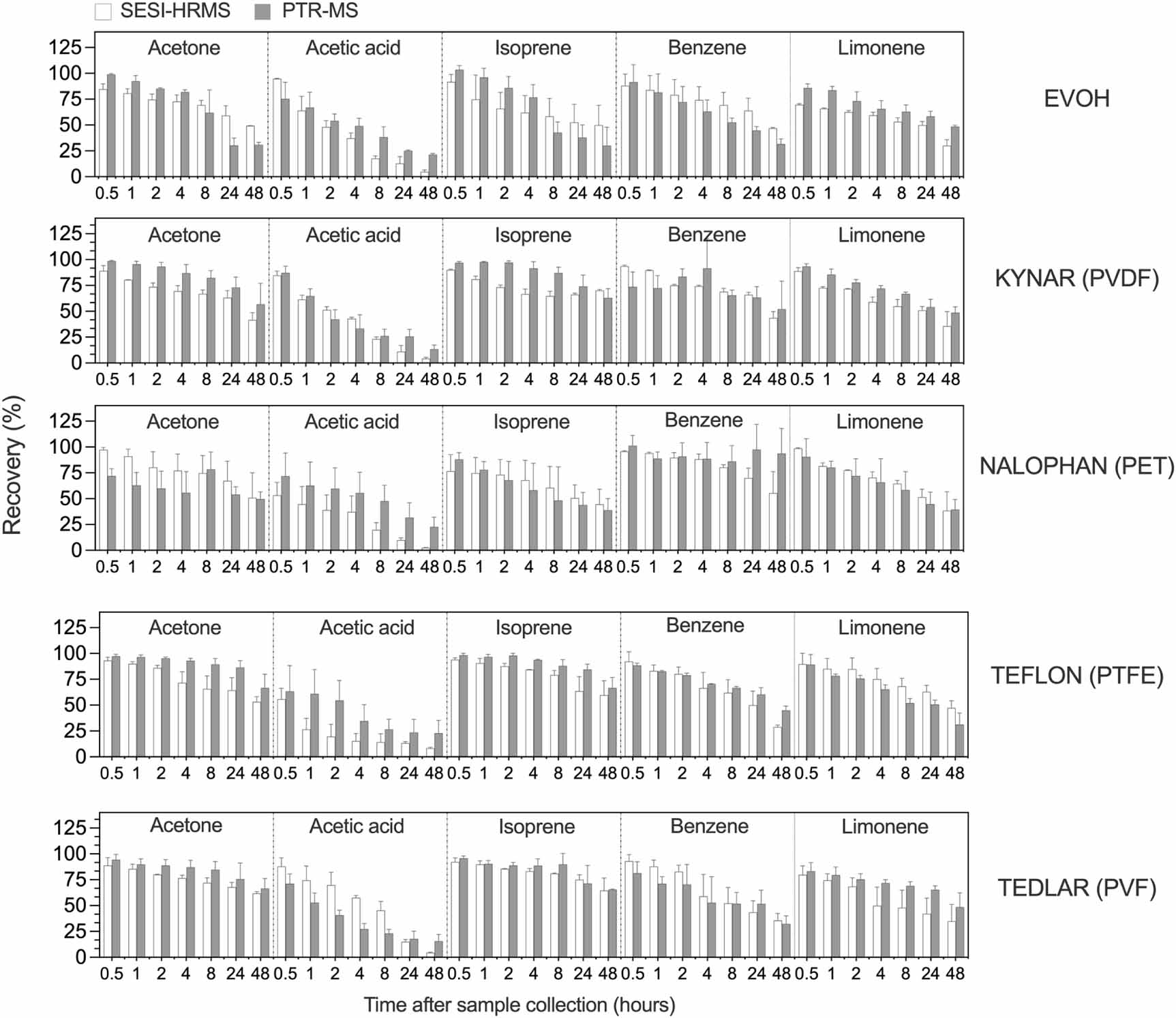
Figure 2. Comparison of targeted breath features (acetone, acetic acid, isoprene, benzene, and limonene) in breath samples stored in five different bag materials, as measured by SESI-HRMS (light bars) and PTR-MS (dark bars). Each bar represents the mean and standard deviation of two (SESI-HRMS) to three (PTR-MS) technical replicates. Consecutive bars represent time points ranging from 0 (measured immediately after sample collection) to 48 h. All intensities for a given analyte were normalized to the initial time point (0).
Download figure:
Standard image High-resolution image 3.1. Recovery of selected VOCs in polymer sampling bagsPrevious works on Tedlar (PVF), Teflon (PTFE), Kynar (PVDF), and Nalophan (PET) have reported varying results in the context of VOC recovery, from good recovery over 48 h [4, 6] to rapid losses, sometimes down to undetectable levels of the analyte [3, 44]. Here we present the relative levels of selected breath constituents coming from samples of real exhaled breath in these bags and in EVOH (new material), analyzed both by SESI-HRMS and PTR-MS.
In our study, we focused on acetone (m/z = 59.0493 [M + H]+), acetic acid (m/z = 61.0285 [M + H]+, isoprene (m/z = 69.0699 [M + H]+), benzene (m/z = 79.0543 [M + H]+), and limonene (m/z = 137.1325 [M + H]+), as these compounds belong to different chemical classes, cover a wide range of concentrations in exhaled breath, and are detectable both in PTR-MS, and SESI-HRMS. The relative abundances of pseudomolecular ions for each of these compounds were traced over 48 h and can be seen in figure 2. Detailed percentage abundances at each time point and their RSD values can be found in tables S3 through S12 (supporting information). To confirm the identities of the compounds in question, we verified the m/z values with tandem MS-MS data-dependent experiments and cross-referenced our findings with the HMDB metabolomic database (table S13 supporting information).
For all bag materials, we observed a steady decline in the relative intensity of all five traced features over 48 h. In our measurements the retrievable concentration of acetic acid decreased most rapidly, followed by benzene and limonene, and to a lesser extent, isoprene, and acetone. In all materials and within all replicates, the retrievable concentration of acetic acid dropped to less than 30% of its initial value after 48 h, as per both SESI-HRMS and PTR-MS. We attributed this to acetic acid's high polarity, resulting in greater affinity to surfaces and likely adsorption on the bag wall.
Overall, we observed large variability between replicates, especially towards the later time points. The new material, EVOH, performed on par with its price-matched alternative, Nalophan. Both Nalophan and EVOH bags were produced in-house.
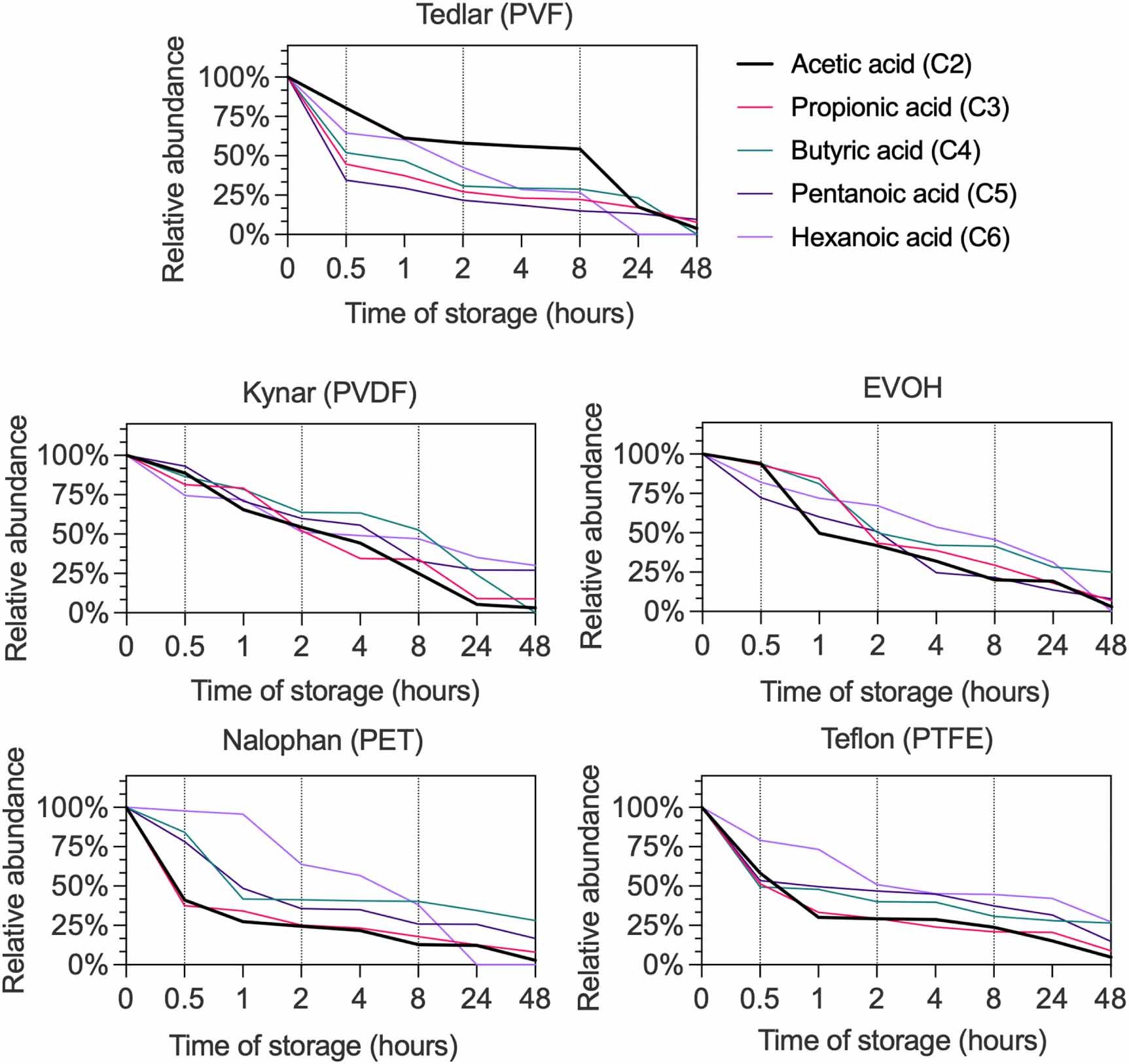
3.2. Investigation of volatile short-chain fatty acids (SCFAs) in gas sampling bagsFollowing the strong loss of acetic acid within all bags after 48 h, we asked the question whether the same occurs for other SCFA compounds (figure 3). SCFAs are essential metabolites produced by gut microbiota during fermentation and play a significant role in many biological processes [15, 45].
Figure 3. Time traces of relative abundance of short-chain fatty acids from C2 (acetic acid) up to C6 (hexanoic acid) found in samples of exhaled breath of a single subject, stored over 48 h in all sampling bags, recorded with SESI-HRMS. Every line represents the mean of two technical replicates.
Download figure:
Standard image High-resolution imageWe observed a rapid decline in the relative abundance of various SCFAs across all bag materials. After 8 h, the concentration of most SCFAs reached approximately 50% of their initial values. Interestingly, we found acetic acid to be a reliable indicator for predicting the overall trend in the intensity of other SCFAs.
3.3. Characterization of EVOH as a new sampling bag materialImpurity release from sampling bag films is a known issue among sampling bag users [4, 6, 44]. This, combined with a gradual loss of retrievability of the analyte molecules, leads to degradation of sample integrity over time. We have found that EVOH is also subject to these problems, maintaining sample integrity on par with other bag materials after 48 h of storage. The overview of the sample contents can be visualized with an intensity matrix (figure 4) of all significant features (i.e., greater than 1000 cps and correlating with the TIC).
Figure 4. (a) Intensity matrix showing changes in absolute intensity (cps) of m/z features found in EVOH sampling bags filled with breath over 48 h. (b) Average mass spectra (positive ion mode) of EVOH immediately (0 h) and 48 h after sampling. The 0 h spectra were nudged upwards and slightly to the right of the 48 h spectra to better distinguish peak intensity.
Download figure:
Standard image High-resolution imageFigure 4 provides an overview of the intensity matrices (heat maps of feature intensity changing over time) in EVOH recorded both by PTR-MS and SESI-HRMS. The most intense features coming from the stored breath sample are acetone and isoprene, whereas the most intense impurities are N,N-dimethylacetamide and phenol.
Figure 4(b) presents the associated average mass spectra at the beginning (0 h) and at the end (48 h) of sample storage. A clear temporal evolution of both sample contents and impurity release can be seen. In PTR-MS the changes are very well visible because considerably fewer m/z peaks are detected, resulting in a clearer spectrum. In SESI-HRMS the spectrum is much more densely populated, especially in the mid-mass region (100–300 Da). The individual rows in the intensity matrix for SESI-HRMS are also narrower and sharper because the mass resolution is greater (140 000 for the SESI-Q Exactive Plus as opposed to ca. 1000 for the PTR-ToF-MS).
3.4. Influence of light exposure on sampling bag materialsHumid samples stored in polymer materials have been suggested to be subject to reactive chemistry inside the bag, especially when exposed to light [25]. We chose to investigate this phenomenon by performing an accelerated aging procedure on bags filled with exhaled breath. For this purpose, we employed two sets of bags: a negative control ('–') set stored in the absence of light in an aluminum-foil-covered enclosure, and a positive test set ('+'), constantly irradiated with ultraviolet light at 365 nm over 48 h. Figure 5 presents the influence of long-term exposure to light simulated by ultraviolet radiation.
Figure 5. Intensity matrices showing square-root transformed intensity changes of features found within EVOH, Kynar (PVDF), Nalophan (PET), Teflon (PTFE), and Tedlar (PVF) sampling bags filled with exhaled breath and measured (a) immediately and (b) after 48 h of storage. '–': no UV exposure, '+': UV exposure (365 nm). (c) Average mass spectra representing a humid sample (exhaled breath) stored in a Nalophan (PET) bag after 48 h of little to no exposure to ambient light ('–'), and UV irradiation ('+').
Download figure:
Standard image High-resolution imageWe found significant bleaching of several features in Nalophan (PET), with lesser impact visible in EVOH, Kynar (PVDF), and Teflon (PTFE), and little to no impact in Tedlar (PVF). Modest changes in intensity matrices of samples stored in materials exposed to and protected from UV light (figure 5(a)) indicate that EVOH, Kynar (PVDF) and PTFE are relatively resistant to light. Significant bleaching was observed for Tedlar after 48 h, though it occurred in both the UV-treated and non-treated samples. However, for Nalophan we observed significant differences between the UV-irradiated and control samples after 48 h, with strong bleaching in several mass regions. Bleaching in the low- (50–90 m/z) and mid-mass regions (90–180 m/z) can be seen for Nalophan (PET).
Upon further inspection, we found some more intense features (e.g. 114 m/z, figure 5(c)) to strongly decrease in intensity while other, less intense features, became more prominent. Further investigation is required to determine if this is a result of the decay or fragmentation of heavier species, or actual chemical reaction products.
3.5. Impact of humidity on sampling bag analysis by PTR-MS and SESI-HRMSHumidity is an important consideration when examining the recovery of VOCs [4, 25]. When analyzing breath samples, high initial relative humidity content of the sample is unavoidable, as breath exhibits nearly 100% humidity (at 32 °C–34 °C). We investigated the influence of humidity on breath samples analyzed by PTR-MS and SESI-HRMS when the samples were rapidly introduced in either dry (<5% RH, relative humidity) or humid (RH > 90%) carrier gas airflow. Previous studies have found either a limited impact of breath humidity levels on the recovery of most sample constituents [6, 11, 26], or higher losses for species heavier than 90 Da [4]. However, humidity is also crucial in the context of the analytical technique. In the case of PTR-MS, the recovery of benzene and toluene, and to some extent isoprene, has been shown to depend on humidity [46]. Therefore, we decided to investigate this effect in the context of sampling bags by combining our sample introduction system with a flow of either dry (<5% RH, relative humidity) or humid (>90% RH) synthetic air (supporting information, figures S14 and S15).
In PTR-MS we found little difference in the distribution of intensity over time between the dry and humid conditions for most analytes. Under humid conditions, we observed, on average, higher relative concentrations of acetic acid in Teflon (PTFE) and Tedlar (PVF) bags, although the difference was not statistically significant (p > 0.01, Tukey's multiple comparisons test, GraphPad Prism 10).
We found much greater variation within SESI-HRMS, especially apparent for acetic acid and benzene. Although not significant due to the large standard deviation, the mean acetic acid recovery within all bags was lower under humid conditions as compared to dry conditions, indicating the influence of humidity on our measurements.
3.6. Differences and similarities between bag materialsTo complement the targeted analyte tracing, we employed an untargeted dimensionality reduction approach to differentiate between bag materials (supporting information, figure S13). Using multiclass LDA, we clustered all 120 measurements (24 per material) recorded with SESI-HRMS based on their feature intensity (438 features). We found the clusters of Kynar and Tedlar to overlap, indicating that the two materials performed similarly in our measurements. The samples stored in EVOH, Teflon, and Nalophan were all successfully separated, indicating fewer similarities between these materials.
3.7. Overview of impuritiesA 5-liter bag of each material was filled with medical air up to 90% capacity and subsequently analyzed over 48 h. For this analysis, the background signal (offline system filled with medical air, without the bag connected) was subtracted from each spectrum. Composite spectra (figure 6) were analyzed to identify peaks emerging during the measurement period and were traced over time. Spectra for all bag materials can be accessed in supporting information (figures S3–S7).
Figure 6. A composite mass spectrum of a Tedlar (PVF) bag, filled with medical air and analyzed over 48 h for release of impurities.
Download figure:
Standard image High-resolution imageThe most abundant ion detected within all bags was identified as N,N-dimethyl acetamide (DMAc, m/z = 88.0757 [M + H]+), which originates from the manufacturing process of bag films, such as Tedlar [26]. Additionally, we found high concentrations of phenol within Tedlar bags, in concert with a distinct smell of phenol that could be detected during the cleaning procedure. In Kynar (PVDF) bags, we also found a prominent acetone peak that was unrelated to the stored breath samples. Acetone has previously been identified as an impurity in Cali-5-Bond (not used in this work) [6].
4.1. Retention of selected analytesThe recovery of acetone, acetic acid, isoprene, benzene, and limonene decayed at a faster rate when compared to some previous studies examining the same or chemically similar analytes [4–6, 25], while others have reported higher losses, e.g., in Nalophan (PET) [9], Teflon (PTFE) and Tedlar (PVF) [3, 44, 47]. However, previous research was largely based on either dry or humid gas standard mixtures, with varying concentrations, and not real exhaled breath samples. Breath is a very complex sample matrix with multiple constituents [48] and techniques such as SESI-HRMS and PTR-MS enable its analysis without any prior sample separation or preconcentration. This is crucial in the context of practical applications (e.g., in the hospital setting) where ease of handling and affordability are a priority, and where polymer bags are sometimes used for sampling [7].
Although the mean relative intensity was significantly lower after 48 h for all the traced analytes, we observed large standard deviations for both analytical methods. This can stem from the fact that all replicates were recorded on separate days or reflect changes in the metabolic composition of the subject's exhaled breath on different days. The gradual loss of sample content in polymer bags is very well known, with chemically different analytes exhibiting varied recovery under specific analytical conditions. Our findings confirm previously reported losses, especially in the context of SCFAs, previously examined as a gas standard mixture [47].
Despite normalizing every measurement to the first time point for both methods, we did not see perfect agreement between SESI-HRMS and PTR-MS. This can result from slight differences in our experimental setups for both the methods (such as differing lengths of the tubing leading to the mass spectrometer) or differences between individual batches of bags from manufacturers (Kynar, Teflon, Tedlar) and those prepared by hand (Nalophan and EVOH).
One key difference might also come from the flow at which samples were introduced into the PTR-MS and SESI-HRMS ion sources. Within the PTR-ToF-1000 the sample content is constantly sucked in through the sampling tube at a rate of about 80 ml min−1 on top of the 5-mbar overpressure provided using our portable sample introduction system. For SESI-HRMS, there is significant backpressure of pure nitrogen used for purging the ion source, that needs to be overcome before introducing the sample into the mass spectrometer. Despite our efforts to minimize the significance of this issue, it might be that the flows have not been identical at each analysis time point, resulting in slightly different amounts of the bag content being ejected. This, in turn, would result in differing surface-to-volume ratios which can influence the recovery of VOCs from bags, as was shown previously [6].
4.2. Long-term storage of SCFAsWe found acetic acid to be a good predictor of the behavior of other SCFAs when traced over time in EVOH, Kynar (PVDF) (PVDF), Nalophan (PET), Teflon (PTFE), and Tedlar (PVF). The rapid drop in the concentration of those compounds is also in concert with previous works on the matter in Tedlar (PVF), Teflon (PTFE), and Nalophan (PET) bags [3, 8, 47]. Therefore, we advise against storing SCFAs in polymer bags, unless the sampling is done for purely qualitative purposes.
4.3. Evaluation of EVOH as a sampling bag candidateTo the authors' knowledge, there is no existing literature on EVOH bags being used for breath sampling. Nonetheless, the EVOH film itself has been proven to have barrier-like properties [24]. It exhibits superior preservation of light molecular weight gasses (such as carbon dioxide, nitrogen, and oxygen) compared to polyethylene terephthalate (PET) and polypropylene (PP) for up to 28 d [49]. Based on our results, the performance of EVOH for breath VOC storage is comparable to other bags and could offer a good alternative to Nalophan with a similar price while offering resistance to light exposure.
4.4. Impact of light on sampling bag storagePrevious studies on light influence on sample bag content have mostly focused on volatile sulfur compounds (VSCs) [5, 11, 50]. Two of the studies found no effect on the recovery of VSCs, and one found significant losses concerning light and humidity. We found that light played a significant role in feature intensity loss in Nalophan (PET), with little impact on other bag materials. This result supports the findings of Le et al. (2015) but may be exaggerated by the fact that Nalophan bags in our study had a relatively lower film thickness than other materials (20 μm compared to 50–110 μm for other bags).
4.5. Influence of humidity on the analysis by PTR-MS and SESI-HRMSOur measurements showed a limited influence of humidity introduced into the mass spectrometer on the recovery of acetone, isoprene, benzene, and limonene, with the greatest difference observable for acetic acid. This can partly explain the anomalous behavior of acetic acid during online measurements, where we found a lower average abundance of acetic acid as opposed to the bags immediately after filling (supporting information, figures S16 and S17). It also coincides with its losses during storage experiments, which may be exacerbated by high humidity immediately after filling and its rapid drop down to ambient humidity levels during subsequent hours [4, 6, 25]. Although most works did not find a significant effect of humidity on sample stability [6, 11, 26], Mochalski et al (2013) found higher losses for heavier species when analyzing humid mixtures. In our case, humidity was an inherent part of the sample matrix, and sample losses resulting from changes in the adsorption properties of analytes as humidity in the bag decreased cannot be excluded. Nonetheless, our results suggest that humidity seems to have no conclusive influence on the analytical properties of either PTR-MS or SESI-HRMS.
4.6. Impurity contentThe most intense impurities detected in sampling bags mainly originate from the manufacturing process and have been identified as N,N-dimethylacetamide (present in all bags), phenol (found most abundantly in Tedlar bags), and acetone (found in Kynar bags). Their concentrations vary among materials but are especially pronounced for Tedlar. As stated previously, Tedlar bags are known for their high background [25, 44]. This aspect needs to be considered by users operating with analytical methods that are sensitive to impurities.
4.7. Other considerationsAside from the characteristics described above, there are other factors influencing the selection of material for a sampling bag. One obvious consideration is the price. Of the popular polymer bags, Tedlar is often the most expensive, followed by Kynar and Teflon. Both the Nalophan and EVOH materials are cheaper, as they are usually sold in film rolls. When purchasing the bag material in this form, sampling bags need to be prepared in-house. Since they require additional fittings and valves, the expenses may rise considerably. Ready-to-use bags, although more expensive, usually come with a fitting attached.
Another criterion to consider is the ease of handling when using a particular bag material for sampling. We found the thin Nalophan film to be extremely fragile during sampling, often breaking and puncturing whenever there was a scratch on the bag film. This has also been reported before by other sampling bag users [6, 25].
Additionally, it is important to consider the ability to clean a bag, and therefore reuse it. For this purpose, researchers have suggested several alternative protocols, including bake-outs, constant heating, and flushing with inert high-purity gases [4–6, 25]. It is worth noting that, contrary to Nalophan, we did not observe any visible scratches on the surface of the EVOH film even after repeated handling, filling, and emptying.
This work presents a thorough comparison of storing humid samples (exhaled breath) in polymer bags of various materials under different storage conditions. We surveyed the recovery of common breath volatiles, found a correlation between acetic acid and other SCFAs, examined the influence of light and humidity on VOC recovery, and provided insight into the most abundant impurities found in all materials.
We showed a new material, EVOH, as a potential affordable alternative to commonly used materials. We determined its capability to retain common VOCs under both dry and humid conditions, and that it is relatively resistant to light, especially when compared to Nalophan, an equally cost-effective alternative.
We presented an accelerated-aging procedure and determined that for most bag materials exposure to light has little influence over sample integrity, except for Nalophan, which we recommend shielding from ambient light as much as possible if it is used for long-term storage of humid gaseous samples.
We also advise against using gas sampling bags made of Tedlar, Teflon, Nalophan, Kynar, or EVOH when sampling SCFAs. The concentrations of acetic acid obtained using SESI-HRMS and PTR-MS showed a rapid decrease, with less than 30% of the initial compound concentration retrievable after 48 h. We further confirmed that the same trend applied to other SCFAs. Thus, when interested in this specific group of metabolites analyzed in exhaled breath, it may be appropriate to consider choosing alternative sampling methods or performing real-time analysis.
We thank Christian Marro and the Departmental Mechanical Workshop for their involvement in constructing the offline system used in this work. We also thank Cedric Wüthrich and Timon Käser for discussions and help with instrumentation. A.T.G. and S.H. acknowledge financial support by Vontobel-Stiftung (1413/2022) and the Swiss State Secretariat for Education, Research and Innovation (SERI) under contract number MB22.00041 (ERC-STG-21 'HEALTHSENSE').
This study was performed in accordance with the Nuremberg Code. Ethics approval for this human study was waived by ETH Zurich Research Ethics and Integrity. All adult participants provided written informed consent to participate in this study.
Comments (0)