
Remember me


The study design was approved in accordance with the Declaration of Helsinki by the University of Health Sciences Gülhane Scientific Research Ethics Committee (no: 2024-328). The sample size was calculated based on a power analysis using G*Power software 3.1.2 (Universitat, Düsseldorf, Germany) with an alpha error probability of 0.05 and a power of 80% (effect size = 0.25) concerning a recent study of similar design [17]. The power analysis showed that a minimum of 15 samples per group and 75 samples were statistically necessary. For all these reasons, 75 bovine mucosa fragments, and 15 single-rooted, single-canal human mandibular premolar teeth with no internal or external resorption, no coronal caries, and restorations, no cracks and fractures, and no previous root canal treatment, which were extracted for periodontal reasons independently of the study, were included in the study. Since a non-destructive method was to be used, the same 15 teeth were used in all experimental groups, following a recent study of a similar design [17], and the obstacle of anatomical variables was avoided.
Periapical radiographs from the buccolingual and mesiodistal angles confirmed that the teeth met the inclusion criteria, had a single root and canal, and were free of internal/external resorption. The included teeth were soaked in 5.25% sodium hypochlorite (NaOCl) (Cerkamed, Cerkamed Company, Stalowa Wola, Poland) for two days to dissolve organic tissue debris. The tissue residues on the teeth were removed with a periodontal curette. The teeth were stored in 0.1% thymol solution until used in the study.
Access cavities were prepared using a high-speed rotary instrument and a diamond bur. After ensuring apical patency with a #10 K-file (Perfect, Shenzhen, China), the working length was determined to be 1 mm short of the apical foramen. At the specified working length, root canals were prepared to X3 using ProTaper Next (Dentsply Maillefer, Ballaigues, Switzerland). All files were utilized at the torque and rpm values specified by the manufacturer. Following each file change, the root canals were irrigated for 30 s with 2 mL of 2% NaOCl (Cerkamed) using a 30G side-vented needle (Ultradent, South Jordan, UT, USA). To standardize the crown and root lengths, the teeth were marked 5 mm coronally and 11 mm apically from the cementoenamel junction and sectioned at these reference points using diamond disc (Bredent, Senden, Germany), resulting in all samples being standardized to 5 ± 1 mm crown length and 11 ± 1 mm root length. In each sample, an artificial pulp chamber, the reservoir area for irrigation solutions, was prepared in the coronal 5 ± 1 mm section of the canal using a diamond bur [14]. The apical opening was designed to be 1.5 mm in size to simulate an immature apex [18]. For this purpose, Gates Glidden (VDW, Munich, Germany) burs from #1 to #6 (1.5 mm) were used. The root canals were irrigated with 2% NaOCl (Cerkamed), 17% EDTA (Cerkamed), and distilled water, respectively.
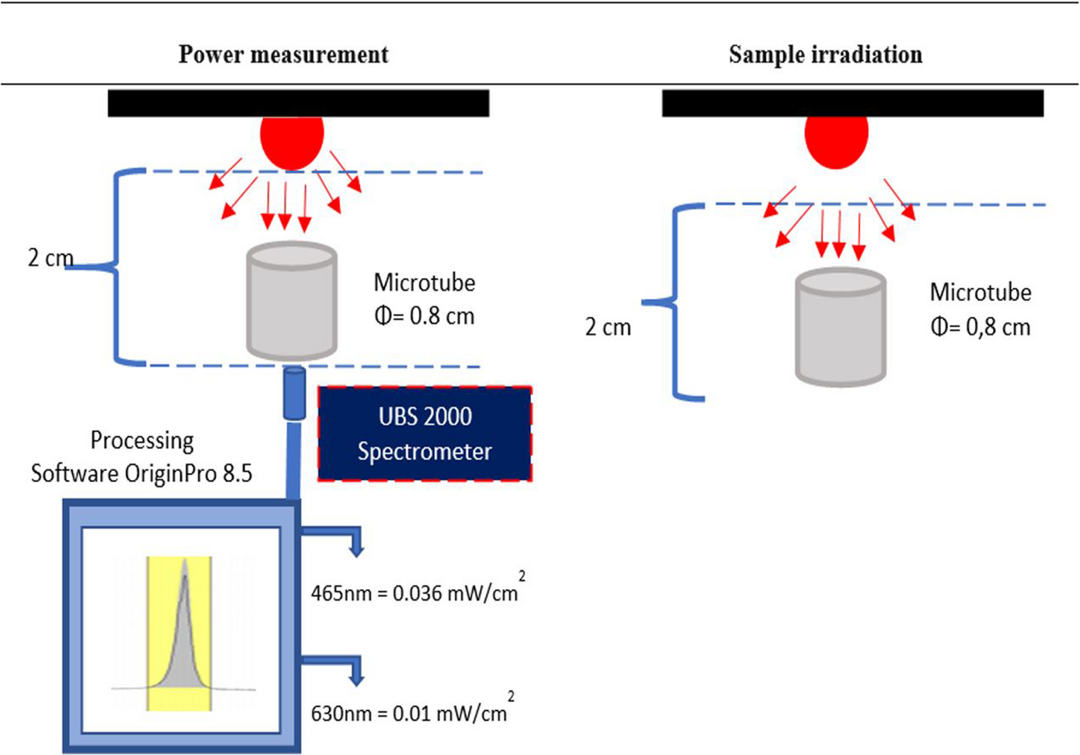
The experimental model (Fig. 1) was prepared concerning the method described by Ribeiro et al. [17]. Two layers of overlapping wax sheets with a diameter of 5 mm and a length of 3 mm were placed on the apical part of the teeth and adapted to the tooth with the help of a heated spatula. The tooth’s root was coated with varnish and immersed in acrylic resin. The experimental setup was placed in ice water until the polymerization was complete to prevent the exothermic reaction from melting the wax. The tooth was marked tangentially to the border of the acrylic. After removal from the acrylic container, the apical wax layer was removed. A second mark parallel to the first mark was drawn 2 mm apically. The tissue for simulating periapical tissues was obtained by removing a full-thickness flap from bovine palates obtained from the slaughterhouse and stored in bovine mucosa − 18 °C until use. During the experiment, bovine mucosa with a diameter of 5.5 mm and a height of 5 mm was prepared for each sample and thawed in saline at room temperature for 30 min. All prepared bovine mucosae were weighed on a precision balance (average 70–80 mg). Bovine mucosae were placed in the experimental model. The bovine mucosae were reduced in size with a scalpel until the second parallel line drawn on the tooth was tangential to the acrylic. Bovine mucosae with a distance of less than 2 mm between the first mark drawn on the tooth and the acrylic were removed from the experiment. The bovine mucosae were dried with blotting papers, weighed 3 times on a precision balance, and averaged (mg). This measurement was recorded as the initial weight. The bovine mucosa was placed in the periapical area created in the experimental model, with the epithelium facing the acrylic and the connective tissue facing the root. The tooth was repositioned using a Universal Tester (Instron Corp, Canton, MA) with a compressive force equivalent to 25 gf until the first drawn line was tangent to the acrylic edge. The acrylic and root interface were sealed with a gingival barrier (OpalDam, Ultradent Products, Inc, USA) to maintain a constant back pressure of the tissue on the apex during irrigation. A #100 plugger (Dentsply Maillefer, Ballaigues, Switzerland) was placed up to the apex to compress the portion of bovine mucosae that had entered the canal [17]. The bovine mucosa samples were numbered from 1 to 75. Randomization was performed using computer-generated sequences (www.random.org), resulting in a table that assigned randomized sample numbers to five groups, each containing 15 samples (n = 15): Standard needle irrigation (SNI), PIPS-flat (F), PIPS-radial (R), SWEEPS-flat (F) and SWEEPS-radial (R).
Fig. 1
(A) An artificial socket model, (B) experimental model
SNI30G side-vented needle (Ultradent) was placed in the root canal 1 mm shorter than the working length. 5 ml of 2% NaOCl (Cerkamed) was applied for 30 s using 3–4 mm amplitude. Thus, the first activation cycle was completed. The same procedure was applied in the second and third activation cycles. After three activation cycles, final irrigation was performed using 5 ml distilled water.
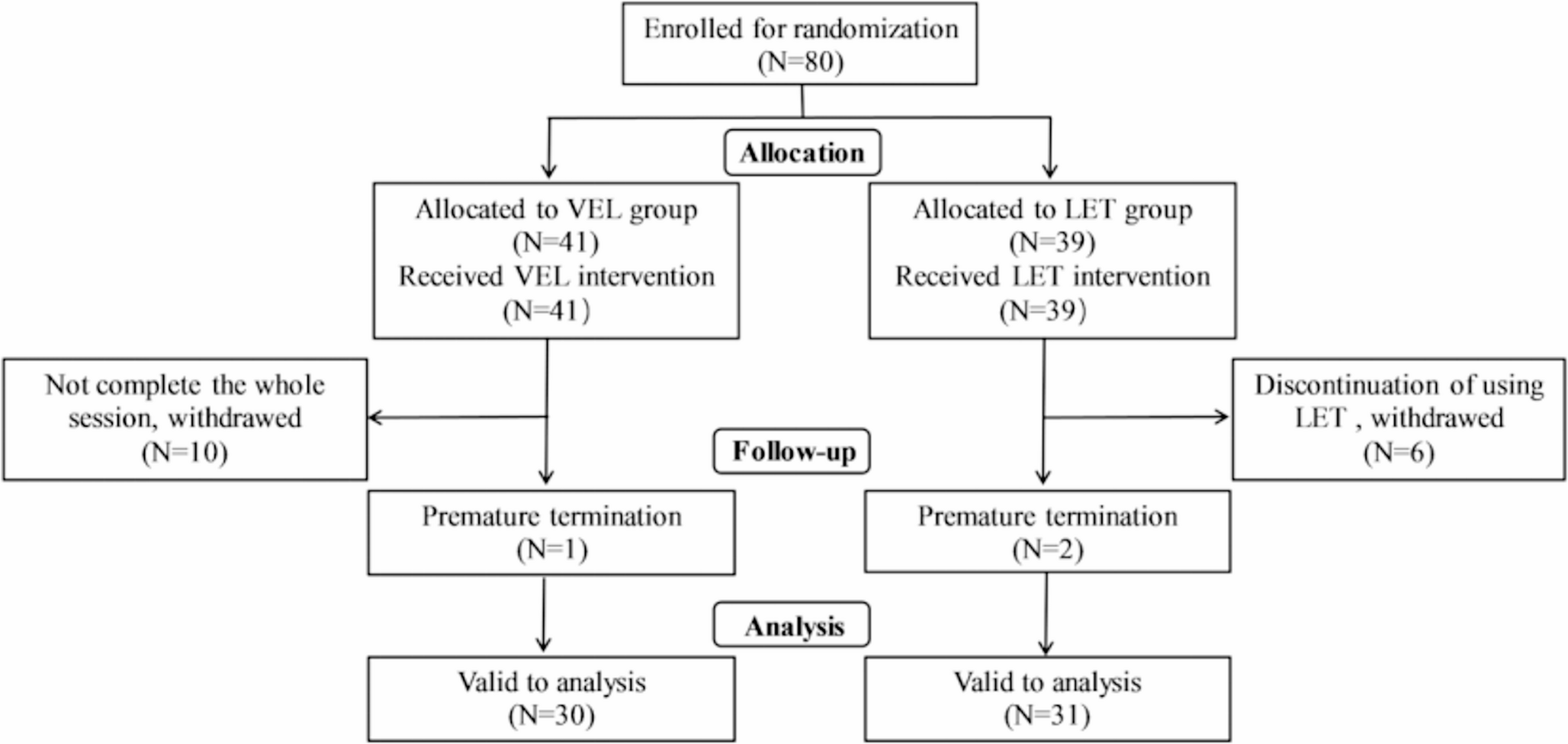
PIPS-FFotona Light Walker Er: YAG laser was set according to the manufacturer’s instructions at 20 mJ, 15 Hz, 0.3 W in SSP (Super Short Pulse) mode with water and air turned off. PIPS (Fotona, Ljubljana, Slovenia) flat fiber tip (400/14) (Fig. 2, A) was placed in the reservoir area. Three activation cycles of 30 s each were performed using the solutions in the order and volume indicated on the SNI.
Fig. 2
Fiber tip types used in LAI, respectively: PIPS-F (400/14) (A), PIPS-R (400/14) (B), SWEEPS-F (300/9) (C), SWEEPS-R (600/9) (D)
PIPS-RWithout changing the parameters, the PIPS radial fiber tip (400/14) (Fig. 2, B) was inserted into the reservoir field, and the final irrigation activation was performed according to the procedure specified in PIPS F.
SWEEPS-FThe Fotona Light Walker Er: YAG laser was set according to the manufacturer’s instructions at 20 mJ, 15 Hz, 0.3 W in auto-SWEEPS mode with water and air off. SWEEPS (Fotona, Ljubljana, Slovenia) flat fiber tip (300/9) (Fig. 2, C) was placed in the reservoir area. Three activation cycles of 30 s each were performed using the solutions in the order and volume indicated on the SNI.
SWEEPS-RThe SWEEPS radial fiber tip (600/9) (Fig. 2, D) was inserted into the reservoir field without changing the parameters. The final irrigation activation was performed according to the procedure specified in SWEEPS-F.
In PIPS-F, PIPS-R, SWEEPS-F, and SWEEPS-R groups, 2% NaOCl (Cerkamed) was continuously added to the reservoir area during activation [19]. After the irrigation activation procedures, bovine mucosae were removed from the experimental model with the help of a fine-tipped tweezer and dried with blotting papers. The dried bovine mucosae were weighed with precision balance three times and averaged (mg). This measurement was recorded as the final weight. The amount (mg) of tissue loss was calculated by subtracting the final measurement from the initial measurement. All procedures were performed by a single endodontist (H.K.).
Statistical analysisShapiro-Wilk test was applied to confirm the normality of the data obtained. Since the data were normally distributed, the weight change was analyzed using One-Way ANOVA and post-hoc Tukey tests. All statistical analyses were performed using SPSS version 23 (IBM, Armonk, NY, USA). p < 0.05 was considered statistically significant.
Comments (0)