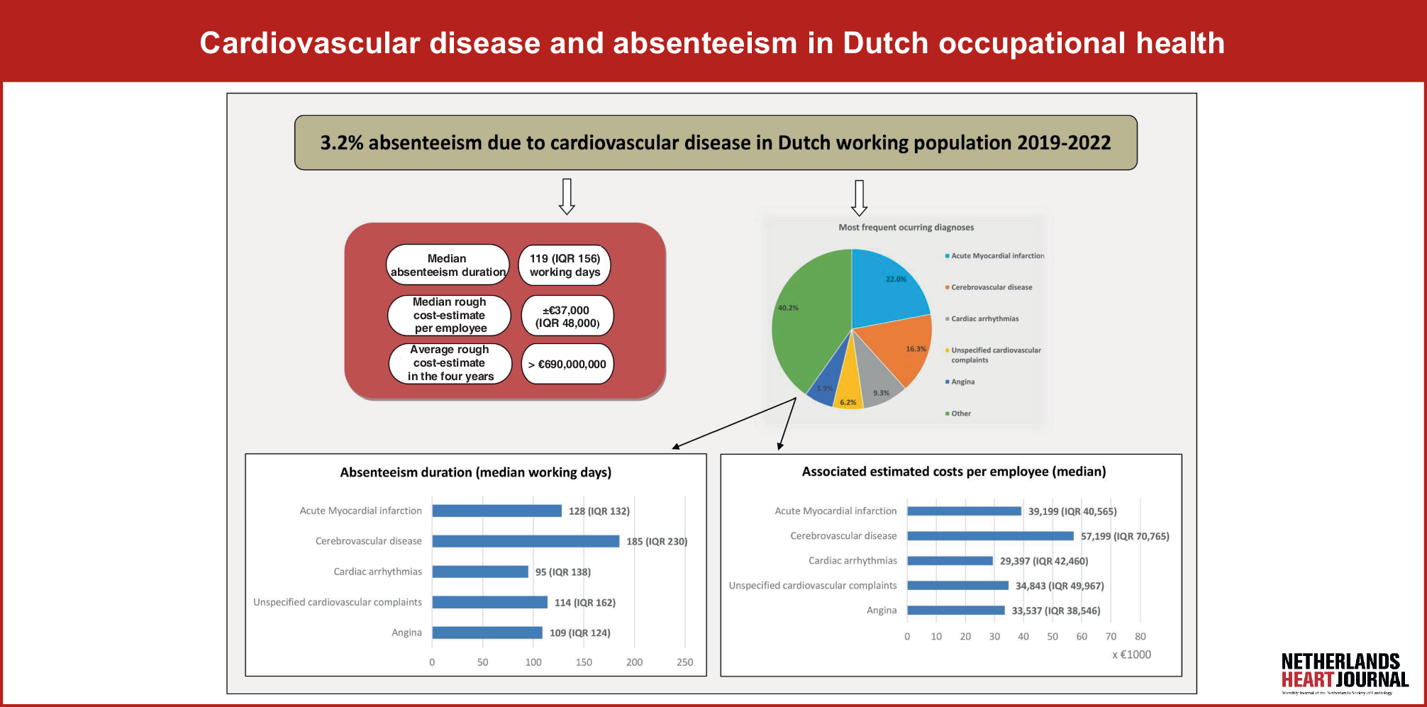
Since 2023, the Netherlands has adopted more stringent guidelines for primary prevention ICD implantation in patients with NICM compared with the ESC guidelines (Fig. 2). Under the Dutch guidelines, patients who meet the criteria for CRT receive a CRT without a defibrillator function (i.e. a CRT-pacemaker). Additionally, patients with known pathogenic arrhythmogenic mutations, such as those associated with HCM or ARVC, are evaluated based on their specific SCD risk models and are considered for ICD implantation only if they have a high risk of SCD [12,13,14]. For the remaining NICM patients, the Dutch guidelines stipulate that the presence of LGE on CMR imaging is a necessary criterion for prophylactic ICD implantation [10].
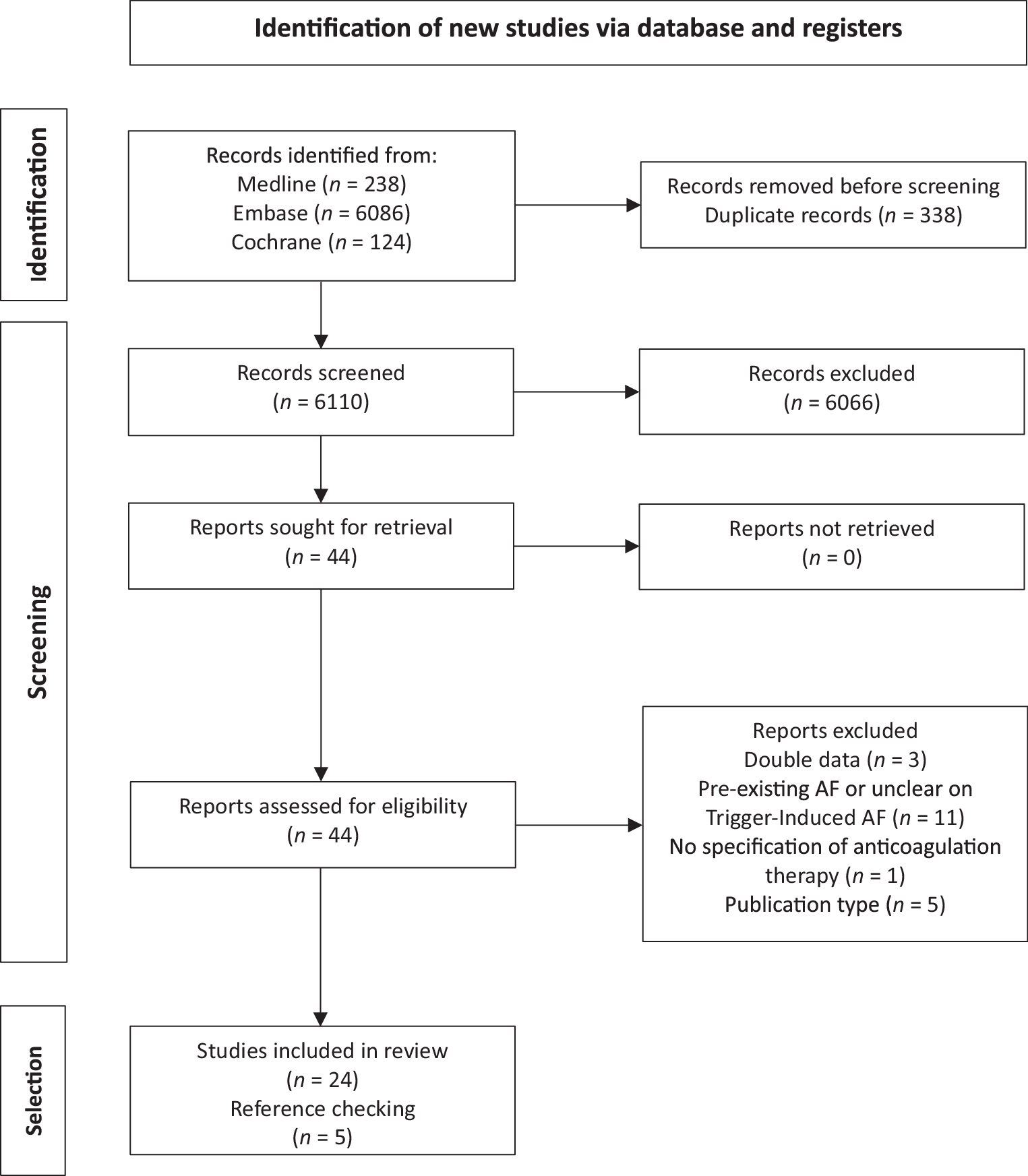
In our cohort of 434 primary prevention NICM patients, 40% did not undergo a CMR scan prior to ICD implantation, thus preventing the assessment of LGE in a substantial portion of the patients. Historically, CMR imaging was not routinely conducted in patients diagnosed with NICM. However, recent guideline updates have increasingly recommended the use of CMR, significantly altering clinical practice [15].
Among the 258 patients who did undergo CMR, nearly half were found to be candidates for CRT, consistent with literature indicating that approximately half of NICM patients qualify for CRT due to significant ventricular conduction delay [16]. Moreover, in the remaining 135 patients who underwent CMR, more than one-third required further evaluation using specific risk models tailored to genetic variations or inflammatory/infiltrative heart diseases. The increased prevalence of genetic testing in recent years has likely contributed to a higher identification rate of individuals with arrhythmogenic mutations.
This trend towards more comprehensive diagnostic assessment, including both CMR and genetic testing, reflects a growing recognition of their importance in refining patient selection for ICD implantation. Among the remaining 85 NICM patients, most of whom had dilated cardiomyopathy of unknown aetiology, half displayed LGE on CMR imaging. Thus, within the group of 258 patients who underwent CMR scanning, only 35% (91/258 NICM patients) met the criteria for ICD implantation under the current, more stringent Dutch guidelines.
In contrast, under the broader ESC guidelines, which allow for ICD implantation in patients without LGE and also include those eligible for CRT‑D therapy, a significantly larger proportion of patients would qualify for ICD implantation.
Appropriate ICD therapy and all-cause mortality in primary prevention NICM patients
In our study cohort, 17% of patients (14 out of 85) received appropriate ICD therapy over a 5-year follow-up period, corresponding to an annual incidence rate of 3%. This rate is relatively modest compared with rates reported in the literature. For instance, a recent pooled analysis of five primary prevention ICD trials, which included patients with both ischaemic cardiomyopathy and NICM, found a 6% annual incidence of appropriate ICD therapy in NICM patients [17]. The lower rate observed in our study may be attributed to the exclusion of NICM patients with an increased risk of ventricular arrhythmia based on specific underlying aetiologies.
In contrast, the more contemporary DO-IT (Dutch Outcome in ICD Therapy) study reported a 5.9% cumulative incidence of appropriate ICD therapy over 2 years in NICM patients, which aligns more closely with our findings. This suggests that our cohort, which excluded higher-risk patients, exhibited an incidence rate that is consistent with recent studies that have employed more stringent patient selection criteria [16].
Our study population also demonstrated a low incidence of all-cause mortality, with an annual rate of 2%. This contrasts with findings from the Danish Study to Assess the Efficacy of ICDs in Patients with Non-Ischaemic Systolic Heart Failure on Mortality (DANISH), which reported an annual mortality rate of 4%. The DANISH study, which involved over 1000 patients with NICM and an LVEF of ≤ 35%, found no significant mortality benefit from the use of ICD therapy compared with medical therapy alone [5]. The implications of the DANISH study are particularly relevant in light of our findings. Given the low incidence of both mortality and appropriate ICD therapy in NICM patients in our study cohort, the potential benefits of ICD implantation may be limited. This suggests that ICD therapy should be considered primarily for carefully selected NICM patients who are at an above-average risk for ventricular arrhythmia. The findings underscore the importance of thorough risk stratification prior to ICD implantation to avoid unnecessary procedures and reduce the risk of adverse events and complications associated with device implantation.
LGE as a risk predictor for ventricular arrhythmias
Previous research has shown that the presence of LGE on CMR is a significant predictor of major ventricular arrhythmic events in patients with dilated NICM [9]. A recent meta-analysis including studies published between 2008 and 2022, with a median follow-up period of 3 years, reported a pooled odds ratio of 3.99 (95% CI 3.08–5.16), highlighting that LGE assessment may be a valuable parameter for primary prevention ICD implantation in NICM patients [18]. However, in our exploratory study, we found no significant association between the presence of LGE and appropriate ICD therapy, potentially due to the limited sample size and low event rate. Approximately half of the NICM patients in our study exhibited LGE on CMR, consistent with existing literature on LGE prevalence in this patient population [18]. Additionally, the prevalence of a midwall septal stripe in the LGE+ group aligned with current findings on LGE distribution in NICM patients [19]. Although specific LGE patterns such as midwall fibrosis have been linked to an increased risk of arrhythmias, our study did not observe a significant association between specific LGE distribution patterns and appropriate ICD therapy [20].
Therefore, our study does not provide conclusive evidence regarding the utility of LGE assessment as a primary risk stratification tool for ICD implantation in patients with NICM. Notably, 14% of patients without LGE on CMR still experienced appropriate ICD therapy, highlighting the necessity of closely monitoring patients who, under the new guidelines, do not receive an ICD. This finding also emphasises the need for large randomised trials to evaluate both the risk of SCD in NICM patients without LGE and the risk-benefit profile of prophylactic ICD implantation in those with LGE. Such studies are essential to establish more definitive guidelines for ICD therapy in this population. Furthermore, we encourage other centres to replicate our analysis to enable pooled data assessments, thereby allowing for a more comprehensive evaluation of arrhythmic risk and the role of LGE in NICM patients.
Limitations
Our study has several limitations. First, in line with the retrospective and exploratory nature of this study on ICD therapy and all-cause mortality in NICM patients, the cohort size and event rate are modest. Second, LGE quantification was not performed because current quantification strategies are inconsistent and lack robust validation [21,22,23]. Moreover, the threshold for LGE extent and its associated risk remains undetermined [24]. Consequently, assessing the presence or absence of LGE visually was deemed practically reasonable, as endorsed by national guidelines for ICD implantation in NICM patients. Thirdly, current guidelines recommend genetic testing for diagnosis and risk stratification in NICM [25]. However, this was not routinely performed in patients included in our analysis. Fourthly, CRT patients were excluded to ensure a homogeneous cohort, as CRT is a heart failure therapy that independently modifies arrhythmic risk and mortality, potentially confounding the analysis of ICD therapy events [26, 27]. Future studies should focus specifically on the CRT‑D population. Finally, heart failure therapy has significantly improved over the years, including the standard use of SGLT2 inhibitors and angiotensin receptor-neprilysin inhibitors, which significantly improve outcomes in heart failure patients [28]. This may have influenced the results of this study significantly.


Comments (0)