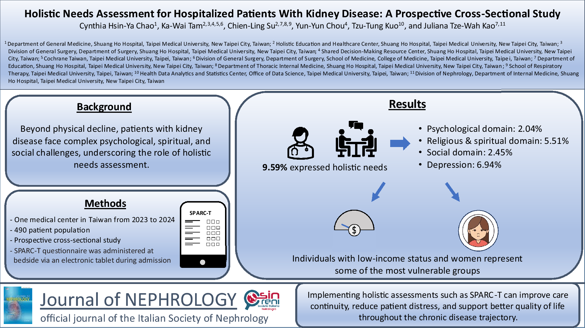
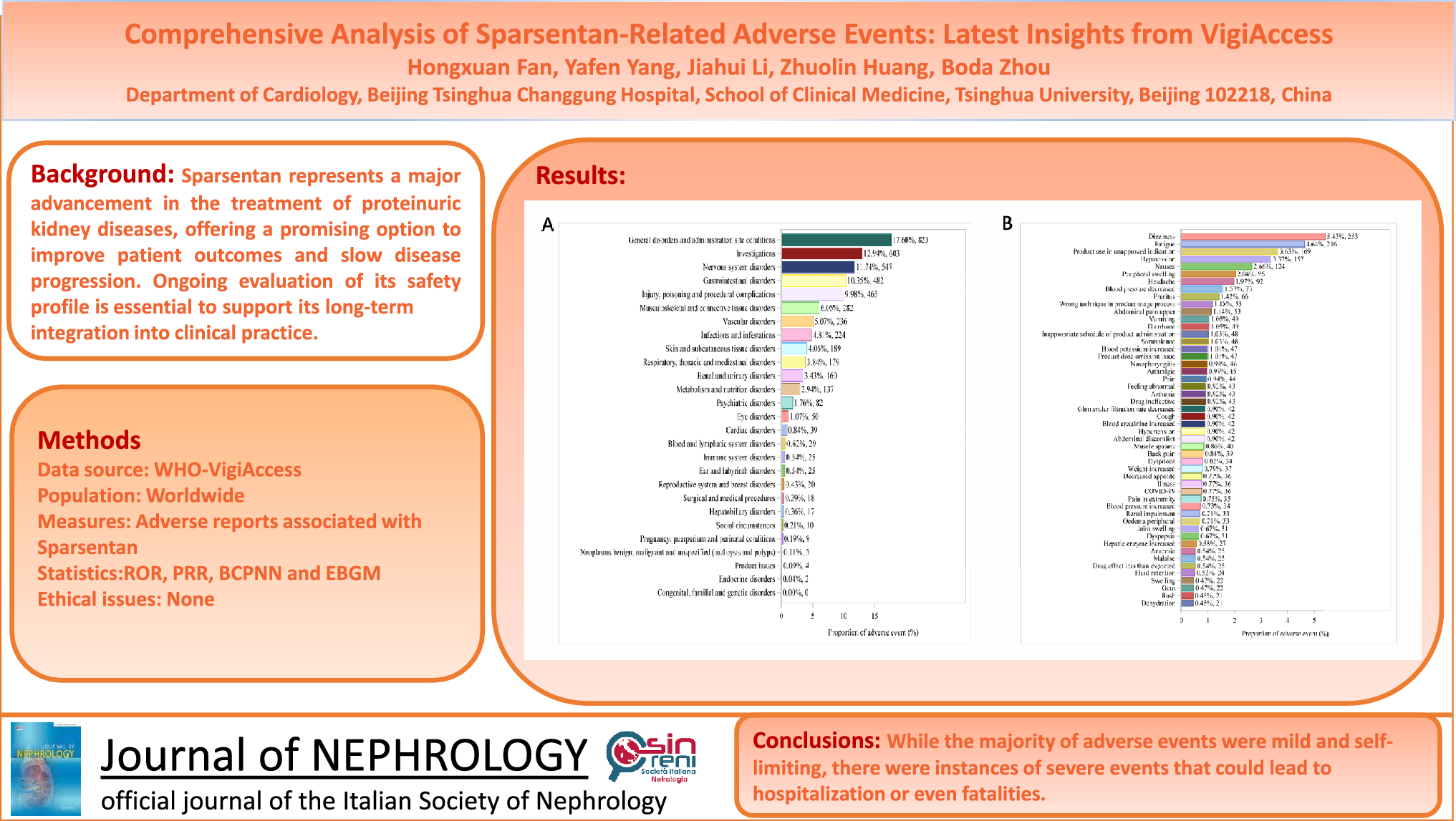
Cardiac hypertrophy in hemodialysis patients
LVH is the most common cardiac abnormality observed in patients with kidney failure and is an independent predictor of survival [1, 2]. Approximately 80% of HD patients exhibit increased LV mass, reflecting the high prevalence of cardiac remodeling in this population [3]. In patients with kidney failure, both pressure and volume overload are prevalent, contributing to an increase in LV mass [13]. Thus, the increase in LV mass in patients with kidney failure may result from wall thickening, LV chamber enlargement, or a combination of both. Chronic pressure overload in dialysis patients leads to an increase in the relative wall thickness resulting in concentric hypertrophy, while volume overload primarily causes LV dilation leading to eccentric hypertrophy [14]. However, accurate classification of LVH as either eccentric or concentric can be challenging in HD patients due to the cyclic fluctuations in extracellular fluid volume and electrolyte balance [15].
Hemodynamic factors, such as arteriovenous fistulas, secondary anemia, and intravascular volume expansion caused by salt and fluid retention are key contributors to volume overload. Moreover, other non-hemodynamic mechanisms may contribute to LV remodeling and hypertrophy. Hyperphosphatemia and vitamin D deficiency may play a role in regulating the growth and differentiation of cardiac myocytes [4]. Additionally, the partial regression of LVH with ACE-inhibitors highlights the involvement of the tissue and cardiac renin-angiotensin system in the development of LVH [16]. Elevated plasma endothelin levels have also been linked to LVH in kidney failure, supported by animal studies showing endothelin receptor antagonists reducing myocardial hypertrophy [17]. Hypoalbuminemia in kidney failure arises from multiple contributing factors, such as diminished liver production of albumin, protein losses during dialysis, inadequate nutritional intake, fluid overload, and other mechanisms related to the chronic kidney disease. Although hypoalbuminemia is often associated with reduced oncotic pressure and thus lower effective circulating volume, in the context of kidney failure it frequently coexists with volume overload and LV dilation [18,19,20,21]. However, the mechanism of the impact of hypoalbuminemia on the LV geometry is still unclear. Hyperparathyroidism, although implicated, remains controversial, as parathyroidectomy has shown minimal effects on cardiac structure and function [22]. Over time, pathological LVH may progress due to collagen deposition, fibrosis, and calcification [2].
Left ventricular hypertrophy is a well-established prognostic factor in patients with kidney failure. A study by Silberberg et al. demonstrated that LVH, as assessed by echocardiography, significantly influences survival outcomes in individuals initiating kidney replacement therapy. Patients in the highest quintile of LV mass index had a 3.7-fold increased risk of both all-cause and cardiac mortality compared to those in the lowest quintile, underscoring LVH as a critical independent predictor of survival in kidney failure [2]. Beyond baseline LVH, changes in LV mass index over time also play a crucial role in patient prognosis. A prospective study involving HD patients, which measured LV mass index twice over an average interval of 18 months, found that fluctuations in LV mass index were strongly associated with adjusted risks for both all-cause mortality and major adverse cardiovascular events [23]. Additionally, the structural geometry of LVH further impacts outcomes. A retrospective study of 175 chronic HD patients revealed that LV geometry and inferior vena cava diameter significantly influence cardiovascular risk and overall survival. Specifically, individuals with a large inferior vena cava diameter and eccentric LVH faced the highest risk of cardiac events, while those with a large inferior vena cava diameter and concentric LVH had the greatest risk of both cardiovascular and all-cause mortality. These findings emphasize the need for close monitoring and targeted interventions to manage LVH and volume status, and to improve survival in kidney failure patients [13].
Several studies suggest that intensive HD may contribute to a reduction in LV mass, potentially lowering the risk of adverse cardiac events. For instance, the Frequent Hemodialysis Network trial, a randomized clinical trial involving 245 patients, demonstrated that both short daily and nocturnal HD schedules resulted in LV mass reductions of 14 g (10%) and 11 g (8%), respectively, compared to the conventional thrice-weekly regimen [24]. Since reduction of LVH is linked to a lower risk of cardiovascular complications such as heart failure, post-myocardial infarction remodeling, and sudden arrhythmic death, further research is needed to explore the potential of intensive HD in minimizing cardiovascular risk [25].
Impact of hemodialysis on left ventricular systolic function
Many studies report that cardiac function is affected by HD. A study by Wang and colleagues on 40 patients showed that the LV end-diastolic volume, LVEF and global longitudinal strain significantly decreased after HD. In this study, LVEF was assessed before and after HD sessions using the Simpson’s method with apical 4- and 2-chamber views, and global longitudinal strain was evaluated using 2D speckle-tracking echocardiography [5]. Another comparative study involving 30 patients with kidney failure and 30 healthy controls demonstrated that two-dimensional speckle-tracking echocardiography is more sensitive than conventional echocardiography in detecting subtle systolic and diastolic dysfunction in LVH patients, even when LVEF appears normal [26]. Moreover, global longitudinal strain is regarded as a more reliable predictor of cardiovascular mortality than echocardiographic LVEF in patients undergoing chronic HD [27].
Kidney failure patients who regularly undergo HD sessions experience acute preload changes, which consequently affect preload-dependent echocardiographic parameters. Deformation parameters have been claimed to be load-independent, however conflicting results have been reported on the changes of strain measurements, such as global longitudinal, circumferential and radial strain of the myocardium after HD. While some studies indicate a significant reduction of these parameters after HD, others showed similar values of global longitudinal strain before and after HD which suggests preload-independence [28,29,30].
Apart from the acute changes induced by HD on LV function, in the long-term HD drives progressive LV remodeling and dysfunction, typically associated with a gradual decline in LVEF. Even a modest drop of LVEF, more than 5% over several years of HD, is linked to higher mortality, while paradoxical small increases—often reflecting volume overload—also predict worse survival. Consequently, serial echocardiographic evaluation of LVEF is indispensable for timely identification and management of cardiovascular risk in this population [31].
Ventricular dysfunction is a known complication of transient myocardial ischemia, even after blood flow is restored. In patients with pre-existing LVH and interstitial fibrosis, HD-induced intravascular volume reduction triggers hemodynamic shifts, reflected in blood pressure and heart rate variations, and activates injury pathways, including high-energy phosphate depletion, microvascular hypoperfusion, impaired neural response, and inflammation [32]. These factors contribute to HD-induced ischemia, or myocardial stunning, leading to cardiac dysfunction. In 2009, Burton and colleagues found that 64% of 70 patients exhibited myocardial stunning during HD, which was defined as a reduction in wall motion greater than 20% from baseline in more than two regions [33]. Moreover, Assa et al. found that 27% of 105 patients undergoing HD developed regional LV systolic dysfunction, with 17 patients showing dysfunction as early as 60 min after dialysis initiation. The key factors associated with this dysfunction were male sex, a higher LV mass index, and pre-existing LV dysfunction. However, dialysis treatment-related factors, such as changes in blood volume, electrolytes, or acid–base parameters, were not linked to LV dysfunction. Furthermore, during follow-up, HD-induced regional LV dysfunction was identified as an independent risk factor for all-cause mortality, as the association remained significant even after adjusting for important prognostic factors, including age, sex, dialysis history, diabetes, cardiovascular risk factors, ultrafiltration volume, LV mass index, and predialysis LV systolic function [34]. These changes suggest that HD in patients with kidney failure may lead to LV dysfunction. This is also supported by the finding of impaired LV systolic function, as assessed by strain measurements, due to acute volume reduction after HD [35].
Impact of hemodialysis on left ventricular diastolic function
Diastolic dysfunction has been shown to provide a significant additional prognostic value in predicting both all-cause mortality and cardiovascular-related death in HD patients [6]. While changes in diastolic function can partly be attributed to preload and afterload reduction, non-volume-related factors may also play a role [36]. Since coronary perfusion occurs during diastole, HD-related diastolic dysfunction may exacerbate myocardial ischemia, creating a cycle of myocardial stunning and further dysfunction [37]. Additionally, electrolyte imbalances—such as fluctuations in calcium, magnesium, and uncontrolled hyperphosphatemia—have been linked to diastolic impairment [38, 39]. A reduction in central aortic pressure during HD may further contribute to the development of diastolic dysfunction [40].
According to the American Society of Echocardiography and the European Association of Cardiovascular Imaging, four echocardiographic variables can be recorded to assess whether LV diastolic function is normal or abnormal. The four variables include mitral annulus e′ velocity (septal e′ < 7 cm/s, lateral e′ < 10 cm/s), average E/e′ ratio > 14, left atrial volume index > 34 mL/m2, and peak tricuspid regurgitation velocity > 2.8 m/s. From these parameters, left atrial pressure can be estimated [41].
In the study by Wang et al., LV diastolic function was evaluated using the aforementioned echocardiographic parameters before and after HD (within 24 h) (Table 1) [5]. This study found a significant reduction in both E-wave velocity and E/A ratio, whereas the decrease in A-wave velocity was not statistically significant. Furthermore, there was a significant decrease of e′ at the septal side of the mitral annulus (6.45 ± 1.88 vs. 5.77 ± 1.63 cm/s, p < 0.001), while the lateral annulus velocities showed no significant difference before and after HD (Table 1). The average value of the septal and lateral e′ also decreased significantly after HD (7.10 ± 2.14 vs. 6.53 ± 1.98 cm/s, p = 0.003). Additionally, the average E/e′ ratio decreased from 12.54 ± 4.08 before HD to 11.28 ± 4.52 after HD (p = 0.049), which was probably due to a greater reduction in E compared to e′ velocity. The decline in both E and e′ velocities suggests that mitral annular velocities are also preload-dependent. In addition, left atrial volume indexed by body surface area declined significantly after the HD session (30.22 ± 9.80 vs 35.55 ± 12.61 mL/m2, p < 0.001). The fourth variable, tricuspid regurgitation velocity, also showed a statistically significant decline after HD session (260.1 ± 36.5 vs. 242.3 ± 32.2 cm/s, p = 0.002) [5].
Table 1 Conventional echocardiography and pulsed Doppler measurements pre- and post-HDRecent studies have highlighted limitations in using mitral annulus velocities, particularly the E/e′ ratio, for assessing LV diastolic function [42]. This method estimates global LV diastolic function based on the assumption that one or multiple measurement sites reflect overall LV relaxation. However, in dialysis patients, significant LV diastolic dyssynchrony is frequently observed, reducing the accuracy of this approach [43]. Additionally, left atrial pressure can influence the early phase of LV filling, further complicating its reliability [44]. To address these limitations, LV global diastolic strain rate measurement using two-dimensional echocardiography speckle-tracking analysis has been demonstrated to be a more effective alternative, particularly in individuals with relatively preserved LVEF or regional wall motion abnormalities [45]. This measurement reflects the overall performance of all LV segments, remains independent of loading conditions, and accounts for initial LV size. For instance, a study involving 77 participants, of whom 59.7% were on HD, found a higher prevalence of diastolic dysfunction (defined by a ratio of diastolic early wave velocity to the global diastolic strain rate during isovolumic relaxation ≥ 236) compared to conventional diastolic dysfunction assessments (48% vs. 39%). These findings suggested that speckle-tracking-derived strain rate analysis might provide a more reliable evaluation of LV diastolic function in this patient population [42].


Comments (0)