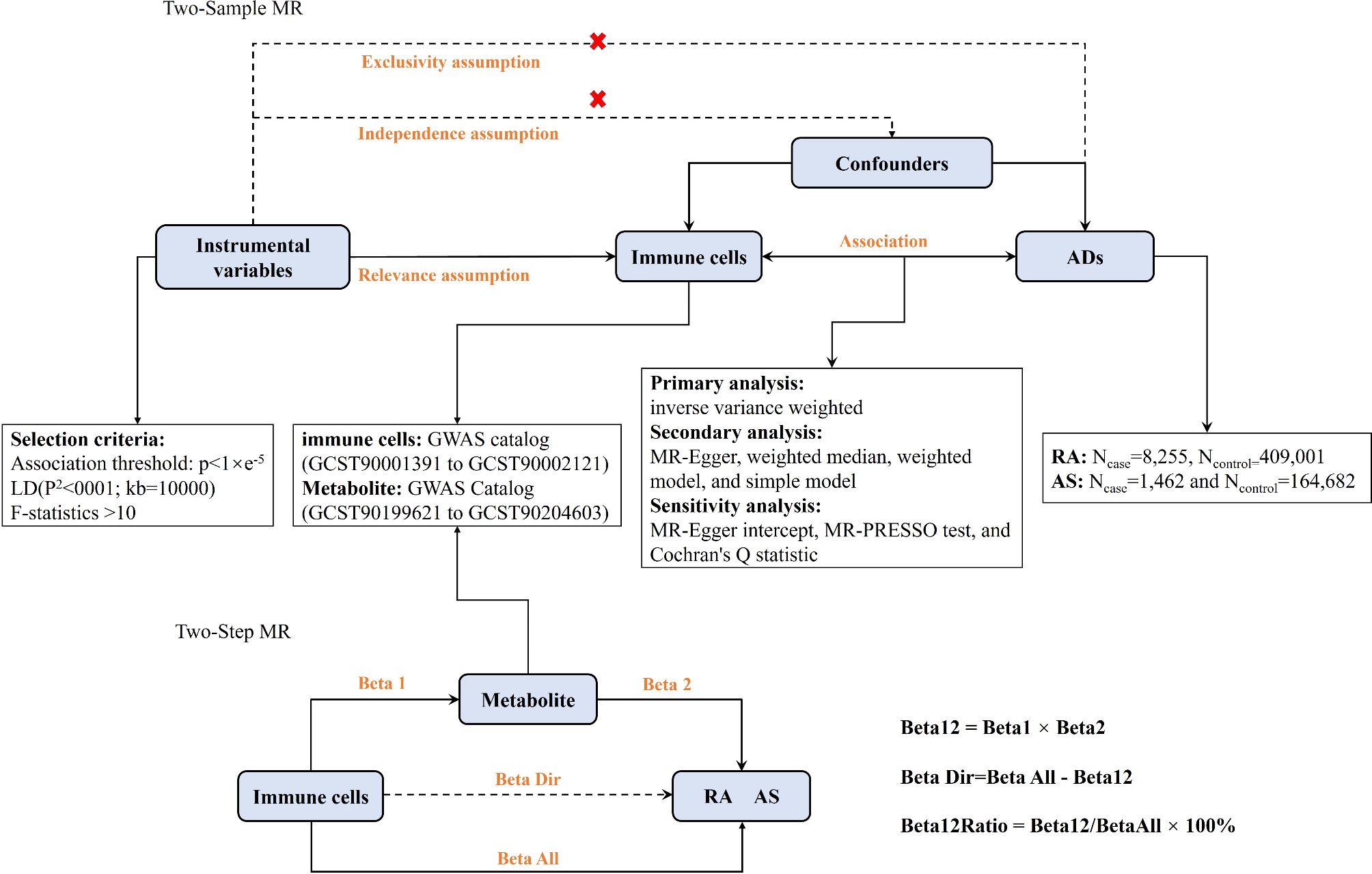
To the best of our knowledge, this is the first study using NODDI models to investigate the microstructural changes of the WM in SLE patients. Furthermore, we compare the discriminative abilities of DKI and NODDI metrics within TBSS clusters and each JHU WM region (totally 20 regions). The main findings of this study are as follows: (i) significantly reduced FA, AD and RK and markedly increased ODI were found in multiple WM regions in non-NPSLE patients compared with controls; (ii) ODI in ATR_R had the greatest discriminative power; and (iii) C3 scores were correlated with mean ODI in F_major.
Our data showed lower FA in non-NPSLE patients mainly in the thalamus and frontal and parietal lobes, which basically corroborated previous DTI studies of non-NPSLE [24,25,26,27]. Reduced FA might reflect damage to the myelin sheath surrounding axons, reduced axonal packing density or enhanced membrane permeability [28]. In addition, reduced AD was mainly found in left frontal and occipital lobes in this study. Decreased AD indicates axonal injury, reduced axonal caliber, or less coherent orientation of axons [28]. Our results about AD metrics are different from findings reported by previous DTI studies on SLE [29, 30]. For example,. Zhao et al. reported non-NPSLE patients had increased AD in bilateral corticospinal tracts and reduced AD in right superior longitudinal fasciculus-temporal terminations [30]. These discrepant results may be explained by the fact that complex cellular components and structures cannot be well described using the DTI model. We also detected reduced radial kurtosis (RK) in patients with non-NPSLE versus healthy controls mainly in the parietal lobe. RK quantifies the limitation of water molecules diffusion, which is affected by cell membranes and myelin [31]. Reduced RK suggests the diffusion of water molecules is closer to the Gaussian distribution and decreased restriction in the diffusion environment [10, 32]. However, no significant differences were observed in RD and AK. We hypothesize that this sensitivity difference may be attributed to specific directional diffusivities and kurtosis. Water molecules exhibit a more uniform diffusion along axonal tracts, resulting in a Gaussian distribution, which can be detected by the axial diffusivity metrics (AD). Conversely, motion is constrained by the complex microstructure in the perpendicular direction, resulting in a non-uniform (non-Gaussian) distribution, and this phenomenon can be detected by radial kurtosis metrics (RK). Therefore, the combined axial diffusivity metrics (AD) and radial kurtosis metrics (RK) can sensitively and accurately reflect changes in the direction of neural fibers. In addition to the DKI results, increased ODI was found in frontal lobe and thalamus, showing a high overlap with the regions of reduced FA while no NDI alterations were detected in this study. Increased ODI indicates the presence of fiber crossing and dispersion [33]. Orientation dispersion index and neurite density are two major aspects of FA [34]. Elevated ODI values within the brain may indicate a change in the morphology of the microstructure rather than in axon density, suggesting that ODI provides more specific information than FA. Therefore, DKI and NODDI metrics were complementary in revealing the underlying mechanism of white matter impairment in SLE. Additionally, the ODI cluster showed the highest statistical significance and the largest AUC in ROC curve analysis, suggesting ODI is more sensitive than DKI metrics. Hence, NODDI could serve as a more sensitive and specific biomarker for the detection of WM abnormalities in non-NPSLE patients that merits further study.
In atlas-based ROI analysis, after FDR correction, only ODI showed a significant difference, indicating that ODI has an advantage in detecting WM fibers over DKI metrics. We found higher ODI values for non-NPSLE patients in bilateral ATR, IFOF, UF and CC (F_major and F_minor). ATR is the predominant component of the anterior limb of the internal capsule (ALIC), which is associated with cognitive functions, including memory encoding and executive function. Mamah et al. revealed asymmetric microstructural changes of the right ALIC in patients with schizophrenia, which correlated with cognitive abnormalities [35]. In addition, our ROC curve analysis showed ODI in the right ATR had the best discriminative ability, indicating that ODI in the right ATR may serve as a potential biomarker of SLE, facilitating early detection and diagnosis. IFOF and UF are association fibers that connect different cortical areas on the same side. IFOF is the main structural pathway for language semantics, and UF is mostly involved in episodic memory, language and socioemotional processing [36]. Comprising commissural fibers, the CC is the largest white matter inter-hemispheric commissure. CC integrity plays a crucial role in sensory-motor functions, attention, language and memory [37]. Previous findings suggested that the morphology of the corpus callosum is associated with several psychiatric disorders, including schizophrenia and bipolar disorder [38]. Abnormalities in ATR, IFOF, UF and CC may account for the manifestation of cognitive dysfunction in SLE patients, including damaged executive function, memory, verbal ability, etc. Future studies combining cognitive and behavioral indicators with fibers in these regions may help explain the NP symptoms of SLE.
In correlation analysis, ODI in F_major was positively correlated with C3 level, as shown above. It was suggested that ODI has a strong correlation with microglial density [39]. The splenium of the corpus callosum (F_major) involves different caliber axonal fibers and the most compact area of glial cells in the CC, affecting language, visual information transfer and behavior, possibly responsible for changes in consciousness. The complement system is involved in the process of neuroinflammation in SLE, and previous studies have demonstrated that low C3 and C4 levels are potential diagnostic markers that could help monitor disease activity in SLE [40]. Additionally, microglia and astrocytes play a protective role in the brain by synthesizing and secreting complement components [41]. Therefore, the detected positive correlation may be because microglial activation in F_major increases the production of complement C3 proteins (higher C3); meanwhile activated microglia reduce the coherence of axonal orientation (higher ODI).
Taken together, TBSS and atlas-based ROI analyses by combining DKI and NODDI metrics can provide a more comprehensive understanding of WM alterations in non-NPSLE patients. ROC curve analysis demonstrated that ODI, rather than DKI, is the most sensitive and specific biomarker. These findings indicate that dMRI is beneficial for identifying subclinical brain structural involvement before the onset of NP symptoms. Consequently, it suggests the necessity for earlier clinical intervention to prevent the progression of brain damage. Furthermore, a deeper understanding of the complex mechanisms of the CNS in SLE helps explain the progression of the disease, facilitating early clinical diagnosis and careful consideration of treatment strategies.
The limitations of this study were as follows. First, due to the absence of NPSLE data, differences among NPSLE, non-NPSLE patients and healthy controls could not be examined. Secondly, we focused solely on the changes of brain WM microstructure in SLE. Future studies should evaluate the gray matter based on the multi-shell dMRI technique. Finally, external validation data were not available to assess the reproducibility and generalizability of the current results.
In summary, microstructural changes in brain WM in non-NPSLE were assessed by analyzing the properties of different biological tissues with the DKI and NODDI models. TBSS analysis revealed reduced FA, AD and RK and increased ODI in multiple WM regions in non-NPSLE patients, which may be attributed to demyelination in myelinated axons and the morphological changes of fibers. Atlas-based ROI analysis reported increased ODI values in 4 fibers, and the right ATR had the highest ability to distinguish non-NPSLE from healthy controls, indicating that ODI in the right ATR may serve as a potential biomarker of SLE. The positive correlation between C3 and ODI in F_major may be associated with microglial activation following WM’s microstructural changes. Our findings provide new insights into CNS microstructural injury and its potential mechanisms in SLE patients, even in the absence of NP symptoms. This contributes to early diagnosis and emphasizes the necessity of early clinical intervention for SLE patients.


Comments (0)