
Remember me


A 2.5-year-old boy with chronic granulomatous disease (CGD) was admitted to Sultan Qaboos University Hospital with a 4-day history of high-grade fevers associated with mild intermittent cough in March 2021. There was no associated coryza, shortness of breath or other symptoms. The patient had no history of previous admissions, travel, sick contacts or animal exposure. Because his older brother had known CGD, he was screened soon after birth, diagnosed with the same condition, and initiated on chemoprophylaxis with itraconazole and cotrimoxazole. Prior to his illness, compliance with chemoprophylaxis was not optimal. His immunizations were up to date except for bacille Calmette-Guerin vaccine, which was avoided because of his abnormal neutrophil function.
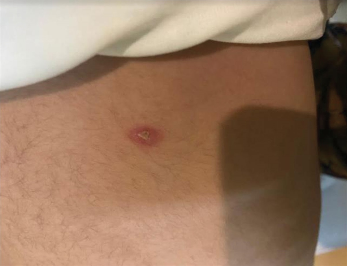
Initial examination revealed a high temperature of 40 °C, associated with tachycardia of 148 beats/minute and tachypnea of 30 breaths/minute. He was normotensive, and his oxygen saturation was 98% in ambient air. He had no signs of increased work of breathing, but had reduced air entry in the left lower lung zone posteriorly on auscultation. His systemic examination was otherwise significant only for a small pustular skin lesion on a red base over the right paraspinal area (Fig. 1). His abdominal and cardiovascular examinations were unremarkable. His initial laboratory investigations revealed a complete blood count with a hemoglobin of 10.8 g/dL, white blood cell count of 24.8 × 10⁹/L, with a differential of 73% neutrophils, 13.2% lymphocytes, 3.4% monocytes and 0% eosinophils and a platelet count of 370 × 109/L. C-reactive protein was 285 mg/L. He had normal liver and renal function tests. His chest radiograph showed left lower consolidation with possible pleural effusion, and chest ultrasound revealed a small pleural effusion with no septations or loculations. He was started on treatment with intravenous vancomycin and meropenem soon after admission, but continued to spike high-grade fevers for more than 1 week. Contrast-enhanced computed tomography (CT) of the chest showed an area of lung consolidation in the left lower lobe with a central area of low attenuation, likely representing necrosis, along with the multiple bilateral pulmonary nodules, mediastinal and left hilar necrotic lymphadenopathy, and minimal left-sided loculated pleural effusion (Fig. 2). A swab collected from the pustular skin lesion on the back revealed the causative pathogen.
 FIGURE 1.:
 Dried pustule on erythematous base in the right paraspinal region.
 FIGURE 2.:
 A and B: Coronal and axial CECT in the mediastinal window show necrotic matted left hilar and mediastinal adenopathy (yellow arrows), along with necrotizing consolidative opacity of the left lower lobe (yellow arrowhead). C: Coronal CT chest in lung window showed variable-sized, slightly irregular-shaped 2 nodules in the right upper lobe (circles).
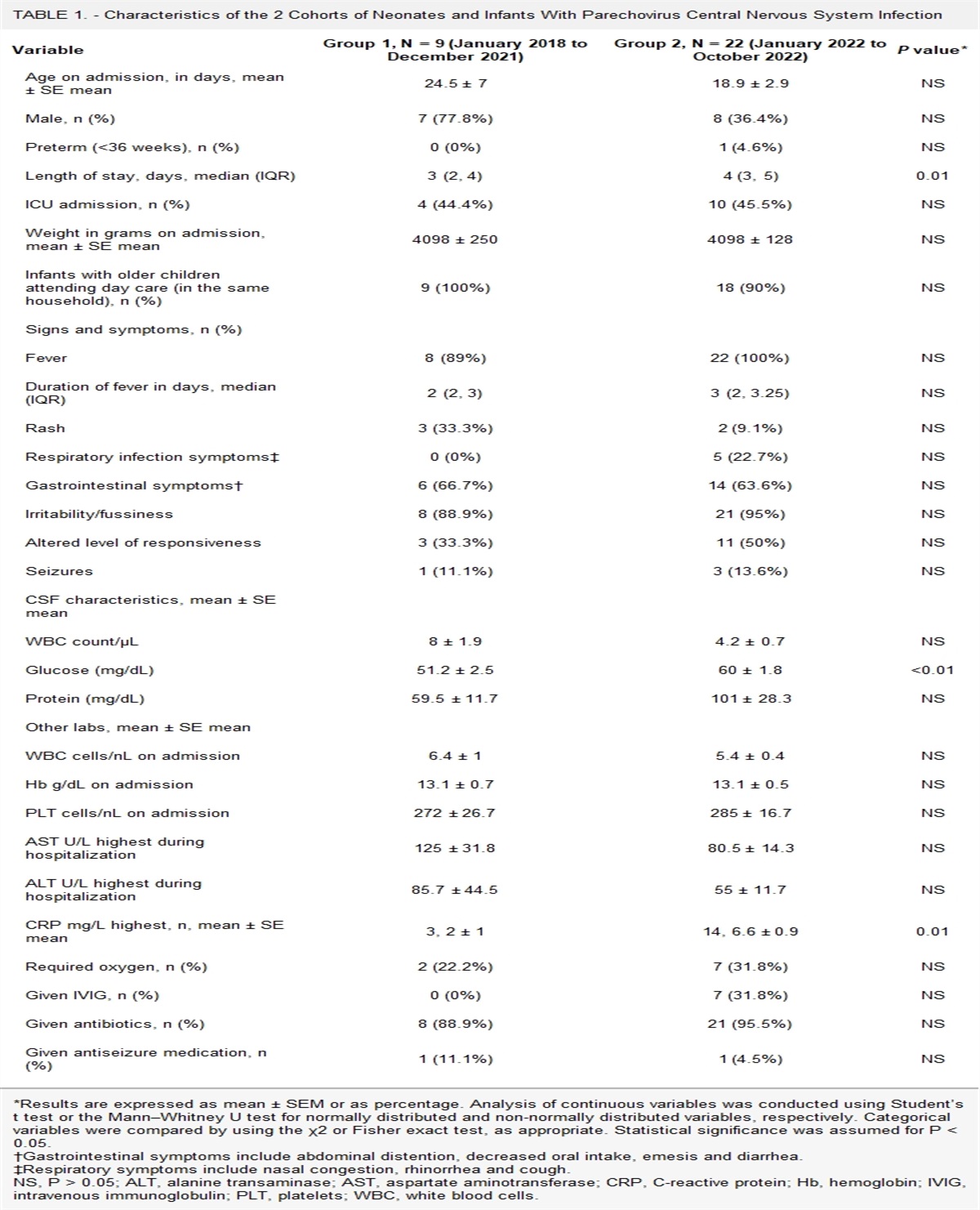
DENOUEMENTOn day 8 of admission, Nocardia cyriacigeorgica was isolated from the pustular skin lesion overlying the right paraspinal area. Intravenous high-dose cotrimoxazole (15 mg/kg/d of trimethoprim component in 2 divided doses) was added to the IV meropenem. Magnetic resonance imaging (MRI) of the brain was performed to look for disseminated disease and revealed a rim-enhancing lesion in the most superior part of the left paramedian parietal lobe with a central enhancing spot, suggestive of central nervous system nocardiosis (Fig. 3). Bronchoalveolar lavage and cerebrospinal fluid specimens were obtained for prolonged cultures. The cerebrospinal fluid specimen was clotted because it was a bloody tap, and it had 2 leukocytes only, with normal protein (0.08 g/L) and sugar. He continued to spike high-grade fever for 4 weeks, and his antibiotic regimen was switched after 2 weeks to IV cotrimoxazole, amikacin (7.5 mg/kg q 12 h), and linezolid (10 mg/kg q 8h) due to concern of resistant nocardiosis. The isolate was sent for susceptibility testing using broth microdilution and was found to be susceptible to amikacin, ceftriaxone, imipenem, linezolid and trimethoprim-sulfamethoxazole (cotrimoxazole). Repeat CT of the chest and MRI of the brain after 6 weeks of treatment showed significant improvement so he was discharged home on oral linezolid and cotrimoxazole. He remained asymptomatic while on oral antibiotics, and repeat imaging after 6 months of treatment showed complete resolution of the brain lesion and near-complete resolution of the chest abnormalities. He completed 12 months of antibiotics in total. Follow-up evaluation at 2 years showed complete recovery with no recurrences.
 FIGURE 3.:
 A contrast-enhanced MRI of the brain in the sagittal plane shows a nodular rim-enhancing lesion in the left parasagittal frontal lobe (double arrows). There was no mass effect of adjacent edema.
Nocardia is an aerobic actinomycete, catalase-positive Gram-positive bacillus. It is a slow-growing, soil-borne bacteria with a branching filamentous form.1,2 Over 100 species have been identified with N. cyriacigeorgica being the most common cause of human infections.3 Disseminated nocardiosis, which has a mortality rate as high as 85%–100%, is mainly seen in immunocompromised children, but has also been reported in immunocompetent children.4 The most common mode of transmission in the immunocompromised is through inhalation, which explains why lung infection is the most common presentation like for our patient.1 Nocardiosis can be chronically progressive and has the tendency to relapse even with adequate treatment.1
Necrotizing pneumonia, multiple cavitary diseases and abscesses, as well as empyema, are the most common manifestations in cases of chest involvement.2 It is a rare infection, even in susceptible patients, with a rate that does not exceed 1% in most groups of immunocompromised patients.5,6
Nocardiosis can potentially disseminate to any organ, especially the central nervous system. The most common involvement of CNS is abscesses and meningitis, and the spinal fluid profile resembles that of meningeal tuberculosis with a lymphocytic pleocytosis and high protein level.4 Our patient had a brain abscess but a normal cerebrospinal fluid examination. Dissemination is more common in immunocompromised patients, especially those with organ transplants, lymphoreticular neoplasms, suppressed T-cell immunity, chronic lung disease and inborn error of immunity like CGD.4 Because our patient had CGD, he was at particularly high risk of infection from catalase-positive organisms like Nocardia spp. Dorman et al7 and her colleagues reported 28 episodes of nocardiosis among 23 patients with CGD managed at the US National Institute of Health between 1973 and 2000. All patients had lung involvement, and 25% had disseminated disease. Fifty-six percent of patients with disseminated disease were not on cotrimoxazole prophylaxis. Patients receiving prophylaxis were less likely to have disseminated disease, and the majority of those who developed disease while on prophylaxis were successfully treated with a sulfonamide-containing regimen.7 Nocardiosis is not usually fatal in patients with CGD if proper treatment is provided.7 It is unclear how many patients in this cohort were children. Extensive literature review on pediatric cases of disseminated nocardiosis resulted in finding of only 8 children with disseminated nocardiosis,4,8–11 One-third (3/8) were not known to have any comorbidities. Males were 4 times more likely to have disseminated disease compared to females. Median age of presentation was 7.5 years. Lungs (7/8) followed by CNS (3/8) and skin (3/) were the most common sites of involvement. Four (45%) of them died.8,10,11,12
Skin involvement in nocardiosis can present as a primary infection, which is more common in immunocompetent patients or secondary to a disseminated disease, which is more common in immunocompromised hosts. Skin involvement in nocardiosis is nonspecific and can have varied appearances. Multifocal skin lesions usually represent dissemination. These lesions can appear as papules, nodules, subcutaneous or muscular abscesses or superficial skin infiltration.6 A skin biopsy should be performed in immunocompromised hosts with any suspicious skin lesions to exclude the possibility of nocardiosis.6 Given that our patient is immunocompromised and has 1 skin lesion which started around the same time with other symptoms and in presence of disseminated diseases to the brain, it is more likely that the skin lesion is secondary to disseminated disease rather than primary infection. The CT chest did not show a clear extension of the skin lesion to the deeper tissue like the underlying muscles.
Diagnosis can be challenging as nocardiosis can mimic many other common infections. Radiological investigations such as radiographs, CTs and MRIs help localize lesions and determine disease extent. In pulmonary cases, the most common radiological abnormality on CT scan is nodular lesions without cavitation, but radiological findings can be nonspecific, leading to delayed diagnosis.1 Microbiological diagnosis with identification to the species level and susceptibility testing is highly recommended for optimal management as Nocardia can be difficult to treat given variable susceptibility profiles among different species.1 Therefore, susceptibility testing should be pursued, especially in patients with extensive or disseminated disease and those who are intolerant to cotrimoxazole. Schlaberg et al13 reported the susceptibility profile of about 1300 clinical Nocardia isolates over a 6-year period, and they found that all isolates were susceptible to linezolid. Cotrimoxazole susceptibility was 98% among tested isolates, but high resistance rates were observed amongst N. pseudobrasiliensis (31%) isolates and members of the N. transvalensis complex (19%). Susceptibility rates to imipenem and ceftriaxone among different Nocardia isolates were variable. An Australian study from Victoria reported excellent susceptibility rates to linezolid and amikacin of 100% and 99%, respectively, among all the reported species in this study of which N. nova and N. cyriacigeorgica were the most common isolates. The lowest susceptibility rate was reported for ceftriaxone (59%) and imipenem (41%). Cotrimoxazole susceptibility was variable among different species.14
There is no consensus on how to manage nocardiosis. Antibiotics remain the mainstay of treatment in these patients, but there is limited literature in this area. Surgical drainage should be considered for large abscesses. Treatment should be guided by the clinical syndrome, the patient immune status and species identification and susceptibility.6 Cotrimoxazole and imipenem-cilastatin are recommended for use for CNS infection, and amikacin can be added in cases of multiorgan involvement. Alternatively, linezolid and meropenem combination therapy can be considered. For brain abscesses in immunocompromised hosts like our patient, it is recommended to give 3 to 6 weeks of intravenous combination therapy followed by combination oral antibiotics for a total of 1 year.6 Shorter durations can be considered for immunocompetent hosts.6
In conclusion, nocardiosis is a rare infection that is difficult to treat. Nocardiosis should always be considered in immunocompromised patients with disseminated disease. Prompt recognition of this infection is vital to avoid adverse outcomes. Species identification and susceptibility testing are highly recommended in these patients for optimal treatment.
REFERENCES 1. Duggal SD, Chugh TD. Nocardiosis: a neglected disease. Med Princ Pract. 2020;29:514–523. Available at: https://www.karger.com/Article/FullText/508717. Accessed December 20, 2023. 2. Lam JC, Chan WW, Walsh JF. Disseminated nocardiosis in an immunocompetent host with occupational exposure. IDCases. 2022;30:e01620. 3. Brown Elliott BA, Zelazny AM, Conville PS. Manual of Clinical Microbiology. ASM; 2023. 4. Ghasemi S, Golnari P, Chehrei A. Disseminated nocardiosis in an immunocompromised child with unknown cause. Iran J Med Sci. 2015;27:147–150. Available at: https://ijms.sums.ac.ir/article_40259.html. Accessed December 20, 2023. 5. Senard O, Blanot S, Jouvion G, et al. Fulminant nocardiosis due to a multidrug-resistant isolate in a 12-year-old immunocompetent child. Pediatrics. 2018;141:e20163131. 6. Margalit I, Lebeaux D, Tishler O, et al. How do I manage nocardiosis?. Clin Microbiol Infect. 2021;27:550–558. 7. Dorman SE, Guide SV, Conville PS, et al. Nocardia infection in chronic granulomatous disease. Clin Infect Dis. 2002;35:390–394. 8. Kouppari G, Zaphiropoulou A, Skandami V, et al. Disseminated Nocardia asteroides complex infection in an immunocompromised child. Clin Microbiol Infect. 2000;6:287–288. 9. Singh NP, Goyal R, Manchanda V, et al. Disseminated nocardiosis in an immunocompetent child. Ann Trop Paediatr. 2003;23:75–78. 10. Carlile WK, Holley KE, Logan GB. Fatal acute disseminated nocardiosis in a child. JAMA. 1963;184:477–480. 11. Tian X, Shi Q, Liu P, et al. Overlapping infection of Nocardia farcinica and Aspergillus fumigatus in a child with X-linked chronic granulomatous disease: a case report. BMC Infect Dis. 2022;22:69. 12. Stadler HE, Kraft B, Weed LA, et al. Chronic pulmonary disease due to Nocardia. AMA Am J Dis Child. 1954;88:485–491. 13. Schlaberg R, Fisher MA, Hanson KE. Susceptibility profiles of Nocardia isolates based on current taxonomy. Antimicrob Agents Chemother. 2014;58:795–800. 14. Yeoh K, Globan M, Naimo P, et al. Identification and antimicrobial susceptibility of referred Nocardia isolates in Victoria, Australia 2009-2019. J Med Microbiol. 2022;71.
Comments (0)