GCA was first recognized by Gagne in 1969 as a tumor of the appendix with histologic carcinoid and adenocarcinoma features [7]. Its classification is currently not defined, and GCA is, therefore, known by several different names [8]. The 4th edition of the WHO Classification of Digestive System Tumors referred to GCA as goblet cell carcinoid and classified it as an appendiceal endocrine tumor and a subtype of mixed adeno-neuroendocrine carcinoma; however, GCAs are more aggressive than typical well-differentiated neuroendocrine tumors of the appendix. The 5th edition of the WHO Classification of Digestive System Tumors gives GCA as the preferred diagnosis, because of the increasing recognition of a frequent co-existing high-grade adenocarcinoma component, and it was classified as a subtype of adenocarcinoma in 2019 [9]. The ambiguous disease classification, however, means that no treatment strategy for GCA has yet been established.
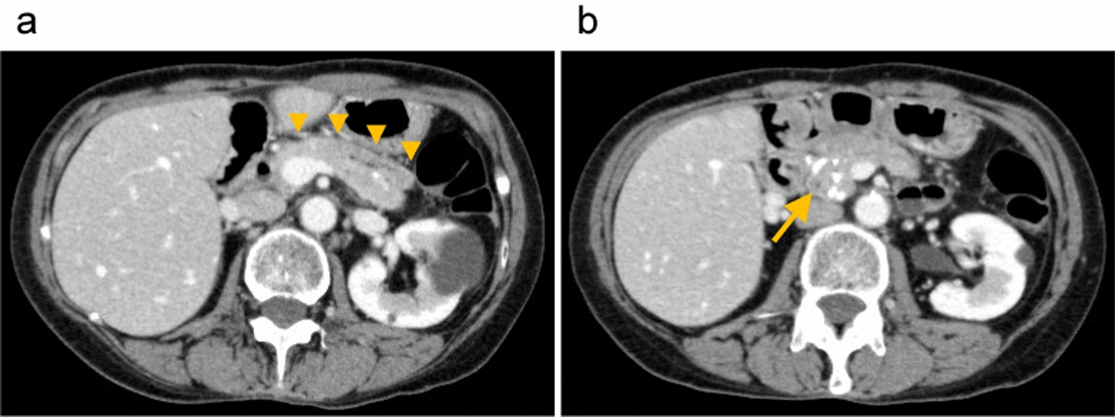
GCA is not usually mass-forming, and the appendix may appear grossly unremarkable [10], and unlike other appendiceal diseases, the CT imaging findings are variable. GCA is, therefore, difficult to identify preoperatively and most cases are diagnosed incidentally after appendectomy. Histologically, appendiceal GCA is an amphicrine tumor composed of goblet-like mucinous cells, as well as variable numbers of endocrine cells and Paneth-like cells, typically arranged as tubules resembling intestinal crypts [11]. All GCAs stain positive with periodic acid-Schiff and Alcian blue for mucin, while immunohistochemical staining shows pronounced expression of the neuroendocrine markers chromogranin A and synaptophysin [12]. In the present case, the tumor grew as a cluster composed of goblet-like mucinous cells with a small number of mixed endocrine cells, leading to the diagnosis of GCA.
The most recent tumor, node, metastasis (TNM) staging system of the combined American Joint Committee on Cancer and UICC classification stages mixed-histology tumors such as GCA using the staging classification for appendiceal carcinomas. The reported 5-year survival rates for GCA Stages I, II, III, and IV are 91.1%, 90.5%, 57.0%, and 18.9%, respectively [13]. The rate of lymph node positivity increases in a stepwise fashion, with lymph node-positivity rates of 1.1%, 2.1%, 9.9%, and 29.1% for T1, T2, T3, and T4 tumors, respectively [14].
The growth pattern of GCA is within the lamina propria and extends through the muscularis propria into the subsequent serosa, with the mucosa remaining intact [15]. GCA has the potential to spread intraperitoneally, even in the absence of nodal metastases [16]. Mcbey et al. showed that only 17% of 224 patients who underwent right hemicolectomy had lymph node metastasis, but 65% of them showed spread through the serosa, invasion of the mesoappendix, or extension to adjacent organs or peritoneum [1]. GCA rarely metastasizes hematogenously to the liver and lungs, despite being widely disseminated on the abdominal surfaces [17], but PNI has been reported in approximately 25% of GCAs and is common among all grades [11, 17]. One study found that PNI was associated with a poor prognosis in multivariate analysis [17]. PNI is often described as skip lesions, which are thought to indicate neoplastic infiltration of a nerve with areas of disease-free nerve [18]; however, some pathologist argue that skip lesions are merely a processing artifact [19]. The existence of skip lesions in cases of PNI makes it difficult to determine the pathological resection margin, leading to the possibility of residual tumor. Thus, even if the appendiceal stump is negative but PNI is positive, as in the current case, additional resection is considered necessary because of the possibility of extension into the cecum.
The most important treatment for GCA is curative resection of the tumor. The American Society of Colon and Rectal Surgeons, the North American Neuroendocrine Tumor Society, and the European Neuroendocrine Tumor Society all recommend right hemicolectomy as the standard surgical treatment for appendiceal GCA of any stage or histology [20,21,22]. In Japan, however, there is currently no consensus on the optimal management strategy for GCA due to the rarity of the disease. It has been reported that 38% of tumors were upstaged following secondary right hemicolectomy, as a result of invasion of lymph nodes in the mesentery [23]. Although lymph node metastasis was negative in the present case, lymph node dissection should be performed because GCA has a high incidence of potential lymph node metastasis to the mesentery.
A German multicenter series reported recurrence rates of 0%–12%, 21%–41%, and 61%–75% for Stages I, II, and III appendiceal carcinoma, respectively [23,24,25], mostly with peritoneal recurrence [26]. Appendiceal neoplasms are frequently associated with perforated appendicitis and the tumor cells are thus likely to reach the serosa because of the thin muscular propria [27]. The value of adjuvant chemotherapy remains unclear, but some reports showed that adjuvant treatment, as for adenocarcinoma, prolonged overall survival in patients with node-positive (Stage III) disease [13, 14]. Although the current patient had Stage II disease, PNI was observed and the risk of recurrence was thus considered to be high, and we, therefore, proposed adjuvant therapy; however, the patient refused because of the risk of adverse events. Adjuvant therapy may help to control peritoneal recurrence and prolong overall survival; however, further studies are needed to investigate the indications for adjuvant therapy.


Comments (0)