The results of the present study indicated that, in comparison to the TPS technique, the PPDS technique achieved a higher success rate in bile duct cannulation and a lower incidence of PEP. However, in instances where the TPS technique was unsuccessful, subsequent incision along the surface of the pancreatic duct stent toward the bile duct using a needle knife resulted in a total success rate comparable to the PPDS technique. Among cases where TPS technique involved immediate placement of pancreatic duct stents, all PEP instances were of mild-to-moderate in severity, and no severe cases were reported, suggesting an overall favorable outcome. The data of the present study exhibited a strong comparability on multiple aspects. Firstly, the two techniques were implemented during distinct periods: the PPDS technique was utilized from April 2019 to December 2021, and the TPS technique was employed from January 2022 to March 2023. Secondly, all patients received NSAID prophylaxis immediately prior to ERCP, the same type of pancreatic stents were placed for all patients during ERCP procedures. Additionally, both groups shared similar baseline characteristics, including age, gender, common bile duct diameter, periampullary diverticulum, and etiologies. Consequently, the results of the present study possess a high level of credibility.
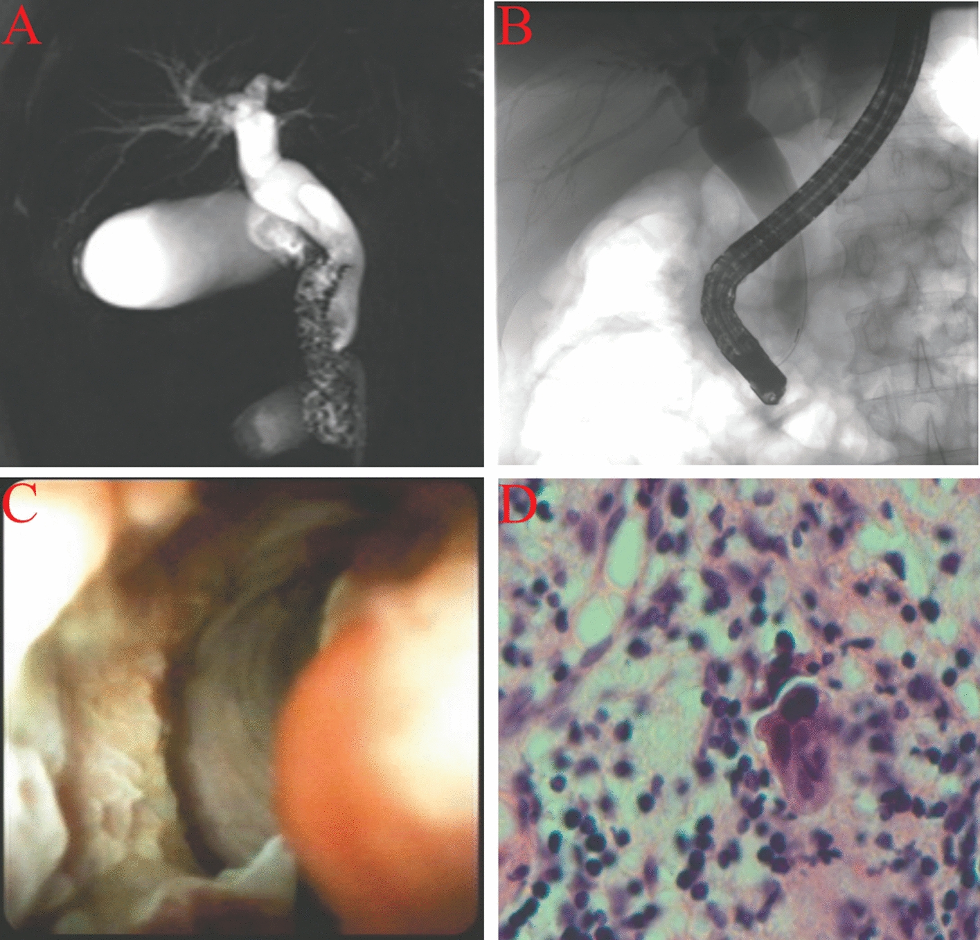
TPS technique involves cutting the septum between the bile duct and pancreatic duct using a sphincterotome, exposing the lower end of the bile duct and aiding in cannulation [14]. TPS is recognized for its simplicity and controlled incision length. However, the procedure may induce pancreatic duct spasms, edema, and increase the risk of PEP [19, 20]. Guidelines suggested placing a pancreatic stent with TPS to prevent PEP [13, 15], while optimal timing remains uncertain. In this study, a pancreatic duct stent was immediately placed after TPS, facilitating uninterrupted pancreatic duct outflow during the entire ERCP procedure. The stent served dual purposes. Firstly, it prevented PEP by maintaining the pancreatic duct unobstructed. Secondly, it acted as a guide to straighten the papilla, preventing guidewire re-entry and promoting bile duct cannulation. In instances of unsuccessful cannulation, mainly due to a long common channel or incomplete septum incision, using a needle-knife over the stent’s surface resulted in the successful bile duct cannulation. While pure TPS has exhibited a moderate success rate, the combined success rate, incorporating needle-knife incision, reached nearly 98%. Importantly, no instances of stent displacement occurred.
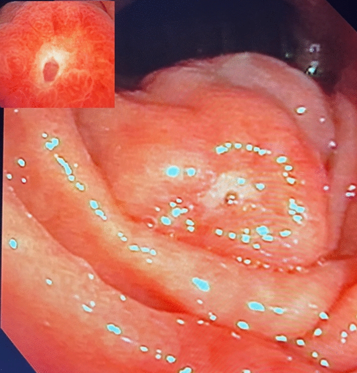
The PPDS technique represents an innovative approach to needle-knife precut sphincterotomy. Traditionally, needle-knife precut sphincterotomy involves two distinct techniques. The first is conventional needle-knife precut papillotomy (NKPP), entailing an incision commencing at the 11 o’clock margin and directs upwards towards the common bile duct (CBD). The second technique is needle-knife fistulotomy (NKF), where the initial incision occurs at the roof of the papilla and is directed either upwards or downwards based on the anatomical considerations. The objective of NKF is to preserve the delicate orifice area, thereby minimizing the risk of pancreatic duct damage related to electrical current and subsequent PEP [21,22,23]. Both techniques necessitate proficient ERCP endoscopists, particularly with NKF requiring a specific anatomical foundation, notably a longer papilla (e.g., papilla type 3) [24, 25]. The implementation of NKF may pose challenges in cases with flat or small papillae, especially those on the inner margins of diverticula (type IIa) [26]. The PPDS technique targets the same incision point as the NKPP technique at the low end of the bile duct. However, its incision method aligns more closely with the NKF technique. Guided by a pancreatic duct stent, the PPDS technique precisely and briefly incises the low end of the bile duct, contrasting with NKPP’s lengthier incision. Importantly, it preserves the pancreatic duct sphincter around the pancreatic duct stent in the lower part of the papilla, reducing adverse events, such as PEP and perforation. Thus, the PPDS technique combines the advantages of both NKPP and NKF techniques while mitigating their drawbacks. The ability of the PPDS technique to preserve the sphincter around the pancreatic duct stent while precisely incising the bile duct sphincter is rooted in the anatomical relationship between the bile duct and pancreatic duct in the papilla. Through extensive practical experience, it is demonstrated that in the endoscopic view, the bile duct lies left, anterior, and upward of the pancreatic duct, while the pancreatic duct is situated right, posterior, and downward under the bile duct. Anatomically, the bile duct runs from the left upper quadrant (at the 11 o’clock position) of the papillary orifice to the mid-point (12 o’clock position) of the papilla’s upper part, and the pancreatic duct extends from the midpoint of the papillary orifice to the upper part of the papilla, spanning the 1–3 o’clock positions. By incising the mucosal and submucosal layers at the joint point using a needle-knife, about 2–3 mm thick, the bile duct sphincter can be identified. Additional incision, typically measuring 1–2 mm in thickness, effectively aids in the cannulation of the bile duct. Our research team’s clarification of this anatomical relationship is groundbreaking, holding the potential to substantially enhance the success rate of both bile duct and pancreatic duct cannulation while reducing the rate of adverse events, pending further confirmation.
In a comparison between TPS and PPDS, the results of two meta-analyses indicated that TPS demonstrated a greater biliary cannulation rate compared with other advanced cannulation techniques, and both early needle-knife and TPS techniques outperformed in reducing the PEP rate [27, 28]. While some experts regarded TPS as a potential alternative for challenging biliary cannulation [7, 29], opinions on its efficacy vary [30, 31]. As a relatively recent needle-knife precut technique, PPDS currently lacks adequate data to assess its comparative effectiveness against other techniques.
According to the findings of this study, the following observations were highlighted. Firstly, PPDS exhibited a higher success rate in bile duct cannulation compared with TPS. However, when TPS was accompanied by needle-knife incision, it could consistently achieve successful bile duct cannulation, resulting in an overall success rate of 97.8%, which is consistent with previously reported results [32]. The final success rate of bile duct cannulation did not exhibit significant differences between the two techniques. The lower success rate with TPS alone (69.6%) was attributed to a limited incision length, especially inadequate for patients with a longer common channel of the bile duct and pancreatic duct. Secondly, TPS was associated with a higher incidence of PEP compared with PPDS. However, the majority of PEP cases were mild, with a smaller proportion being of moderate severity, and the overall outcomes remained satisfactory. Thirdly, PPDS may encounter challenges in cases of a deviated papilla, such as those within the inner margins of the diverticulum (type IIa), while TPS proved to be more versatile for all papilla types. Fourthly, TPS is a relatively simple procedure, eliminating the need to exchange the sphincterotome for a needle knife, thereby reducing costs. Consequently, PPDS may be more appropriate for high-risk PEP patients [33], including female patients with normal liver function and those with dysfunctional Oddi sphincter.
This single-center retrospective study with a relatively small sample size underscores the need for future multicenter prospective studies to validate the findings.
In conclusion, when encountered with difficult biliary cannulation and accidental guidewire insertion into the pancreatic duct, both PPDS and TPS followed by immediate pancreatic duct stent placement, are viable options. TPS stands out for its simplicity and cost-effectiveness, while PPDS is more appropriate for patients who are at a high-risk of developing PEP.


Comments (0)