
Remember me


Breast cancer is the most common cancer in women [1] and is currently one of the most frequently diagnosed cancers and the fifth cause of cancer-related deaths with an estimated number of 2.3 million new cases worldwide according to the GLOBOCAN 2020 data [2].
According to the literature, tumors located in the retro-areolar region account for 5 to 20% of breast cancer [3, 4].
Patients with centrally located breast cancer were used to receive radical mastectomy because of safety and cosmetic problems, but recently breast-conserving therapy in combination with postoperative radiotherapy has been shown to have an equivalent survival rate to mastectomy and has emerged as the most effective method of treating breast cancer [5].
The development of oncoplastic surgery in recent years has allowed for an improvement in the treatment of breast cancers. It maintains the same level of oncological safety as radical surgery while enabling the performance of a massive glandular excision with a satisfactory aesthetic outcome.
In 1993, Galimberti et al. described an oncoplastic procedure (Grisotti flap) for the management of retro-areolar breast cancers. He described the technique as excision of the NAC, directly over the site of the tumor, extending down to mobilize a dermo-glandular flap which is then de-epithelized in order to reshape the breast and recreate an areola [6].
In tumors involving lactiferous ducts but 1 cm away from the areola, we tried to perform nipplectomy with areola preservation and central quadrantectomy to avoid losing the normal color and sensation of areola and keep the normal anatomical configuration of the breast.
In our study, we will compare the Grisotti technique versus nipplectomy and central quadrantectomy with areola preservation, as in malignancies extending to the nipple, more than 1 cm away from the areola.
Aim of workTo compare nipplectomy and central quadrantectomy with areola preservation as a new reconstructive oncoplastic technique Versus Grisotti flap mammoplasty in central malignant tumors of the breast extending to the nipple, in terms of time procedures, breast symmetry, patient satisfaction, postoperative complications, and local recurrence.
Patients and methodsThe present study was a single-blind, prospective, randomized, controlled single-center trial that was conducted at University Hospitals' breast surgical unit from May 2018 to May 2023. This trial followed the CONSORT guidelines and involved 40 female patients who had centrally located breast carcinoma extending to the nipple. Twenty patients underwent nipplectomy and central quadrantectomy with areola preservation, and the other 20 patients underwent Grisotti flap mammoplasty. The citations, references, and in-line citations were not modified, and the American English spelling, specific terms, and phrases were strictly adhered to.
Inclusion criteria (1)Female Patients above 18
(2)Patients with proven breast cancer involving lactiferous ducts but at least 1 cm away from the areola.
Exclusion criteria (1)Candidates with ages below 18 years old.
(2)Patients with proven breast cancer involving lactiferous ducts less than 1 cm away from the areola Patient refusal.
All individuals (as shown in Table 1) were initially evaluated during the preoperative period, following the confirmation of their diagnosis via biopsy. A multidisciplinary team consisting of a surgeon, oncologist, pathologist, and radiation therapist determined whether to proceed with upfront surgery or neoadjuvant therapy. The same surgeon performed all operations. For each case, the status of the lymph nodes was assessed through either a complete lymph node dissection or a sentinel lymph node biopsy with frozen section analysis. Depending on the lymph node status, a complete axillary lymph node dissection might be performed. After tumor resection in the central quadrant, an intraoperative frozen section analysis is conducted to verify clear margins.
Table 1 Detailed descriptions of patient tumor characteristicsRandomization and blindingThe randomization process was carried out the day before surgery, and patients were assigned to either experimental Group I for nipplectomy and central quadrantectomy with areola preservation or experimental Group II for Grisotti flap mammoplasty using a computer-generated randomization code. The two groups were balanced at a ratio of 1:1 to ensure that the study was conducted under single-blind conditions. This study was a randomized control trial of parallel design, with a sample size of 20 patients in each group, resulting in a total sample size of 40 patients. The sample size was estimated using the online software "Riskcalc®" and the study was a non-inferiority clinical trial with a least allowable clinical difference of 15%. The study patients were included and randomized via block randomization, where the first twenty-eight patients who met the eligibility criteria underwent axillary lymph node ultrasound-shear dissection, while the next twenty-eight patients underwent conventional radio frequency electric dissection.
Pre-operativeThe pre-operative investigationsLaboratory tests, including a routine complete blood count, liver and kidney profiles, coagulation profile, blood sugar level, and comprehensive virology screening, were performed. Additionally, bilateral mammography and ultrasound exams (for breast and axillary exploration) were conducted, with optional bilateral breast magnetic resonance imaging (MRI) if necessary. Biopsies were taken from the tumor, followed by histological and immunohistochemical analysis, including the examination of hormone receptors, Ki67 proliferation index, and HER2 status. ECG and echocardiography were performed upon request by the anesthesiologist when indicated, along with stress ECG. Finally, patient counseling and consent were obtained.
Operative detailsAll surgical procedures were carried out by a single team under general anesthesia. Prior to the start of the operation, all patients received a single dose of 1g of a third-generation cephalosporin intravenously. The breast surgery team, consisting of multiple specialties, discussed and planned the surgical scar resection area, tumor size and location, and axillary dissection. The reduction pattern and tumor location were marked on the patient's standing breast, followed by prepping and draping. The operation began with a sentinel lymph node biopsy in the ipsilateral axilla, which was subjected to frozen section analysis. The tumor was then removed, weighed, and sent for mammographic and pathological evaluation. After the tumor was removed, tissue extensions were taken from all tumor bed dimensions, and surgical clips were used to mark the tumor bed margins to aid in the location of the original tumor bed for the expected radiation boost. The contralateral breast underwent adjustment using the superior-medial pattern of breast reduction. Finally, the patient was positioned upright for the final assessment of symmetry, flap placement, and breast shape. Drainage was assessed in each breast using the oncoplastic technique, and if necessary, two drains were inserted (one in the contralateral breast and one in the axilla after axillary dissection). The incisions were then sutured, with suturing done in stages to ensure that the deeper glandular layers were positioned as far forward as possible to completely fill the central defect and create a satisfactory cone. After the subcutaneous layers were closed, cutaneous closure was achieved using continuous intradermal sutures. The surgical scars were then covered with strips and gauze pads, and a comfortable sports bra was fitted over the entire operated field.
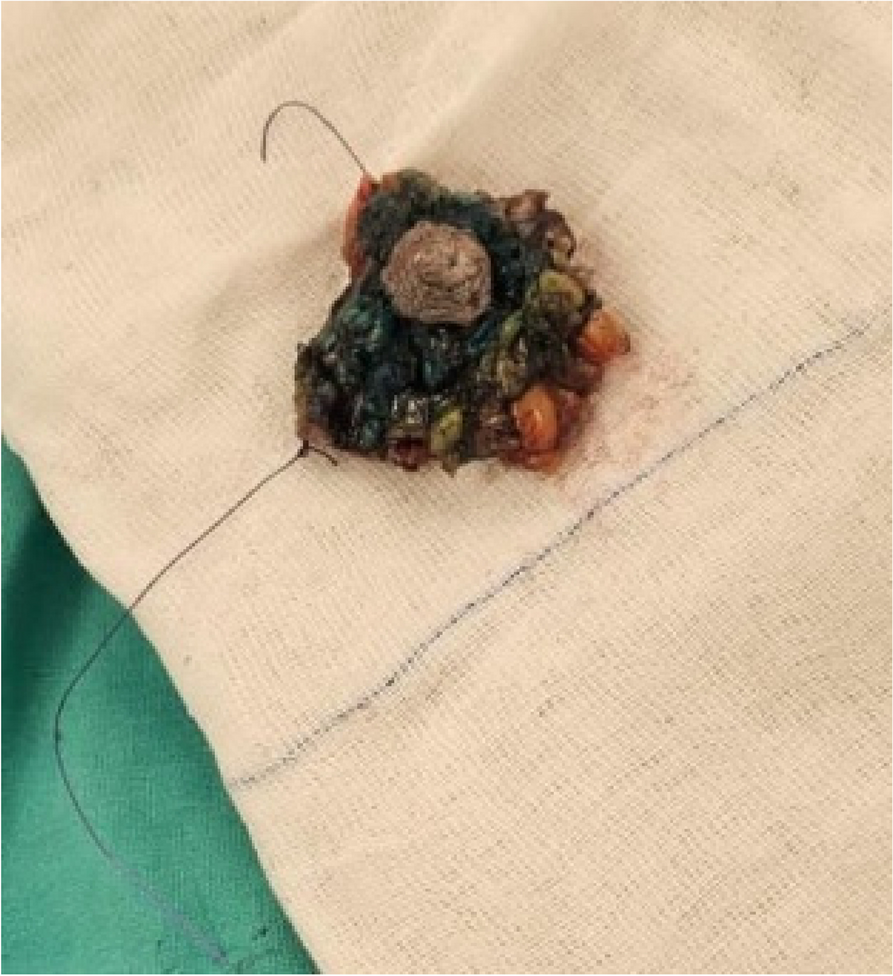
Group (I) underwent nipplectomy and central quadrantectomy with areola preservationA periareolar round incision and a cut around the nipple were used to perform a wire-guided lumpectomy, reaching the depth of the resected breast cone tissue and preserving the areola. Figure (1).
After the tumor had been removed, the orientation of the breast defect determined the positioning of the glandular pedicle and the tissue rearrangement. The pedicle stub was obtained along with additional glandular tissue from the remaining breast, which was then repositioned in the central area of the breast to achieve better projection. Fig (2).
Fig. 1 Fig. 2
After closing the subcutaneous layers in nipplectomy and central quadrantectomy with areola preservation with the drain
Group (II) underwent Grisotti flap mammoplastyThe preoperative steps involve marking the inframammary line, NAC outline, new nipple and areola position below the NAC, and two vertical lines to mark the pillars of the flap, starting from the most lateral points of the old NAC and extending longitudinally down to the previously drawn inframammary line (Fig. 3).
Next, a central quadrantectomy including the NAC and tumor with a cone of tissue from the subcutaneous layer down to the pectoral fascia was performed with ample safety margins (Fig. 4).
Then, complete de-epithelialization of the flap was carried out, excluding the new areola (Fig. 5).
A caudally located, de-epithelialized pedicled flap was created, including a skin island for restoring the areola (Fig. 6).
After carefully and meticulously mobilizing the flap from the lateral pillars and dissecting down to the pectoral fascia, the flap was easily rotated upward to fill the empty central quadrant, with the skin island replacing the removed nipple-areola complex (Fig. 6).
Fig. 3
Marking breast for Grisotti Flap mammoplasty
Fig. 4
Central quadrantectomy including NAC and tumor with a cone of tissue
Fig. 5
Complete de-epithelialization of the flap
Fig. 6
After closing the subcutaneous layers in Grisotti Flap mammoplasty
During the year following surgery Fig. 7, patients underwent monitoring and management of potential complications, including wound infection, seroma, and recurrence. Oral antibiotics were administered for a week after the procedure, and wound dressing was performed on the second day postoperatively. All patients were instructed on the wound dressing process, and follow-up appointments were scheduled every two weeks until the wound was fully healed. Furthermore, follow-up appointments were held every two months for a period of two years after complete healing.
Fig. 7
During early follow-up for the Grisotti Flap Mammoplasty group
Statistical analysisThe researchers utilized IBM SPSS Statistics version 26 to gather, process, and analyze the data. They transformed qualitative data into numerical values and percentages, while quantitative data was presented as means, standard deviations, and ranges with a normal distribution. For comparing two groups with qualitative data, the chi-square test was employed, and if any expected count in any cell was less than 5, the Fisher exact test was used. For comparing two independent groups with quantitative data and a normal distribution, the independent t-test was used, and for more than two groups, One-way ANOVA was applied. The confidence interval was set at 95%, and the margin of error accepted was 5%. Therefore, the p-value was considered significant as follows: P > 0.05: Not significant (NS), P < 0.05: Significant (S), and P < 0.01: Highly significant (HS).
Comments (0)