We herein report a patient previously undiagnosed with lupus nephritis, who developed PRES presenting sudden onset of visual impairment, headache, and hypertensive emergency. Her PRES-associated neurological symptoms and brain-MRI abnormalities were improved following anti-hypertensive treatment with calcium channel blocker. A kidney biopsy showed a class IV-G (A) lupus nephritis with vasculitis and the immunosuppressive therapy with intravenous mPSL pulse followed by oral PSL, MMF, and intravenous belimumab, attenuated SLE-associated clinical manifestations including butterfly rush, edema, renal dysfunction, and proteinuria.
PRES has been well documented in patients with established diagnosis of SLE and lupus nephritis. However, as far as we are aware, there were only four reported cases with undiagnosed lupus nephritis who developed PRES as summarized in Table 1 [9,10,11,12]. The age at the onset of PRES was relatively young and ranged from 21 to 37 years. All cases were female and had severe hypertension. Most patients had renal insufficiency and massive proteinuria, one patient required hemodialysis, and one patient received plasma exchange. Three out of four patients underwent kidney biopsy and were diagnosed with lupus nephritis, including one class IV and two class V cases. Most cases received immunosuppressive agents, such as glucocorticoids, MMF, azathioprine, and cyclophosphamide, and hydroxycholoroquine. Taken together, age, sex, presence of hypertension, and renal insufficiency and massive proteinuria with lupus nephritis (class IV), and immunosuppressive treatment, in this case were overall similar to those in previous cases, and these clinical features are generally consistent with risk factors of PRES in patients with established diagnosis of SLE [6, 7].
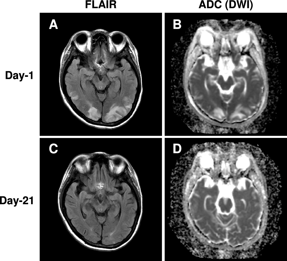
Table 1 A summary of previous PRES cases in undiagnosed lupus nephritisAlthough the overall prognosis of PRES is favorable, permanent neurological impairment and death can occur [1, 2]. Previous studies have shown that intracranial hemorrhage and brainstem involvement are major risk factors of poor clinical outcome of SLE-associated PRES, such as incomplete recovery and death [13]. More importantly, PRES is associated with a high mortality rate in SLE patients [14]. Our patient significantly improved PRES-associated clinical symptoms and brain-MRI lesions during hospitalization. Nonetheless, given the presence of subcortical hemorrhage at the diagnosis of PRES, additional follow-up brain imaging and neurological evaluation should be considered important in our case.
The underlying pathogenic mechanism of PRES remains unclear, but could be explained by rapid elevation of arterial blood pressure above the upper limit of cerebral autoregulation, leading to cerebral hyperperfusion and vasogenic edema [1, 2]. Supporting the notion, our patient developed hypertensive emergency around the time of PRES onset and anti-hypertensive therapy led to resolution of PRES-associated clinical symptoms and MRI abnormalities. SLE and lupus nephritis likely contribute to the development of hypertension through multiple complex mechanisms including chronic inflammation, oxidative stress, endothelin, renin–angiotensin system, and metabolic derangement in our case [15]. In addition, we cannot exclude the possibility that low-estrogen oral contraceptive use may have affected onset of hypertensive emergency [16]. Another possible mechanism of PRES involves endothelial damage due to systemic and local inflammatory responses associated with SLE and subsequent increased blood–brain barrier (BBB) permeability [17]. Indeed, previous studies have shown that SLE is an independent risk factor for the development of vascular inflammation and endothelial dysfunction [18, 19]. Unfortunately, we did not directly evaluate markers of systemic and local endothelial dysfunction such as circulating concentrations of tissue-type plasminogen activator, and von Willebrand factor, flow-mediated dilatation (FMD), carotid-intima media thickness, arterial stiffness and ankle-brachial index (ABI) [20]. However, given the presence of biopsy-proven renal vasculitis, our patient could have had endothelial damage in multiple organs including brain. Consistent with our findings, PRES is associated with vasculitis involving kidney including ANCA-associated vasculitis [21] and anti-glomerular basement membrane disease [22]. In addition, uremic toxins caused by renal insufficiency, a key risk factor of PRES [23], could trigger endothelial damage in our case [24]. We did not detect thrombocytopenia or pathological changes in the kidney biopsy indicating the development of thrombotic microangiopathy (TMA). Although we cannot rule out a possible sampling error associated renal biopsy, these findings indicate that TMA may not be a major contributor to systemic or renal endothelial damage in our patient. Nonetheless, elevated anti-cardiolipin antibodies may induce locally prothrombotic effects and brain endothelial dysfunction, leading to disruption of BBB [25]. Taken together, hypertension and endothelial dysfunction, two distinct but interrelated pathogenic mechanisms associated with lupus nephritis, could synergistically contribute to the development of PRES in our patient.
Recent studies found that 4–28% of patients with lupus nephritis eventually develop end-stage renal disease (ESRD) [26] and that hypertension at the onset of lupus nephritis predicts the development of CKD and ESRD [27, 28]. In addition, our patient had other risk factors of ESRD associated with lupus nephritis such as race (nonwhite), eGFR at baseline (36.6 mL/min/1.73 m2), hypocomplementaemia (C3 26 mg/l, C4 4 mg/l, and CH50 < 14 U/mL) and renal histology (class IV) [26, 29]. Given that our patient did not achieve remission of proteinuria following immunosuppressive therapy, long-term careful monitoring and management of disease activity in lupus nephritis are considered to be particularly necessary and important.
In conclusion, we present a case of PRES in a patient previously undiagnosed with SLE and lupus nephritis. There are various neuropsychiatric syndromes in SLE patients who may have significant overlap in neurological symptoms with PRES such as headache, seizures, epilepsy, and reduced consciousness. Thus, our case highlights the need to consider PRES as an initial clinical presentation of lupus nephritis and provide the early diagnosis and timely treatment to achieve a favorable outcome.


Comments (0)