
Remember me


A 14-year-old girl came to our attention, accompanied by her parents, complaining the appearance of painful vulvar lesions.
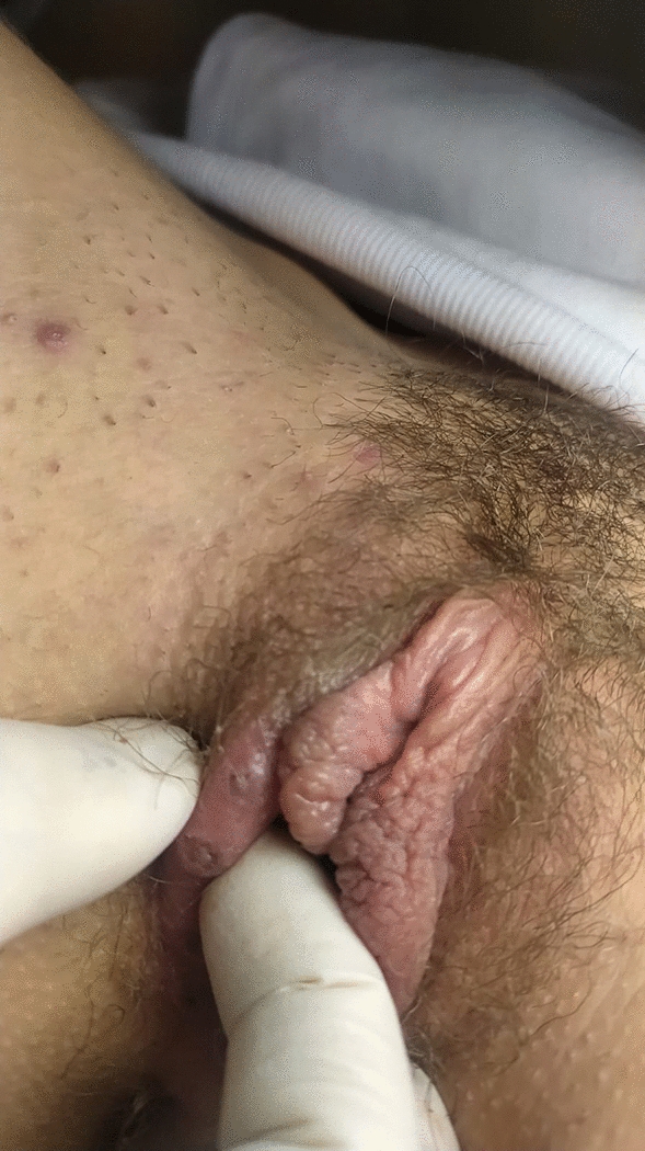
During the gynaecological examination, the presence of multiple ulcers localized on the labia majora and labia minora, < 1 cm in size, in different developmental states, was noted. These ulcerative lesions appeared greyish-white in colour, markedly painful, with a fibrinous background. Upon palpation, they had a soft consistency. Leucorrhoea is absent. A mild lymphopathy was appreciated in the groin. No signs of trauma is observed (Figs. 1, 2).
Fig. 1
Lipschütz ulcer: right labia majora
Fig. 2
Lipschütz ulcer: left labia minora and vestibule
Absence of vesicles in the vulvar and anal area.
The patient also reported asthenia, fever, physical discomfort and headache.
Virgo patient. The absence of oral lesions in the anamnesis do not allow the diagnosis of Behçet’s disease. Concomitant modest pharyngotonsillitis and mild pharyngeal erythema. In the immediate medical history, a very recent hospitalization for Mycoplasma pneumoniae pneumonia emerged (positive throat swab, positive ELISA test). Despite this, the patient underwent blood chemistry tests, oropharyngeal swab and genital swab.
White blood cells were normal except for mild lymphocytosis in percentage terms (66%, normal range 19–45%).
Light increase in transaminases: Alanine aminotransferase (ALT) (56 U/I, normal range < 40 U/I), lactate dehydrogenases (LDH) 318 U/I (normal range 0–248 U/I).
Antibodies for hepatotropic viruses: Hepatitis B surface antigen (HbsAg): negative; antibodies to Hepatitis C virus (anti-HCV test): negative; Hepatitis A immunoglobulin IgM/IgG (anti-HAV): negative.
Haemagglutination Treponema pallidum (TPHA): negative; Venereal Disease Research Laboratory test (VDRL): negative.
Immunoglobulin M (IgM) for Epstein–Barr Virus (EBV): positive.
SARS-Cov-2 oropharyngeal swab: negative.
SARS-Cov-2 quantitative reverse transcription polymerase chain reaction (RT-qPCR): negative.
Immunoglobulin M (IgM), Immunoglobulin G (IgG) for Herpes Simplex Virus 1(HSV-1) and Herpes Simplex Virus 2 (HSV- 2): negative.
Urethral and vaginal swabs for Bacteria, Candida, Trichomonas, Chlamydia trachomatis, Neisseria gonorrhoeae research: negative.
Swabs performed on ulcerative lesions: negative.
Considering the anamnestic data, the oropharyngeal swab, the blood chemistry tests and the objective examination, the diagnosis of EBV infectious mononucleosis was made and then the diagnosis of Lipschütz ulcer was made. The patient underwent symptomatic therapy. The longitudinal clinical control performed at 14 days showed complete healing of the lesions and confirmed the previously made diagnosis.
Comments (0)