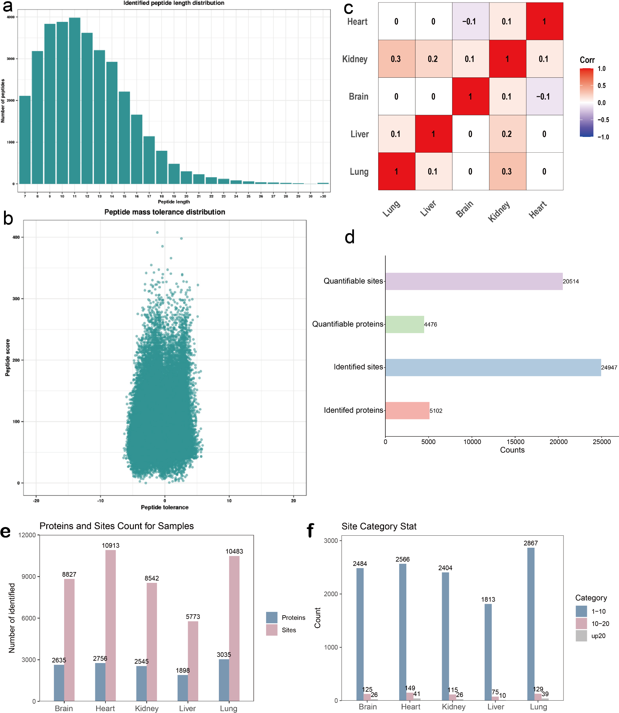
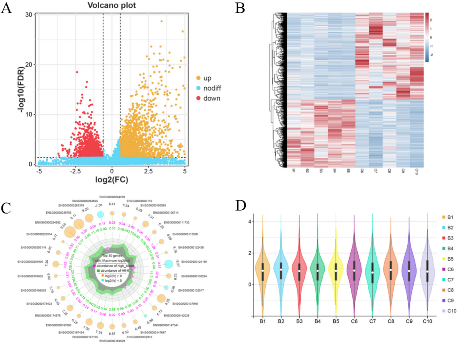
In recent years, septic AKI accounts for 45–70% of all critically ill AKI cases, and its mortality rate remains high. Proteomic analysis can enable us to understand the molecular mechanisms of disease occurrence and progression, provide new ideas for identifying potential diagnostic biomarkers, and can also be a potential target for the treatment of septic AKI. In this study, a rat model of sepsis AKI was established. Heat map (Fig. 2C) shows the standardized expression of DEPs in sepsis. Irf7 is highlighted for its significant upregulation, suggesting its role in inflammation and immune regulation in septic AKI. Ifit2, Ifit3 and Ptx3 are noted for their involvement in innate immunity and inflammatory responses, which align with the pathophysiology of sepsis-induced AKI. Cyp27b1 and Neil3 are discussed for their potential roles in cellular stress response and DNA repair mechanisms, which may contribute to kidney injury during sepsis. Ten key proteins with interaction degree, such as Isg15, Irf7, Oasl2, Ifit3, Apob, Oasl, Ube2l6, Ifit2, Ifih1 and Ifit1 were identified. We demonstrated that Irf7 was significantly up-regulated in rat renal tissues.
Irf and its effectors are closely related to the balance of immunosuppression, and Irf is a key mediator of signal transduction related to host immune response and immune regulation. Immunosuppression also plays a key role in sepsis induced organ failure. Irf is an important regulatory protein in TLR and IFN signaling pathways. Studies have shown that Irf7 is a key regulator of type I interferon against pathogen infection. Various pathogen-associated molecular patterns (PAMPs) and damage associated molecular patterns are sensed by innate pattern recognition receptors, including TLRs, RIG-I-like receptors, C-type lectin receptors and NOD-like receptors [21]. Our KEGG analysis also confirmed that Irf7 plays an important role in TLR (map04620), RIG-I-like receptor (map04622) and NOD-like receptor (map04621) signaling pathways. Irf also contributes to the action of TLR. After PAMPs bind to TLRs or IFN binds to IFN receptors, a signaling cascade causes Irfs to activate and relocate to the nucleus, where they activate gene expression. As such TLR4 downstream signaling pathways either work in a manner dependent on the universal adapter protein called MyD88, or in a MyD88-independent way. In the MyD88-dependent group of TLR4 signal transduction, NF-κB is able to translocation and induce the expression of proinflammatory cytokine genes, such as TNF-α and IL-6 [10, 22].
Thus, blocking Irf-DNA binding with Irf-specific or pan-Irf inhibitors is a promising tool for the treatment of sepsis. Indirect regulation can be achieved by targeting known activators and regulators of Irf expression and key pathways downstream of Irf. The significant upregulation of Irf7 suggests that it could be a potential biomarker for early diagnosis of septic AKI. Future studies could further validate its functional mechanisms in inflammation and kidney injury by knocking down or overexpressing Irf7 in animal and cellular models. Design experiments prove transcriptional upregulation of Irf7 is regulated upstream by phosphorylation of various signaling proteins. Identify the cell type responsible for Irf7 upregulation and validate its signal transduction pathway. The diagnostic predictive ability of sepsis can also be evaluated by clinical detection of Irf7 levels in the blood, peritoneal fluid, or urine of patients with sepsis. At the same time, the development of specific intervention strategies targeting the Irf7 signaling pathway is expected to provide a new direction for the treatment of septic AKI.
Type I IFN induces the expression of more than 500 genes, which are collectively known as IFN-stimulating genes (Isgs). IRF closely controls transcriptional activation of Isg. Interferon stimulating gene 15 (Isg15) is the earliest interferon-induced gene. The free Isg15 protein synthesized by the Isg15 gene is coupled to the cellular protein after translation and secreted by the cell into the extracellular environment. Type I interferon stimulated the activation of more than 2000 Isg transcripts. Ube2l6 (Isg15/ ubiquitin E2 binding enzyme) is one of the key enzymes of Isg. We now know that the occurrence of sepsis AKI is closely related to autophagy, and the autophagy pathway is up-regulated under cellular stress [23]. Our data show that Ube2l6 and Isg15 protein expression is significantly upregulated in septic AKI renal tissue. Studies have confirmed that the expression level of Isg15mRNA is positively correlated with the occurrence of sepsis and septic shock [24]. Recent studies have shown that Isg15 overexpression can interact with HDAC6 and p62 to promote aggregate formation and thus promote autophagy [25]. Therefore, we hypothesize that decreasing the expression of Isg15 and Ube2l6 may reduce autophagy to alleviate sepsis AKI.
The interferon-induced tetrapeptide repeating protein (Ifit) gene is a prominent Isg. Ifit proteins are involved in a variety of biological processes, including host innate immunity, antiviral immune response, virus-induced translation initiation, replication, double-stranded RNA signaling, and PAMP recognition [26]. Although Ifit protein was originally studied as an antiviral protein, recent studies have shown that its expression in the context of sepsis also has important implications. In this study, Ifit3, Ifit2, and Ifit1 were significantly up-regulated in septic AKI. Alexandra Siegfried’s research showed that Ifit2 is a key signaling medium in LPS-induced septic shock, and the expression of Ifit2 is significantly up-regulated under LPS stimulation in IFN-a receptor and Ifit9-dependent manner, and Ifit3 and Ifit1 are also up-regulated accordingly [27].
In addition, Ifit1, Ifit2, and Ifit3 combine into a stable trimer complex in humans, where Ifit3 enhances and regulates the central hub of Ifit1RNA binding, while Ifit2 also promotes apoptosis. Ifit2-mediated apoptosis acts through the mitochondrial pathway, in which the balance between pro-apoptotic and anti-apoptotic Bcl-2 family proteins regulates the permeability of the mitochondrial outer membrane, and overexpression of Ifit2 leads to activation of Caspase-3 and disruption of plasma membrane asymmetry, which is characteristic of apoptotic cell death [28, 29]. In overexpression studies, Ifit3 co-expression has been shown to improve Ifit2-dependent apoptosis. In addition, Ifit2 is involved in the negative regulation of inflammation by down-regulating Toll-like receptor 4 response and regulating reactive oxygen species production. Ifit family members participate in a variety of pathophysiological processes in the body, regulate the homeostasis and differentiation of a variety of cells, including immune cells, and are closely related to a variety of autoimmune diseases, which is expected to become a new therapeutic target [30]. Therefore, Ifit1, Ifit2 and Ifit3 may be the key nodes in the pathogenesis of sepsis AKI.
In this study, a peritoneal injection of lipopolysaccharide sepsis AKI model was established. The degree of AKI infection in this model can be controlled by the dose of lipopolysaccharide, and the model is stable and reproducible [31]. However, there are some limitations in this study, and our hypothesis based on database calculation needs to be further verified. In future studies, we plan to expand the sample size and validate it in conjunction with other animal models, such as different species or methods of sepsis induction. In the future, we will increase cell experiments and clinical trials to verify various aspects. In this study, proteomics was used to analyze the molecular mechanism of sepsis AKI, which should be combined with genomics, metabolomics, and bioinformatics to construct a global molecular network of sepsis AKI.


Comments (0)