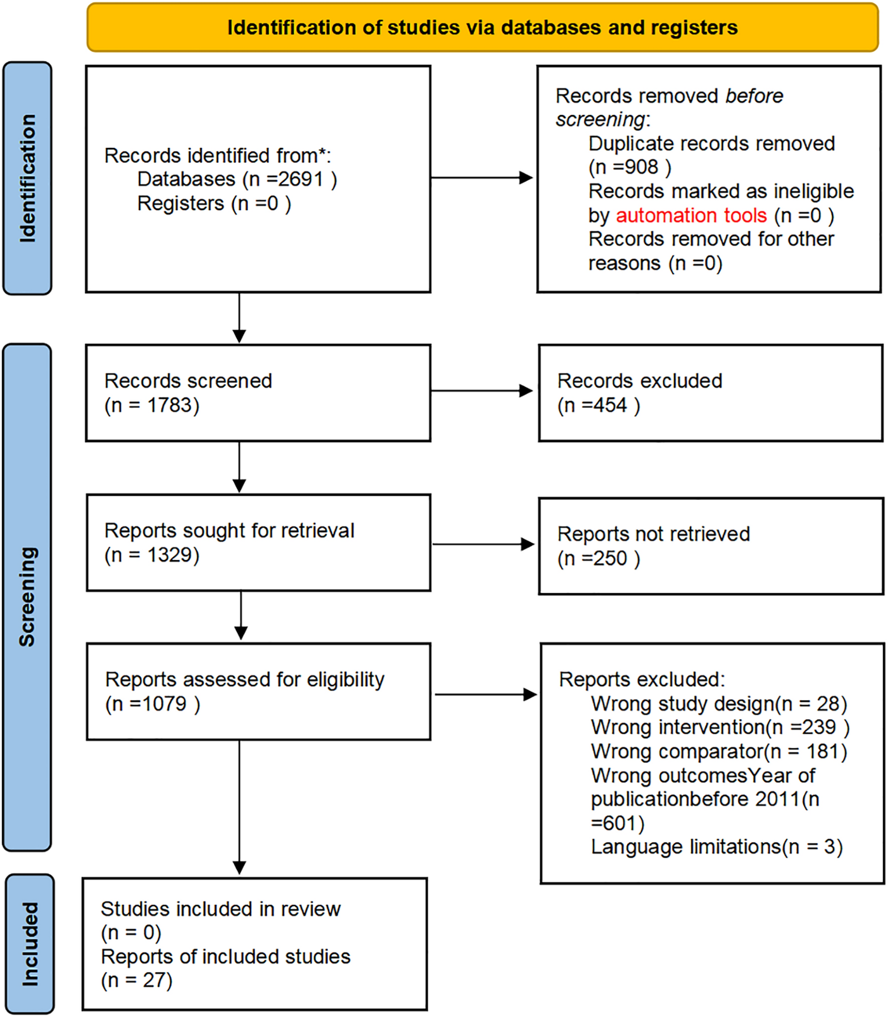
In this retrospective study of full-thickness degenerative rotator cuff tears, we found that both obesity and preoperative nutritional status were associated with rotator cuff muscle degeneration. Patients with obesity (BMI ≥ 28 kg/m²) had a higher prevalence of muscle atrophy and fatty infiltration compared to non-obese patients. Similarly, indicators of malnutrition—including lower PNI, serum albumin, and hemoglobin levels—were significantly associated with the presence of rotator cuff muscle atrophy and fatty infiltration. Logistic analysis adjusted by age suggested that nutritional factors (e.g. low PNI and albumin) remained predictors of muscle degeneration, underscoring the role of systemic health in rotator cuff pathology. Overall, our results support the concept that modifiable patient factors, such as body composition and nutritional status, can influence the extent of muscle atrophy and fatty change in chronic rotator cuff tears.
Obesity is a well-established risk factor for a wide range of metabolic, cardiovascular, and even psychological disorders [19]. In recent years, it has also been increasingly recognized as a contributing factor to the progression and poor prognosis of rotator cuff disease [11, 20,21,22]. Previous studies suggest that obesity may induce a state of chronic low-grade inflammation, which promotes muscle degeneration through inflammatory mechanisms [23]. The release of pro-inflammatory cytokines has been shown to impair cellular regenerative capacity and accelerate apoptosis [24, 25]. A growing body of evidence supports the role of macrophage recruitment and activation in adipose tissue as a key driver of this inflammation, suggesting that obesity may be best characterized as a chronic inflammatory disease originating in fat depots [19, 23]. Under this persistent inflammatory condition, structural and functional changes occur in musculoskeletal tissues through pathways involving oxidative stress, mitochondrial dysfunction, and programmed cell death [19, 24]. This inflammatory milieu ultimately triggers programmed cell death pathways described in apoptosis reviews [26]. These processes collectively contribute to muscle atrophy and fatty infiltration, key features of rotator cuff muscle degeneration. Consistent with previous findings, our study demonstrated that obesity may increase the likelihood of muscle atrophy and fatty infiltration in patients with degenerative rotator cuff tears. Given that modifiable risk factors like obesity are associated with impaired tendon healing and worse postoperative outcomes after rotator cuff repair, our results emphasize the potential value of preoperative intervention. Targeting this modifiable metabolic state—through weight reduction, dietary optimization, or inflammation control—may help slow disease progression and improve surgical prognosis in patients undergoing treatment for rotator cuff pathology. However, previous study has shown that triglycerides and total serum cholesterol(lipid markers typically elevated in obese patients) have not been significantly associated with rotator cuff tears [27]. Accordingly, metabolic management should not rely solely on lowering these lipid levels but instead adopt a broader strategy that targets multiple aspects of the patient`s metabolic profile. Equally important, recent evidence further suggests that body-mass index is socially patterned: patients in the most deprived tertile showed a mean BMI of 32.2 kg/m² compared with 28.1 kg/m² in the least deprived group [28]. This socioeconomic-obesity link may partly explain why disadvantaged individuals experience more profound rotator-cuff muscle degeneration
Nutritional Status, Laboratory Markers, and Rotator Cuff Muscle Degeneration
Our study underscores that poor preoperative nutritional status—particularly as measured by the PNI—is independently associated with more severe rotator cuff muscle degeneration, including both atrophy and fatty infiltration. Nutritional status is closely related to the immune system and the local cellular microenvironment [12, 29]. Tendinopathy, as a condition intimately linked to immune mechanisms, involves various immune cells and activated tenocytes that produce multiple inflammatory cytokines and chemokines [30]. These mediators contribute to, and exacerbate, adverse changes in the microstructure and composition of the tendon, and are considered hallmarks of tendinopathy [31, 32]. Additionally, the involvement of certain neuropeptides may also play a role in the pathogenesis of tendinopathy [33, 34]. Studies have shown that tenocytes exposed to glutamate exhibit reduced cell viability, decreased COL1A1 expression, and increased ACAN expression, potentially aggravating tendon degeneration [33]. These observations raise the question: could tendinopathy be associated with patients’ nutritional status? In our retrospective analysis of indicators reflective of nutritional condition, we found that markers such as PNI, serum albumin, and hemoglobin levels may be related to muscle atrophy and fatty infiltration observed in tendinopathy. The PNI, which combines lymphocyte count and serum albumin, is recognized as an integrated index reflecting both nutritional and inflammatory status [13]. Previous studies have demonstrated that PNI effectively reflects host nutritional and immune function, and it has been validated as a predictor of both short- and long-term prognosis [35]. Patients in a state of malnutrition and chronic inflammation may undergo a pathological process similar to that induced by obesity [19]. However, these findings still require further validation through additional studies. According to our results, PNI represents a simple and widely accessible metric that may be useful for the preliminary assessment of nutritional status in patients with tendinopathy. Preventive interventions based on nutritional evaluation may potentially delay tendon degeneration and improve therapeutic outcomes in the future. Functional improvement’s closer correlation with structural healing than kinematic measures [36] underscores that nutritionally optimizable tissue integrity outweighs isolated biomechanical restoration in clinical importance. Serum albumin, as one of the most commonly used indicators of nutritional status, indirectly reflects circulating plasma protein content [29, 37]. It is also widely used in screening for malnutrition. Tendinopathy, being a degenerative condition, is closely associated with aging—a trend also observed in our study [30]. However, the influence of nutritional status on tendinopathy progression with aging has been largely overlooked. In our analysis, serum albumin was identified as a potential contributing factor to muscle degeneration and fatty infiltration. However, this difference did not reach clinical significance, and our findings can only suggest a potential association between nutritional status and rotator cuff degeneration. Future research combining animal studies and prospective clinical trials could leverage telemedicine advantages [38], particularly for populations with limited healthcare access, to investigate mechanistic relationships between patient nutritional status and rotator cuff muscle degeneration
Our study may indicate that a holistic, patient-centred approach could extend beyond surgical technique. For example, a meta-analysis of 1,644 patients found no significant outcome differences between arthroscopic and mini-open repairs [39], while suggesting patient factors might predict re-tear risk. Since procedural choices may offer comparable results, optimising modifiable systemic factors could become pivotal for success.
This study has several limitations. First, the sample size was relatively small and derived from a single center, which may limit the generalizability of our findings. The number of obese patients was particularly low, potentially reducing statistical power and contributing to unexpected results that require validation in larger cohorts. Second, the retrospective, cross-sectional design precludes causal inference. Associations between obesity, nutritional markers, and muscle degeneration may be influenced by unmeasured confounders such as tear duration, physical activity, or comorbidities. Thirdly, region-specific BMI thresholds (≥ 28 kg/m²) may limit international comparability. Subjective MRI grading of muscle atrophy and fat infiltration lacks quantitative validation. Additionally, patients with symptom durations under 6 months may demonstrate distinct disease characteristics from chronic cases [40]. Additionally, tendon imaging may not correlate with pathology or symptom resolution [41, 42]. Finally, nutritional status was assessed solely through serum biomarkers and the PNI, without direct analysis of dietary intake or body composition. Consequently, this approach may not capture the full complexity of patient nutrition and its effects on muscle health, given that additional factors such as non-protein nitrogen [43], fasting plasma glucose levels [44] and hyperlipidemia [45] can independently influence rotator cuff tear risk.


Comments (0)