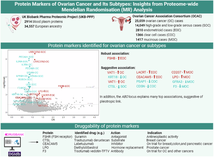
In this study, we conducted a comprehensive proteome-wide MR analysis to identify potential protein markers and druggable targets for epithelial OC and its various subtypes. We found robust evidence of an association between plasma FSHB (and more notably, FSH) levels and endometrioid OC, suggesting that FSHB, more relevantly FSH, may serve as a potential serum marker for early detection. Importantly, we report that the downstream target of this protein, the FSH receptor (FSHR), has been suggested as drug target for multiple chemicals, including approved receptor agonist drugs primarily used for managing female infertility and an investigational receptor antagonist, suramin, for anticancer activity [25]. We also identified eight other proteins associated with epithelial OC, and nine proteins (six of which overlapped with OC) associated with serous OC. However, these associations were attributed to pleiotropic variants primarily from the ABO and MAPT-AS1 loci, regions that were also identified as OC risk loci in OC GWAS.
Our finding for a link between FSHB and endometrioid OC is consistent with the hypothesis suggesting that conditions such as late menopause, ovulation, and infertility therapy, which lead to excess exposure of the ovarian surface epithelium to gonadotropins (FSH and luteinizing hormone (LH)), increase the risk of OC [26, 27]. This is in line our prior machine learning-based study, where oral contraceptive use and higher parity (conditions that lead to lower ovulations) were observed to be protective against OC, while later age at menopause was associated with a higher risk of clear cell and endometrioid OC [28]. The identification of FSHB as a shared risk-associated gene for endometriosis and endometrioid OC in cross-trait meta-analyses, alongside evidence of a genetic correlation and causal link between endometriosis and OC subtypes, particularly endometrioid and clear cell OC [29], support the importance of the FSHB genes and its encoded protein in the progression of both endometrioid OC and endometriosis. FSH is a glycoprotein composed of two subunits: FSHB which is specific to FSH, and CGA, which is nonspecific and shared with other hormones such as LH, thyroid-stimulating hormone, and human chorionic gonadotropin [27]. Consistent with the effect of FSHB, we also found suggestive evidence for an association between CGA and higher risk of OC, including serous OC, and clear cell OC subtypes. However, as there was only a single instrument available for this protein, we were unable to test the robustness of this association. Through binding to the FSHR on ovarian follicles, FSH regulates sexual development, reproduction, follicular development, and ovulation [27]. FSHR is present in ovarian granulosa cells and has also been found to be expressed in normal ovarian surface epithelial cells as well as in epithelial OC cells [30]. FSH binding to FSHR leads to upregulation of multiple signalling pathways, including the Notch signalling, protein kinase C and sphingosine kinase pathways, which results in the proliferation of OC cells and the progression of the cancer [31, 32]. Furthermore, ascites, a common condition during the late stage of OC, contains cellular aggregates detached from the primary tumour, known as spheroids [31]. These spheroids express FSHB mRNA and secrete FSH, further promoting cancer cell proliferation and metastasis through activation of the FSHR in the tumour cell [31]. The majority of approved drugs targeting the FSHR mimic the function of FSH and are commonly used for diagnosing and treating infertility [25]. We identified one FSHR antagonist, suramin, which was originally developed for treating African trypanosomiasis [25]. Suramin’s antineoplastic effects have been tested in clinical trials for a variety of human cancers, including prostate cancer, OC, breast cancer, bladder cancer, brain and central nervous system tumours, multiple myeloma, and plasma cell neoplasm, although it has not yet been approved for treatment of any cancer [25, 33].
In addition to the robust association observed for FSHB, we identified 11 proteins with suggestive evidence of associations with OC or its subtypes, most of which have been identified as druggable targets by approved or investigational drugs. Among these, CTSL is a lysosomal cysteine protease primarily involved in the terminal degradation of intracellular and endocytosed proteins [34]. This protein has been found overexpressed in OC, with its expression level correlated with disease progression and metastasis, while downregulation was shown to inhibit OC cell growth and migration [34]. We found approved drugs (such as fostamatinib [for chronic immune thrompocytopenia], trastuzumab deruxtecan [for breast cancer] and bortezomib [for multiple myeloma]) and investigational drugs (such as Felbinac and) that target CTSL, indicating its potential role in cancer treatment. In our study, however, we found an inverse association between CTSL and serous OC, which requires further investigation.
CEACAM5, originally known as a gastrointestinal oncofetal antigen, is now recognised as being expressed in the majority of carcinomas of the gastrointestinal, respiratory, and genitourinary systems [35]. CEACAM5 is a cell surface glycoprotein that plays a role in cell adhesion and intracellular signalling, and is highly expressed in tumours of epithelial origin including OC, particularly in mucinous and endometrioid tumours [35, 36]. Our study found that higher plasma levels of CEACAM5 are associated with higher risk of endometrioid OC, with a potential link also with a higher risk of mucinous OC, although the confidence interval crossed the null, possibly due to limited power (n = 1417 cases). CEACAM5 is being investigated as a potential drug target for various agents due to its role in cancer progression and metastasis [36]. Potential drugs such as labetuzumab, a humanised IgG1 monoclonal antibody, through directly binding with CEACAM5, inhibits tumour growth [37]. Given the higher expression of CEACAM5 in tumour compared to healthy cells, CEACAM5 can facilitate the delivery of cytotoxic agents to tumour cells as “antibody-drug – conjugates” [38], such as the delivery of SN-38 (a topoisomerase I inhibitor) to CEACAM5-expressing tumour cells as SN-38-labetuzumab conjugates (labetuzumab govitecan). Labetezumab and its conjugate have been investigated for the treatment of breast cancer, colorectal cancer, lung cancer and pancreatic cancer and could also be explored as a potential treatment for OC including endometrioid and mucinous OC [39].
F3, also referred to as coagulation factor III, tissue factor, thromboplastin, or CD142, is a transmembrane glycoprotein that plays a crucial role in the activation of the coagulation extrinsic cascade [38]. F3 is overexpressed in tumour cells and is involved in tumour growth and metastasis mainly by promoting inflammation, and angiogenesis by increasing expression of vascular endothelial growth factor (VEGF) [40]. It has been targeted by tissue factor-directed antibodies such as tisotumab vedotin, antibody-drug conjugates comprised of anti-tissue factor human IgG1-kappa antibody conjugated to monomethyl auristatin E, a cytotoxic agent that disrupts the microtubule of the tumour cells [41]. This drug is used to treat recurrent and metastatic cervical cancer and is being investigated in clinical trials for its potential use in treating multiple cancers, including OC, fallopian tube cancer, endometrial cancer and prostate cancer [42]. In our study, we found a lower risk of mucinous OC with higher plasma F3 levels, which warrants further investigation given the unexpected directionality of the association. However, population-based studies have shown that plasma F3 levels are elevated in patients with OC, contributing to the higher risk of venous thromboembolism in these patients [40]. Our study also identified other protein markers, including KRT18, CDSN, LPO, and GFRA3, as druggable targets, and their potential use in OC treatment should be explored in future studies.
The primary strength of our proteome-wide MR analyses lies in the large-scale design, which allowed us to screen known and potential novel drug targets and biomarkers using a hypothesis-free approach. By including both cis-pQTLs and trans-pQTLs, we were able to test for horizontal pleiotropy, and demonstrate that many apparent blood protein signals for OC or subtypes were driven by the ABO locus. Our approach allow us to re-evaluate the previously identified 26 proteins associated with invasive epithelial OC in the INTERVAL study, which included 1329 proteins from 3301 healthy participants [8]. However, this earlier study employed only a single instrument for each protein and did not include pleiotropy tests. Of the 26 identified proteins, we tested 16 proteins that were available in our dataset for their association with OC, but found evidence suggesting that the associations in each case were driven by pleiotropy. These associations were mostly driven by the ABO locus, suggesting that the observed associations in the prior MR study are likely attributable to pleiotropic relationships rather than causal links. Indeed, the ABO is a well-known pleiotropic locus that was associated with over 300 proteins in the latest proteome GWAS [10] and it was also identified as a risk locus in the OC GWAS conducted by the OCAC [9]. Furthermore, blood groups defined by the ABO locus differentially associate with the risk of OC, particularly high-grade serous OC, with the variants linked to the “A1” blood group antigen (rs507666) associated with a higher risk, while variants indexing the “O” blood group antigen (rs687289) linked with a lower risk. Although our blood protein panel includes ABO, we did not observe an association between ABO protein abundance and OC. This may reflect an importance of the blood type itself, rather than the abundance of ABO protein, in OC risk. An additional strength of our study was that we accounted for potential protein assay effects by replicating the analysis with data from SomaScan [17].
However, our study also has some limitations. First, despite including a large set of proteomic markers from the largest population cohort available at present, due to the limited number of OC cases and its subtypes among UK Biobank participants with proteomic data, our study was unable to conduct and confirm the predictive and prognostic impact of the identified protein markers in the observational analysis. Second, some of our MR analyses, particularly those involving OC subtypes such as endometrioid, clear cell, and mucinous OC, were likely underpowered due to limited number of cases (n < 2810). This limitation may have been further exacerbated in the leave-one-out analyses by the exclusion of key pQTLs that contribute substantially to the heritability of protein expression. Mucin-16 (MUC16), also known as CA125, a well-known OC tumour marker, was initially associated with a higher risk of OC and its subtypes in our study at the nominally significant threshold. However, after excluding rs62193070 (GAL3ST2), a trans-pQTL that explains 1.8% of the variability, the association was lost (power dropped from 0.96 to 0.45 for detecting OR = 1.22). Similarly, of the top signals identified at FDR threshold, the lack of association involving CD34, IL3RA, PECAM1, ROBO4, PTPRM, ROBO4 and SELE after excluding the pleiotropic variants might reflect lack of power (Supplementary Table 10). Third, the lack of a robust association with established OC markers, such as CA125 and the latest approved Human Epididymis protein 4 (HE4, also known as WAP four-disulfide core domain protein 2 or WFDC2) [43], could be partly attributed to the use of genetic variants derived from a sex-combined population, as the female-specific summary results were unavailable. Fourth, while our study encompassed 2337 proteins with at least one instrumental variable, it is important to acknowledge that certain relevant plasma proteins may not have been captured. This limitation underscores the need for future research employing broader and more comprehensive proteomic platforms to ensure a more exhaustive representation of the plasma proteome. Fifth, it is also important to note that our proteome-wide analysis focuses on protein abundance measured in plasma rather than tissue-specific proteins, that are often more directly relevant to disease progression and therapeutic targeting. For instance, a previous study identified an expression quantitative trait locus (eQTL) for OC associated with the expression of LRRC37A2 in 47 tissues, including the endometrium and ovary [44]. In our study we observed an association between LRRC37A2 and OC, in cis-pQTL but not trans-pQTL analyses (Supplementary Tables 7 and 8), which is relevant as cis-pQTL are more likely to reflect tissue-specific expression differences in the ovary compared to trans-pQTL which typically capture pleiotropic influences. Further studies incorporating tissue-specific protein data are warranted to provide more comprehensive insights. Sixth, we applied FDR correction to account for multiple testing across 2337 proteins and five outcomes, and also reported nominally significant signals as ‘suggestive associations, with related caution required when interpreting these findings. Seventh, we did not observe strong evidence for colocalisation between blood FSHB level and endometrioid OC association (PPH4 = 0.65), although this represents suggestive support. Eighth, although we replicated the association between FSHB and endometrioid OC with an independent data measured with the Olink platform, which has demonstrated superior protein target specificity compared to SomaScan [45, 46], we were unable to replicate this finding using SomaScan-based FSHB data. Future replication using mass spectrometry-based protein measurement, an orthogonal method to affinity-based platforms, may therefore be warranted. However, for FSH, the clinically relevant and predominant form of the protein complex containing FSHB, we found consistent supporting evidence for an association with endometrioid OC across three independent cohorts. In light of replication and the biological relevance of FSH in the ovarian tissue, we believe this finding is likely to be robust. Nineth, given that our analysis was restricted to a European ancestry population, the findings of this study may not be representative of other population groups.
In conclusion, our study identified association between plasma FSHB, along with 11 other proteins, with OC or its subtypes. These findings suggest that these serum biomarkers could be useful for the early detection of OC. Notably, most of these proteins have already been recognised as drug targets, underscoring the need for further investigation into their roles in OC development and treatment.
Reporting summary
Further information on research design is available in the Nature Research Reporting Summary linked to this article.


Comments (0)