
Remember me


A 29-year-old female was referred from the intensive unit care (ICU) due to sudden bilateral vision loss over four days. She received the Human Papillomavirus vaccine one month prior, subsequently developing fever and symmetrical annular erythema(Fig. 1A) on her trunk and limbs ten days later. Laboratory tests revealed Increased systemic inflammatory markers and liver and kidney damage: white blood cell count of 14,030/µL, with 31% cytotoxic T cells and 23.4% B cells. D-dimer levels were at 9.55 mg/L, IL-6 at 30 pg/mL, aspartate aminotransferase at 82 U/L, and creatinine at 198 µmol/L. She had no previous health issues and was diagnosed with DRESS syndrome twenty days post-vaccine.
Fig. 1
First Presentation of Bilateral Purtscher-Like Retinopathy in a 29-Year-Old Female. A: A photo of the patient’s skin. B: Bilateral fundus photographs and OCT
At the first presentation, her visual acuity was finger count at 10 cm OU, with normal intraocular pressure. The anterior segment was normal, and the vitreous was clear. Dilated fundus examination revealed numerous cotton-wool spots and retinal hemorrhages in the macular and peripapillary regions bilaterally. Optical coherence tomography (OCT) showed severe macular edema and “middle limiting membrane sign” in both eyes. Aqueous humor tapped for next-generation sequencing yielded negative results for pathogens (Fig. 1B). A diagnosis of Bilateral Purtscher-like retinopathy was made, and intravitreal injection of Ranibizumab (IVR) were administered. Systemic treatment in the ICU included the administration of steroids, anticoagulants, plasma exchange, and supportive therapies.
A, Photos of the patient’s skin shows the typical skin manifestations of DRESS syndrome. B, Bilateral fundus photographs showing intraretinal hemorrhages and numerous cotton wool spots.Bilateral OCT B-scans (24 × 20 mm, BM-400 K TowardPi) showing severe macular edema and “middle limiting membrane” sign.
One month post-IVR, the patient was discharged from the ICU with controlled systemic conditions. Her best-corrected visual acuity; improved to 0.12 OD and 0.01 OS. Fundus examination showed significant improvement but residual thinning of the inner retina (Fig. 2).
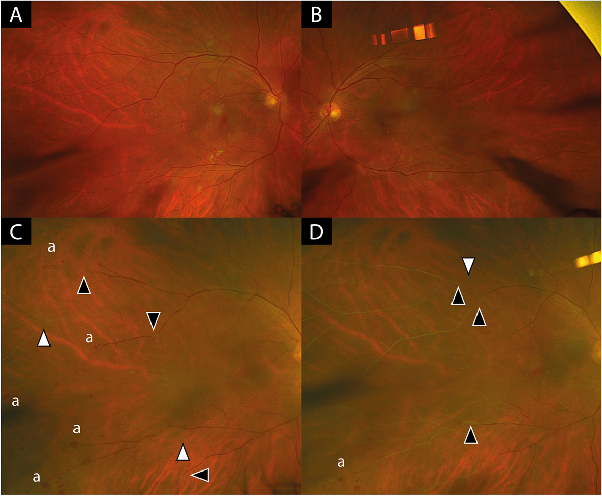
Fig. 2
One-Month Follow-up of Bilateral Purtscher-Like Retinopathy in a 29-Year-Old Female
Bilateral fundus photographs showing much improvement of the retinal lesions.OCT B-Scans (512 × 128 volumes scans, Cirrus HD5000, Zeiss)) showing subtle subretinal fluid and thinning of the inner retina bilaterally.
Comments (0)