Construction of CD90 adeno-associated virus (AAV) knockdown mice
Twelve female BALB/C mice (100 ± 20) g aged 6–8 weeks were purchased from Beijing VitalRiver Laboratory Animal Technology Co., LTD. All mice were kept in a shielded environment at constant temperature of 22–24℃, constant humidity of 55–60%, and light/dark condition of 12h/12h, After purchasing AAV-shCD90-GFP and AAV-GFP, transfect mice with adeno-associated virus (AAV-shCD90-GFP) to knock down CD90, with AAV-GFP mice as controls. Specifically, 100 μL of adeno-associated virus was injected into the mouse via the tail vein.
Establishment of mouse rib injury model
Mice anesthetized with 3% isoflurane were placed in a supine position, stratified incision of the skin, subcutaneous tissue and muscle separation, stripping of the periosteum to expose the middle rib of the chest, transverse incision of the middle rib, and then stratified counterposition suture.
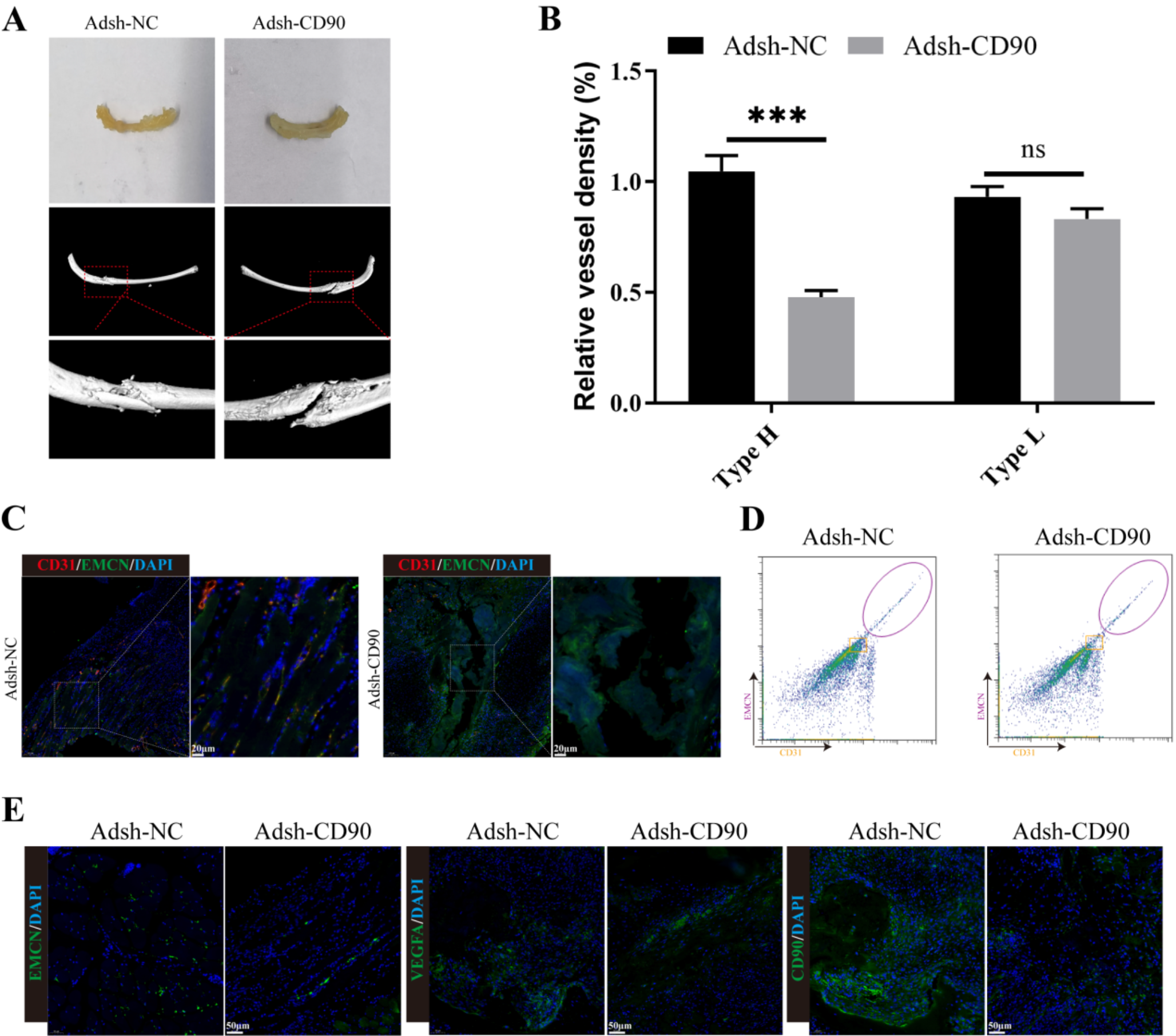
Observation of bone regeneration in mice by micro-CT
In this study, mice were divided into the following two groups: Adsh-NC and Adsh-CD90. After four weeks of mouse model preparation and culture, we used X-ray technology to observe bone healing.
Cell culture
Both bone endothelial cells, perivascular cells and MC3T3-E1 cell were cultured in MEM-α culture medium(Gibco, USA) containing 10% FBS, and cells were passaged every 2–3 days to avoid cell contamination. Subsequently, cells in the logarithmic phase were selected for the experiment.
Cell grouping
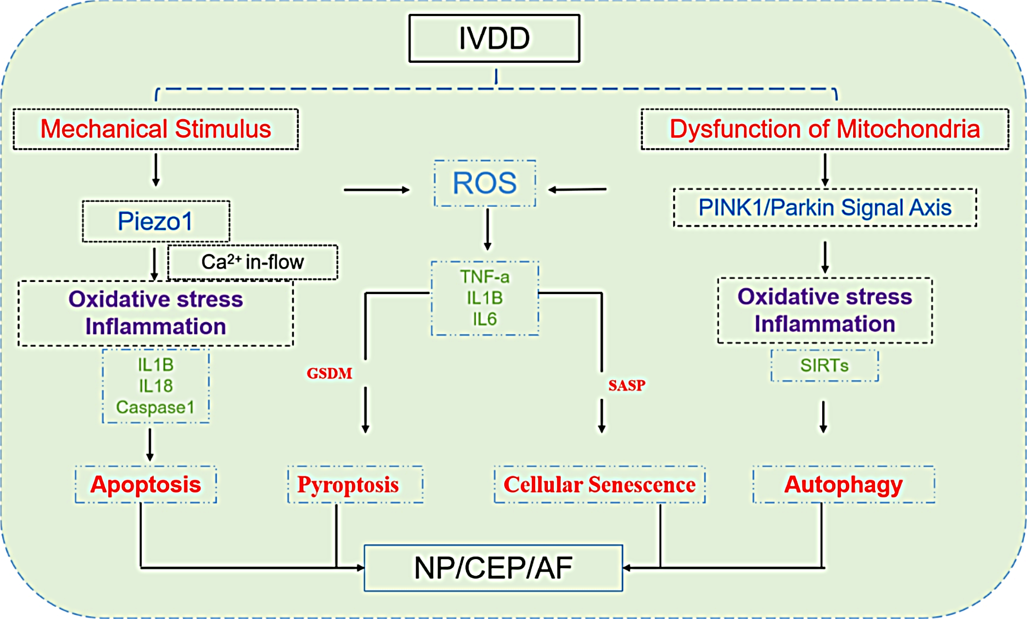
The periosteal stem cells treated with r.DLL4 and transfected with shCD90 were incubated for 24 h, centrifuged at 3000×g for 5 min, and filtered through a 0.3μm filter to obtain the cell supernatant, which was then co-cultured with MC3T3-E1 cells. The periosteal stem cells were co-cultured with MC3T3-E1 cells in four groups: shNC + MC3T3-E1, shCD90 + MC3T3-E1, rdll4 + shNC + MC3T3-E1, and rdll4 + shCD90 + MC3T3-E1. In addition, bone microvascular endothelial cells (BMEs) were co-cultured with periosteal stem cells using cell culture dishes and Transwell culture dishes, respectively. The cells cultured with bone microvascular endothelial cells and transfected CD90 bone membrane stem cells were divided into the following eight groups: NC + BMEs, OE-CD90 + BMEs, shNC + BMEs, shCD90 + BMEs, r.Dll4 + NC + BMEs, r.Dll4 + OE-CD90 + BMEs, r.Dll4 + shNC + BMEs, r.Dll4 + shCD90 + BMEs. Transfection of CD90 and Notch1 overexpression vectors into bone microvascular endothelial cells and co-culture with CD90 periosteal stem cells was divided into 8 groups: NC + BMEs, OE-CD90 + BMEs, shNC + BMEs, shCD90 + BMEs, Notch1 + NC + BMEs, Notch1 + OE-CD90 + BMEs, Notch1 + shNC + BMEs, and Notch1 + shCD90 + BMEs. The mechanism of this study was shown in Fig. 7.
Cell transfection
First, periosteal stem cells were selected based on the markers PDPN + CD146-CD73 + CD164 + , and then transfected with NC, OE-CD90, shNC, or shCD90. The periosteum stem cells transfected with plasmid were mixed with MC3T3-E1 cells, and the co-cultured cells were incubated with R.DELL4 protein, and transfected with shNC and shCD90. In addition, after the periosteal stem cells transfected with CD90 were transfected with the Notch1 overexpression vector, the follow-up experiment was performed. The transfection procedure was as follows: the recombinant plasmid and liposome were mixed by Turbofect (Thermo Scientific, R0533), then gently added into the cells, and the cells were incubated in the incubator after standing at room temperature for 20 min.
RT-qPCR
Rib injury tissues of mice in each group and periosteum stem cells in each transfection group were collected, and Trizol reagent was added to extract total RNA, the concentration of total RNA was detected by ultraviolet spectrophotometer, and template cDNA was synthesized according to reverse transcription kit, using GAPDH as the internal reference. The transcriptional levels of RUNX2, OCN, VEGF, HIF-1α, OPN, Dll4, Notch1, Notch2, Notch3, Notch4, Hes1, Hes5, Hey1, and Efnb2 were detected with 3 parallel pores in each group. The amplification conditions for PCR are as follows: pre-denaturation at 94 °C for 5 min; denaturation at 94 °C for 15 s, annealing at 62 °C for 30 s, extension at 72°C for 30 s, and 35 cycles in total, with a final extension at 72 °C for 5 min. The relative expression levels were detected by the 2−ΔΔCt method. The sequence and length of primers were shown in Table 1.
Immunofluorescence
The damaged rib tissues of mice and periosteum stem cells of each transfection group were collected and inoculated on the crawling plate, soaked with PBS 3 times, fixed with paraformaldehyde for 15min, soaked with PBS and added with 0.5% Triton X-100 at room temperature for 15min, cleaned with PBS, and sealed with goat serum for 30 min. The primary antibody [Dll4 (GB11322-100, Servicebio, 1:200), Notch1 (ab52627, 1:500), CD90 (ab92574, 1:500), VEGF (ab32152, 1:500), HIF-1α (Catalog# ER1802-41, Huabio, 1:100), CD31 (ab9498, 1:500), and Endomucin (GB112648-100, Servicebio, 1:600)] was added and incubated overnight, the fluorescent secondary antibody was added to avoid light, incubated for 1.5 h, soaked with PBS, and then DAPI was added to avoid light and incubated for 5 min. The tablets were sealed with a sealing solution containing fluorescence quencher, and the acquired images were observed under a fluorescence microscope.
Flow cytometry
CD31 and EMCN dual staining in single cell suspensions from rib fracture sites were detected by Flow cytometry. The specific operation should be carried out according to the instructions of Annexin V-FITC apoptosis detection kit (Shanghai Beyotime Biotechnology Co., Ltd). Collect the supernatant of the rib fracture from each group of mice stained with CD31 and EMCN, and mix with an appropriate amount of PBS. Then, centrifuge for 5 min, aspirate the supernatant, and resuspend in serum-free basal culture medium to prepare cell suspension. Add 1μL of flow cytometry antibody (1 × 10⁶ cell/100μl) to each tube and incubate at room temperature in the dark for 20 min. Subsequently, wash with PBS, centrifuge, resuspend in 200 μL of PBS, and finally detect using flow cytometry.
Western blot
Periosteal stem cells of each transfection group were collected, cleaned with PBS for 3 times, and total protein was obtained by adding cell lysate containing protease inhibitor, the protein content was determined by BCA kit (Shanghai Beyotime Biotechnology Co., Ltd), and then denatured at 100℃ for 5 min. After separation by SDS-PAGE gel electrophoresis, protein transfer was performed. The protein membrane was enclosed in 5% BSA at room temperature for 2 h, and the corresponding primary antibodies [RUNX2 (ab76956, 1:1000), OCN (ab133612, 1:1000), CD31 (ab9498, 1:5000), HIF-1α (ab308637, 1:2000), OPN (ab75285, 1:1000), CD90 (ab92574, 1:2000), Notch1 (ab52627, 1:1000), Notch2 (ab307700, 1:1000), Notch3 (ab300527, 1:1000), Notch4 (ab184742, 1:1000), Hes1 (ab119776, 1:1000), Hes5 (ab194111, 1:500), Hey1 (ab154077, 1:500), Hey2 (ab167280, 1:500), and Efnb2 (ab131536, 1:500)] were added. The cells were incubated at 4℃ overnight, and the secondary antibodies were incubated for 1 h. Finally, the luminescent solution was added for gel imaging for exposure and photography. ImageJ software counted the gray values of proteins in each transfected histone cell.
ALP staining
After co-culturing the transfected plasmid bone marrow mesenchymal stem cells with MC3T3-E1, wash three times with PBS, fix with 4% paraformaldehyde for 20 min, and wash three times with PBS again. The alkaline phosphatase staining working solution was prepared according to the instructions of the staining kit. Add an appropriate amount of staining working solution to the cells, then incubate at room temperature in the dark for 20 min. The staining solution was removed and washed with distilled water to stop the color reaction, and finally observed the staining and capture relevant images.
Alizarin red staining
The culture medium of periosteal stem cells in each transfection group was discarded, 400 μL PBS was added to each well, the cell surface was cleaned 3 times, and 300 μL fixing solution was added to each well for 10 min. The fixing solution was discarded, and 500 μL PBS was added to each well to clean the cell surface, and 300 μL staining solution was added to each well to stain the cells at room temperature for 15 min, and PBS was added to clean the cells for 3 times. The formation of mineralized nodules was observed under optical microscope and images were collected.
CCK-8
CCK8 cell viability assay kit came from Shanghai Beyotime Biotechnology Co., Ltd. The cell suspension was prepared by co-culture of periosteal stem cells and MC3T3-E1 cells in logarithmic growth stage. A total of 4 × 10³ cells per well were inoculated on 96-well plates, and after 48 h of culture, 10 μL of CCK-8 solution was added to each well, and incubated in a 5% CO2 cell incubator at 37℃ for 4 h. The light absorption value was detected at 450 nm by enzyme-labeled instrument, and cell viability was calculated.
Angiogenesis formation assay
Matrige was diluted and placed it in a 24-well plate. A total of 150μL diluted Matrige was added to each well and incubated at room temperature to solidify into a gel. Bone microvascular endothelial cells transfected with CD90 and Notch1 overexpression vector were co-cultured with CD90 periosteal stem cells, and cells were seeded at a density of 5 × 10⁴ cells per well in 24-well plates coated with Matrigel. After 24 h of culture, the tube formation was observed by optical microscope, and 5 fields were randomly selected to count the number of tubes formed under the field.
Statistical analysis
Data analysis was performed using SPSS23.0 software, and statistical graphs were drawn using GraphPad Prism 8.30 software. All experimental data were presented in the form of mean ± standard deviation (x ± s). Data comparisons were made using one-way analysis of variance (ANOVA), and LSD-t test was used to compare two-by-two data within each group. P < 0.05 was considered statistically significant.


Comments (0)