
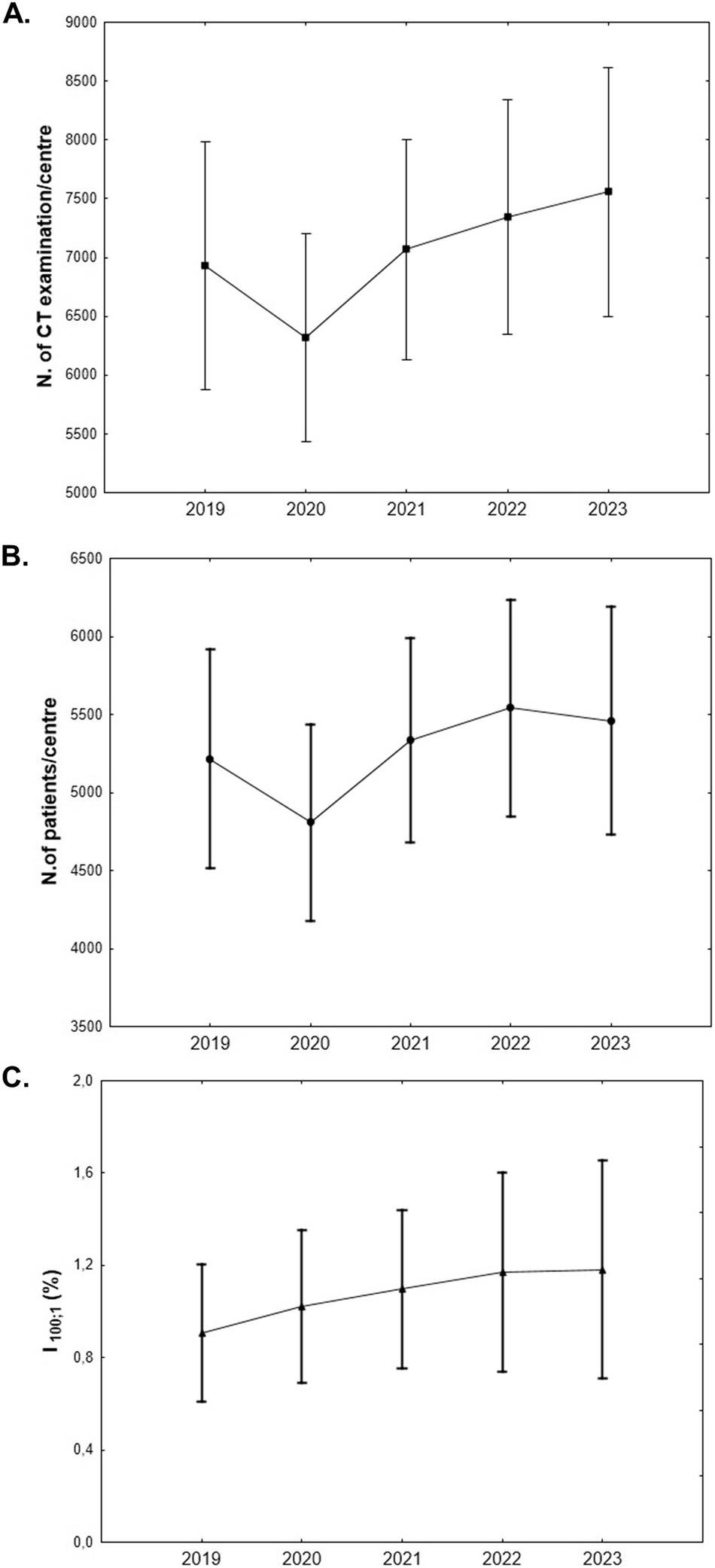
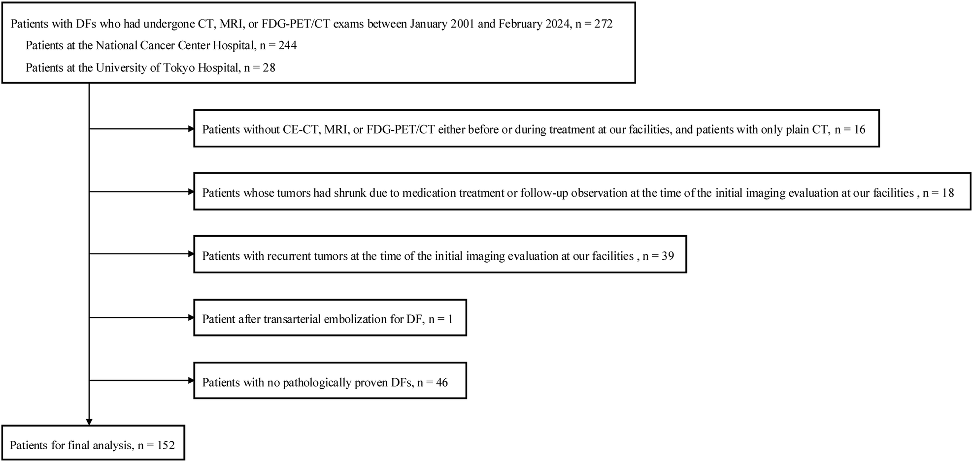
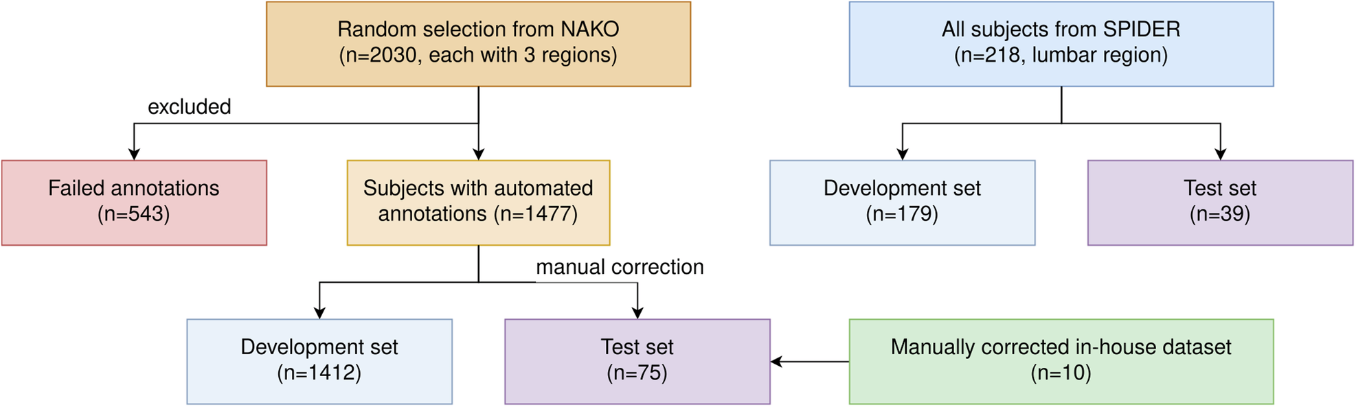
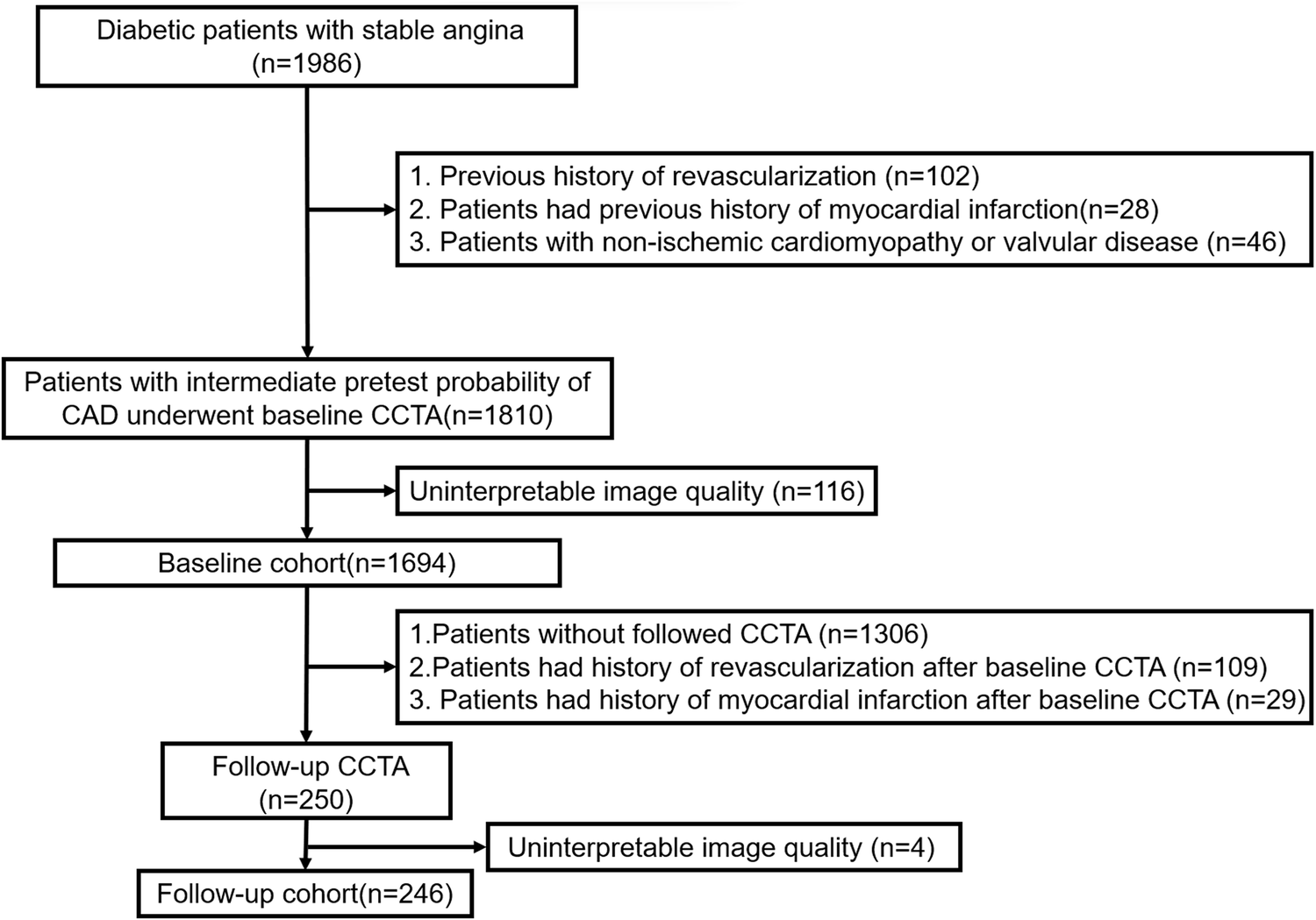
Remember me


Baseline patient characteristics are summarized in Table 1. The cohort was balanced with 59 (49.2%) female and 61 (50.8%) male patients. The mean age was 62 ± 13.3 years (range 17–88). The most frequent tumor type was uveal melanoma (28.3%). The mean time between baseline imaging used for segmentation and SIRT was 19 ± 8.9 days. Most patients underwent one 90Y-radioembolization session (n = 72, 60%) and received resin-based microspheres (n = 78, 65%) (Table 1).
Table 1 Baseline patient characteristicsCorrelation of CT-based sarcopenia with morphometric parametersSeventy patients (58.3%) were diagnosed with sarcopenia based on CT measurements. The comparison of baseline characteristics between patients with and without sarcopenia is shown in Table 2. The mean value of BMI and SMRA was significantly lower in patients with sarcopenia compared with those without sarcopenia. Of note, the perfused tumor volume was comparable between groups (272.9 ± 287.8 vs. 293.3 ± 354.9 cm3, p = 0.73). The mean SMI value in the whole cohort was 44.2 ± 8.7 cm2/m2. The SMI in male was significantly higher than in female patients (49.8 ± 8.2 vs. 38.4 ± 4.4 cm2/m2, p < 0.001). In the correlation analysis, the SMI was positively correlated with BMI and VATI while negatively correlated with VFRA (Fig. 2).
Table 2 Comparison of demographics and body composition parameters between patients with and without CT-based sarcopenia (n = 120)Fig. 2
Correlation analysis of SMI with BMI, VATI, and VFRA. SMI, skeletal muscle index; BMI, body mass index; VATI, visceral adipose tissue index; VFRA, visceral fat radiation attenuation
The multivariate regression analysis demonstrated that male sex, lower BMI, lower SMRA, lower SFRA, and higher VFRA were associated with the risk of sarcopenia with the odds ratio (OR) of 8.81 (95% CI, 2.09–37.1, p = 0.003), 0.64 (95% CI, 0.48–0.85, p = 0.002), 0.79 (95% CI, 0.69–0.91, p = 0.001), 0.84 (95% CI, 0.76–0.93, p = 0.001) and 1.23 (95% CI, 1.06–1.42, p = 0.006), respectively (Fig. 3).
Fig. 3
Forest plot showing factors associated with the risk of sarcopenia. BMI, body mass index; SMRA, skeletal muscle radiation attenuation; SFRA, subcutaneous fat radiation attenuation; SATI, subcutaneous adipose tissue index; VATI, visceral adipose tissue index; VFRA, visceral fat radiation attenuation; IMATI, intramuscular adipose tissue index; BMI, body mass index
According to our criteria for classifying BMI, 73, 33, and 14 patients were classified with normal/underweight, overweight and obesity status, respectively. The incidence of sarcopenia in the three groups was 64.4% (47 of 73), 63.6% (21 of 33), and 14.3% (2 of 14), respectively. The incidence of sarcopenia was significantly lower in the obesity group than in the other two groups (p < 0.01) (Fig. 4A). In addition, the median OS of these three groups was 16.5 months (95% CI, 7.8–25.4), 13.9 months (95% CI, 9.1–18.7), and 17.5 months (95% CI, 14.0–21.0) for normal/underweight, overweight and obesity groups, respectively (Fig. 4B). There was no statistically significant difference among these three groups (p = 0.201).
Fig. 4
A The incidence of sarcopenia was significantly lower in the obesity group than in the other groups. B Survival analysis with Kaplan–Meier curves showing median OS among normal/underweight, overweight and obesity groups. **p < 0.01
In addition, IMATI, VATI, and SATI were strongly positively correlated with BMI, while VFRA was negatively correlated with BMI (Supplementary Fig. 1).
Survival and prognostic analysis in the whole cohortBy March 2022, 98 patients (81.7%) had died. The median follow-up duration was 71 months (IQR 49.6–98.3). The median OS of the whole cohort was 15.4 months (95% CI, 10.8–20) (Fig. 5A). There was no significant difference in median OS between patients with and without sarcopenia (15.7 vs. 14.8 months, p = 0.773) (Fig. 5B). The OS of patients with various primary malignant tumors was significantly different (p = 0.032). The median OS was 23.3 months (95% CI, 4.9–41.7), 23 months (95% CI, 16.3–29.7), 11.3 months (8.8–13.8), 7.9 months (95% CI not reached–18.3), and 7.4 months (95% CI, 0.3–14.5) in patients with neuroendocrine tumors, uveal melanoma, other malignant subtypes, colorectal cancer, and intrahepatic cholangiocarcinoma, respectively (Fig. 5C).
Fig. 5
Survival analysis. A Overall survival in the whole cohort. B Overall survival in sarcopenic vs. non-sarcopenic patients. C Overall survival of patients with various primary malignant tumors significantly differed among groups
In the whole cohort, age, SMI, and tumor subtypes were independent prognostic factors for OS, with HR values of 1.03 (95% CI, 1.01–1.05, p = 0.01), 0.92 (95% CI, 0.86–0.99, p = 0.021), and 2.09 (95% CI, 1.31–3.33, p = 0.002), respectively (Fig. 6).
Fig. 6
Forest plot of factors associated with overall survival. BMI, body mass index; SMRA, skeletal muscle radiation attenuation; SFRA, subcutaneous fat radiation attenuation; SATI, subcutaneous adipose tissue index; VATI, visceral adipose tissue index; VFRA, visceral fat radiation attenuation; IMATI, intermuscular adipose tissue index; BMI, body mass index
Survival and prognostic analysis per tumor typeIn patients with intrahepatic cholangiocarcinoma, the median OS of patients without sarcopenia was significantly longer than those with sarcopenia (25.9 versus 6.5 months, p = 0.029). Scatter plots were used to investigate relationships between the morphometric parameters and time to death. In patients with cholangiocarcinoma, statistically significant relationships were found between time to death and the following body composition variables: VFRA (p = 0.002, R = −0.715) and SFRA (p < 0.001, R = −0.779) (Supplementary Fig. 2). VFRA and SFRA were still negatively correlated with survival after adjusting for age, sex, and BMI, with the R-values of −0.665 (p = 0.013) and −0.713 (p = 0.006), respectively. No statistically significant correlation was found between survival and morphometric parameters for the other tumor types.
Comments (0)