Simultaneous occurrence of epidural, subdural, and subarachnoid hemorrhages in the spinal canal: a rare case report
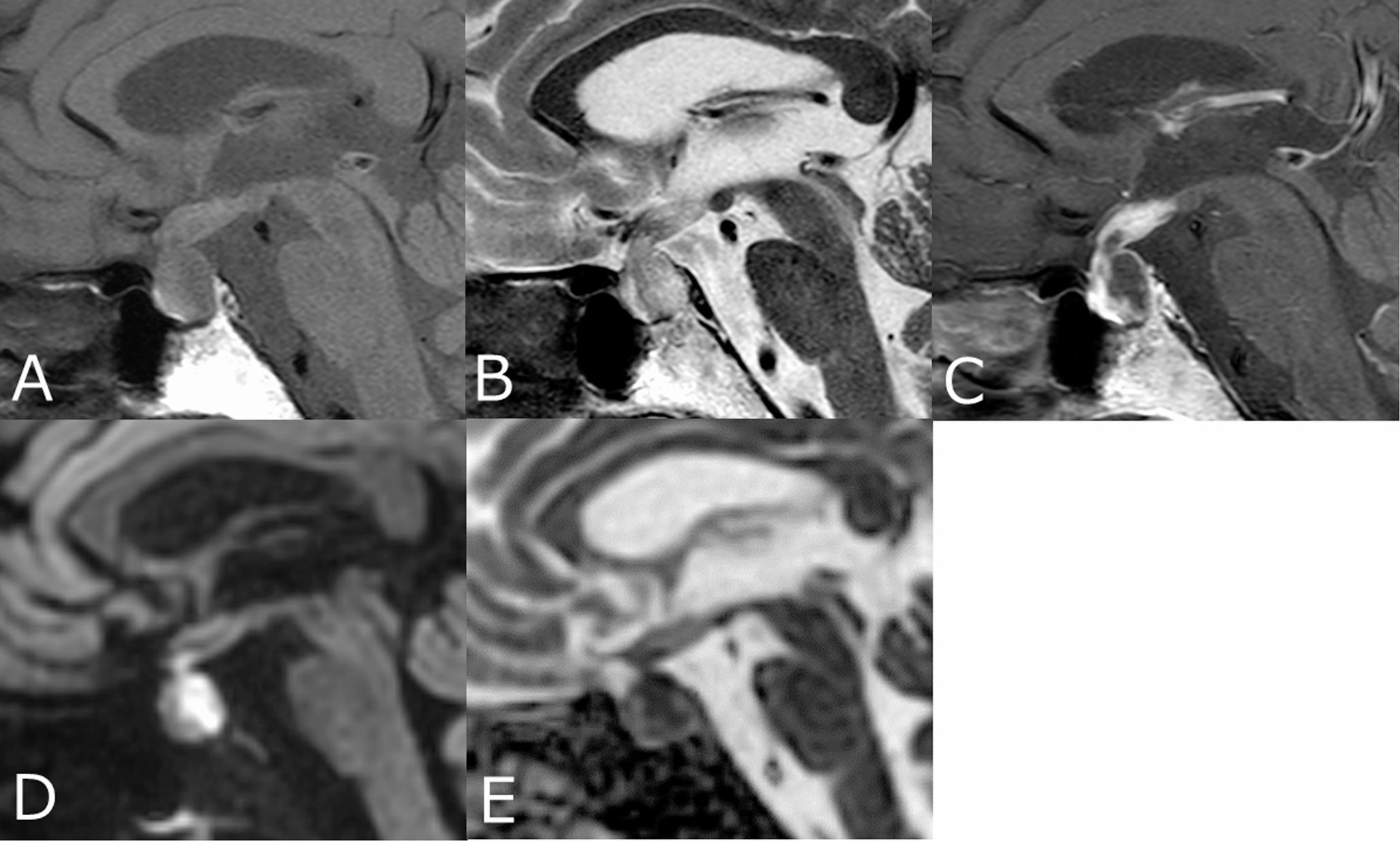
MRI plays a pivotal role in diagnosing spinal hemorrhages. MRI provides detailed information about the anatomical location of the hemorrhage, the affected structures, and the stage of the hemorrhage [2]. The signal characteristics of the hematoma help determine whether the hemorrhage is in the hyperacute, acute, early/late subacute, or chronic stage. Due to MRI’s high soft tissue resolution, the anatomical compartment where the hemorrhage occurs, the potential cause, and the affected structures can be accurately visualized [2, 5].
Epidural hematomas are located in the epidural space between the dura mater and the vertebrae. They create external compression on the thecal sac and displace the dura mater toward the spinal cord. The loss of normal epidural fat signal is an important indicator of an epidural hematoma [2, 5]. In our case, the anterior epidural fatty tissue could not be distinguished due to the presence of the epidural hematoma.
Subdural hematomas occur in the potential space between the dura mater and the arachnoid membrane. In subdural hematomas, the epidural fatty tissue is preserved, and inward displacement of the dura mater is not observed [2, 5]. In our case, MRI clearly demonstrated the subdural hematoma, which narrowed the spinal canal and exerted compression on the spinal cord.
Subarachnoid hemorrhages occur into the CSF between the arachnoid membrane and the pia mater. Subarachnoid hemorrhage spreads longitudinally due to CSF flow, and hematoma formation is typically not observed [6]. T2 hyperintense hemorrhage or clot formation may be seen around the spinal cord and between the cauda equina fibers. Aggregation of cauda equina fibers may occur at the lumbar level due to compression [2, 5]. In our case, MRI clearly demonstrated subarachnoid hemorrhage leading to aggregation of the cauda equina fibers.
In our case, spinal MRI revealed clear separation of the hemorrhages into three compartments, as well as cord compression and clumping of the cauda equina fibers in the lumbar region. The rapid diagnosis facilitated by spinal MRI allowed for timely surgical intervention, which was crucial in preventing further neurological decline.


Comments (0)