
Remember me


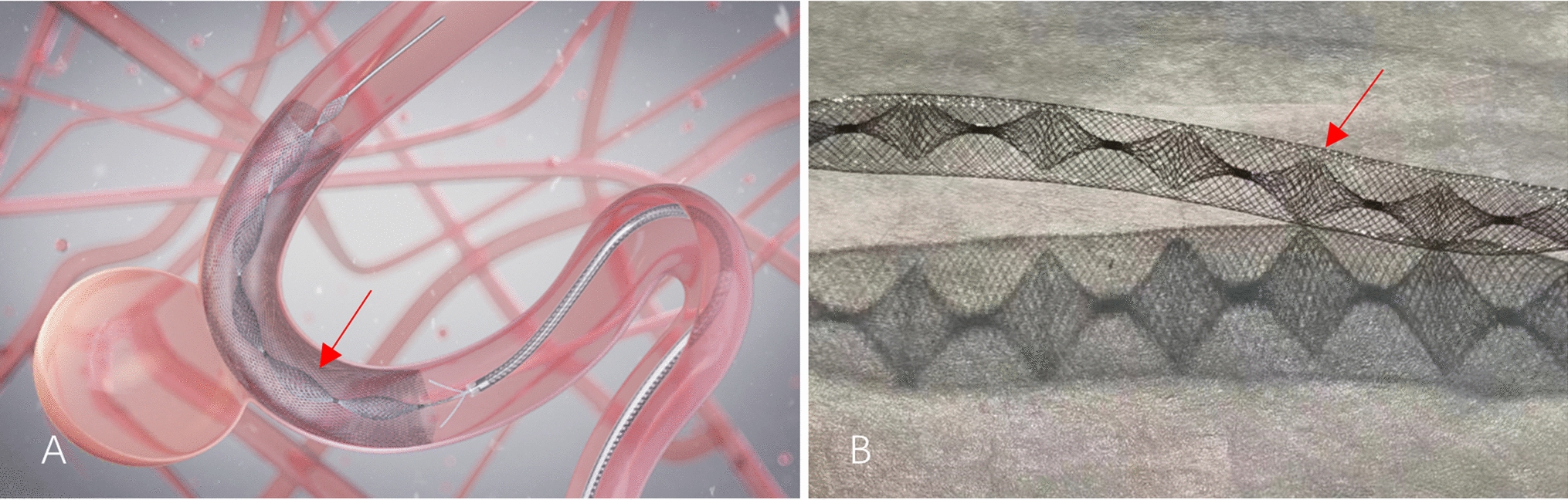


The model creation process involved the following steps: Under fluoroscopic guidance, a puncture point located in the middle of the stomach wall was selected. A 21-gauge puncture needle (Merit Medical, USA) was then used to puncture both the gastric wall and the anterior wall of the jejunum at the designated point. Following successful puncture, a 0.018-inch guidewire (Merit Medical, USA) was inserted into the jejunum through the 21-gauge needle. The guidewire was retained while the needle was withdrawn. A 6F sheath (Merit Medical, USA) was then advanced over the guidewire into the jejunum, and its position was confirmed by imaging. Subsequently, an anchor (self-made, the diameter is 0.018 inches) was inserted through the 6F sheath into the jejunum, followed by the insertion of a 0.035-inch guidewire (Terumo, Japan) through the 6F sheath while retaining both the anchor and guidewire. The 6F sheath was withdrawn, and the anchor was pulled to appose the jejunal wall to the gastric wall. Finally, a 10-F drainage tube (EVD Medical, China) was inserted into the jejunal lumen over the guidewire, and the tube was looped (Fig. 3).
Fig. 3
Fluoroscopically guided percutaneous jejunostomy: The drainage tube has been fixed in a loop (yellow arrow) and its position has been determined by contrast. The red arrow indicates the anchor
This ex vivo model does not fully replicate clinical conditions, as neither glucagon nor the amount of air typically used in percutaneous jejunostomy procedures in patients or in vivo studies were applied.
Comments (0)