
Remember me


This retrospective study involved 65 patients diagnosed with LDH who underwent either ECCNL or IECCNL at the First People’s Hospital of Changzhou City from April 2021 to April 2022(Table 1). The study received approval from the Ethics Committee of Changzhou First People’s Hospital and was conducted in accordance with the principles of the Declaration of Helsinki. Out of these patients, 30 received ECCNL, and 35 underwent IECCNL. All participants were followed for a duration of 24 months (Fig. 1). All patients were followed up through outpatient revisits and telephone follow-up. Patients who missed one outpatient revisit or three telephone follow-up were considered lost to follow-up. Those who underwent other surgical procedures during the follow-up period were excluded from the data. In Group A, 1 case was lost to follow-up, and 2 cases underwent other surgical treatments. In Group B, 4 cases were lost to follow-up. Inclusion criteria included: (1) established diagnostic criteria for LDH [2]; (2) age between 20 and 70 years; (3) indications for collagenase chemolysis [9]; (4) patient consent. Exclusion criteria comprised: (1) severe organic diseases associated with significant organ dysfunction; (2) severe coagulation disorders; (3) severe lumbar spinal stenosis or lumbar spondylolisthesis; (4) history of lumbar spine surgery; (5) mental disorders or inability to cooperate with surgical procedures. Surgical procedures and image interpretation were conducted by physicians holding vice senior or higher professional titles.
Table 1 Baseline demographics and subject characteristicsFig. 1 Patients Preparation
Prior to treatment, all patients underwent comprehensive diagnostic evaluations, including magnetic resonance imaging (MRI), computed tomography (CT), axial and lateral X-rays of the lumbar spine, complete blood counts, and biochemical tests. Informed consent was obtained from all participants, who were also apprised of potential preoperative risks and adverse reactions. Prophylactic antibiotics were administered before surgery, and continuous electrocardiographic monitoring was ensured during the procedure.
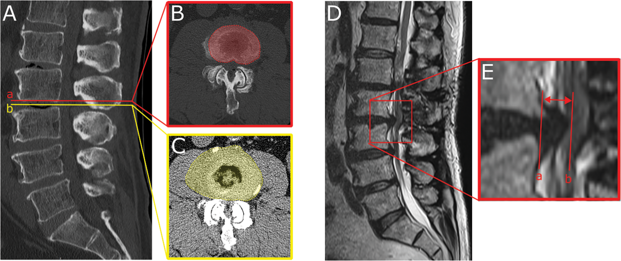
Surgical procedureThe patient was positioned prone with an abdominal pillow. The positioning grid was placed at the midline of the spinal segment corresponding to the target intervertebral disc and the targeted intervertebral disc was localized under CT guidance using an Aquilion One TSX-301 A CT simulator(Fig. 2A). Marking the needle trajectory on the plain scan slice(Fig. 2B). The surgical site was disinfected with iodine and prepared with sterile draping. Anesthesia was administered via layer-by-layer infiltration using a 0.5% lidocaine hydrochloride injection (batch number 42021839, Hubei Tiansheng Pharmaceutical Co., Ltd). For the extrapedicular puncture approach, a FLEX radiofrequency puncture needle (Boston Scientific, USA) was inserted through the marked posterolateral safe triangle on the affected side. Under CT non-contrast scan guidance, the needle was carefully advanced while avoiding critical tissues and blood vessels, with continuous adjustments until reaching the external opening of the intervertebral foramen. (Fig. 2C).Next, the FLEX electrode was inserted, ensuring its tip reached the anterior epidural space (Fig. 2D). The puncture needle was then advanced an additional 0.3 cm to bluntly dissect the herniated material before withdrawing the electrode. The needle tip was confirmed to be positioned at the internal opening of the intervertebral foramen (Fig. 2E). Subsequently, 1 mL of 0.5% lidocaine was injected as a local anesthetic test. After a 15-minute observation period, 600 IU of collagenase(collagenase for injection, Batch: JZA211202; Manufacturer: Shanghai Qiaoyuan BioPharm, China) was administered. During the intradiscal puncture procedure, the FLEX radiofrequency puncture needle was advanced via the posterolateral safe triangle approach. Under CT non-contrast scan guidance, the needle was adjusted to reach the center of the disc. Upon confirming the correct position, the FLEX electrode was inserted to perform blunt dissection of the nucleus pulposus tissue.Omnipaque (Iohexol Injection, Batch 15770905, GE Healthcare Shanghai) was then injected for contrast imaging, confirming that the contrast agent could diffuse outward through the fissure(Fig. 2F). Subsequently, 200 IU of collagenase was administered into the disc. Throughout the procedure, continuous communication with the patient was maintained, with close monitoring of pain levels and lower limb function.
Fig. 2
Fig. 2A and B are puncture positioning diagrams. The needle was adjusted to position the external opening of the intervertebral foramen in Fig. 2C. The FLEX electrode was positioned to extend the front end into the anterior epidural space in Fig. 2D. The needle was then advanced by 0.3 cm to exit the electrode in Fig. 2E. Figure 2F shows the discography image
Postoperative managementRoutine anti-infection, dehydration, and analgesic treatments were administered. Patients receiving ECCNL were required to maintain strict bed rest for 3 days. Patients undergoing IECCNL were prescribed strict bed rest for 7 days. All patients were instructed to wear a lumbar brace when ambulating. Back muscle rehabilitation exercises were initiated after 1 month, and physical labor was prohibited for 3 months postoperatively.
Outcome assessmentThis study primarily utilized the Numeric Rating Scale (NRS) [10] to assess pain severity at preoperative, 3 days, 1 month, 3 months, 6 months, and 1 year postoperative intervals, categorized as follows: 0 (no pain), 1–3 (mild pain), 4–6 (moderate pain), and 7–10 (severe pain). Lumbar spine function was evaluated using the Japanese Orthopedic Association (JOA) score [11], which encompasses three dimensions: main signs (9 points), clinical symptoms (6 points), and daily life restrictions (14 points). The efficacy was evaluated at 3 months, 6 months, 12 months, and 24 months postoperatively using the modified MacNab criteria [12], with outcomes categorized as excellent, good, fair, or poor. The excellent-good rate (number of excellent + good cases / total number of cases) and the excellent rate (number of excellent cases / total number of cases) were calculated. Pain relief rate was used to assess the difference in postoperative pain relief duration between the two groups (pain relief defined as > 50% reduction in pain intensity). At the final follow-up, the recurrence rate and reoperation rate were calculated. Additionally, the occurrence and number of severe adverse events and complications were recorded.
Satistical analysisData analysis was conducted using IBM SPSS Statistics 27.0. Measurement data adhering to a normal distribution were presented as mean ± standard deviation (x̄ ± s), whereas non-normally distributed data were represented by the median and interquartile range [M(IQR)]. For normally distributed data, analyses were performed using analysis of variance and the two independent sample t-tests. The Wilcoxon test was applied to non-normally distributed data, with further pairwise comparisons corrected using the Bonferroni method. Categorical data were expressed as frequencies or proportions, and the significance of differences was assessed using Pearson’s chi-square test or the continuity-corrected chi-square test. A P-value of < 0.05 was considered statistically significant.
Comments (0)