Spike-shaped ossification of the posterior longitudinal ligament combined with dural ossification: a case report and literature review on imaging diagnosis and pathogenesis
AbstractSection
Background
Ossification of the posterior longitudinal ligament (OPLL) combined with dural ossification (DO) significantly increases the technical difficulty and risk of complications in spinal canal decompression surgery. The pathological mechanisms underlying DO remain incompletely elucidated, and standardized imaging diagnostic criteria are currently lacking.
AbstractSection
Case presentation
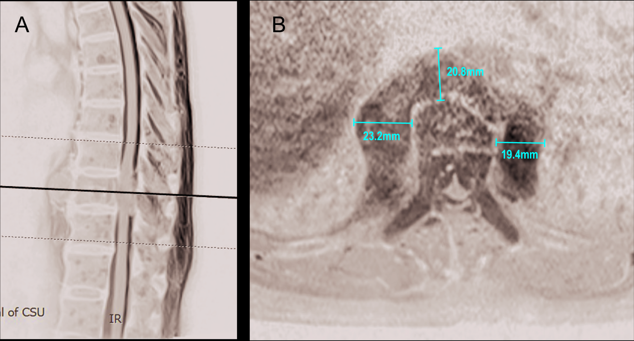
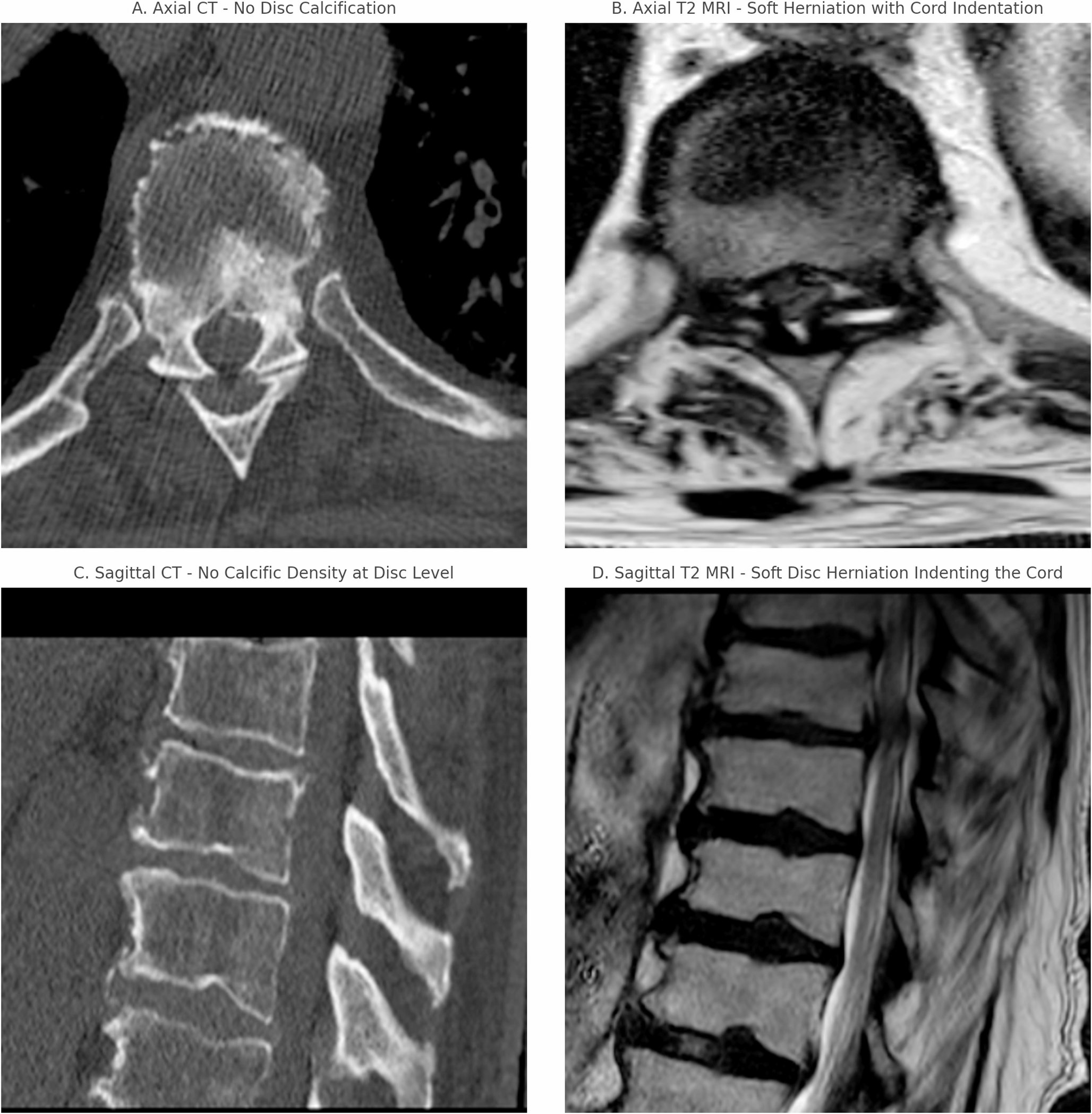
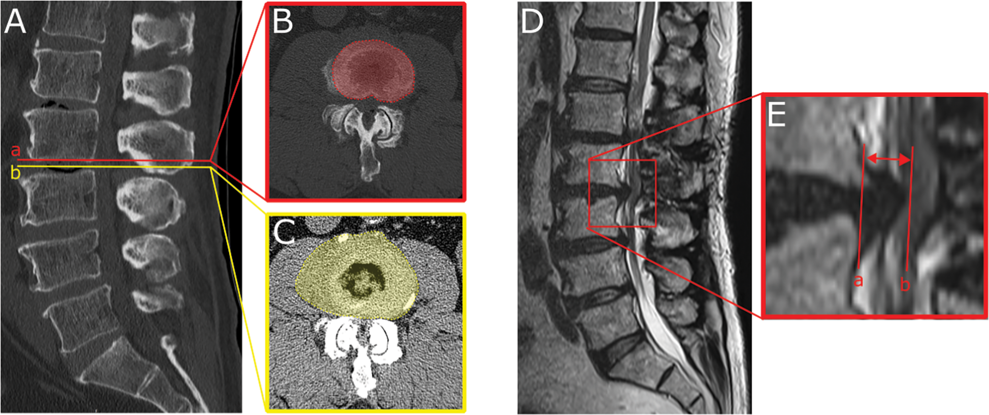
This study reports a 59-year-old female diagnosed with mixed-type cervical spondylopathy, OPLL at C5/6 and C6/7 levels, and hypertension. Preoperative computed tomography (CT) revealed flat-type OPLL at C5/6 and spike-shaped OPLL protruding into the spinal canal at C6/7. Intraoperative exploration identified regional plate-like hardening of the dura mater at C6/7. Following anterior cervical discectomy combined with ossified dura mater floating, the patient exhibited significant neurological improvement without postoperative complication.
AbstractSection
Conlusion
CT demonstrates diagnostic value for mature-stage DO but lacks sensitivity in early ossification phases. Spike-shaped OPLL may promote dural ossification by inducing mechanical stress-mediated transfer of osteogenic factors. Future research may need to explore artificial intelligence-based novel imaging biomarkers to enhance early diagnostic accuracy for DO.


Comments (0)