In this study we present our updated figures for ITx for advanced FAP-related desmoid disease [11]. Our center has been performing transplantation for this indication since 2008 but there has been a significant increase in activity recently due to accrued experience leading to earlier referral from the main national FAP center (St. Mark’s in London, UK).
We found three main indications in our cohort:
1/Large, fistulating desmoid with difficult to control or recurrent abdominal sepsis.
2/Patient with recurrent desmoid disease after previous major intestinal resections that would lead to ultrashort gut syndrome.
3/Patients requiring foregut resections (gastric or duodenal resections) due to large adenomas at high risk for malignant degeneration but who lack sufficient remnant bowel length to complete a reconstruction.
The first category represents the largest group of patients which often have a very poor quality of life due. Furthermore, even though desmoids are considered benign and do not metastasize, they can lead to life-threatening complications. Large series have consistently demonstrated poor outcomes with up to 14–30% mortality [21, 22]. In these patients, ITx allows for maximal desmoid resection although the main aim is source control with removal of all the affected mesentery and associated collections.
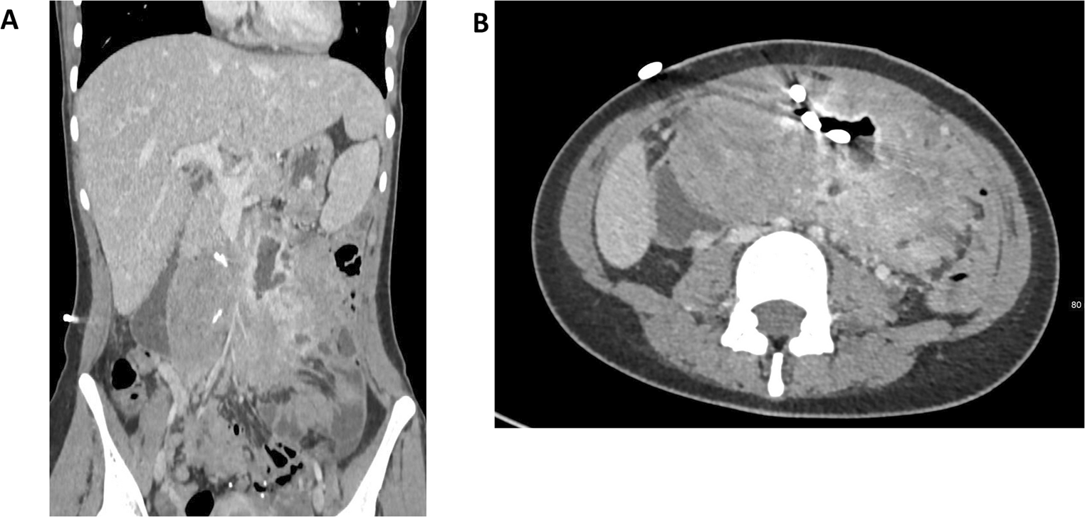
Pelvic disease is frequently encountered and leads to specific surgical issues like ureteric, J-pouch or vascular involvement. Almost half of our patients presented with ureteric obstruction, which is a recognized complication in advanced desmoid disease [23]. Many patients presented with chronic ureteric obstruction with stents or even nephrostomies in situ. Often the distal ureters are drawn into the desmoid mass and therefore have to be meticulously resected off in order to spare them. Due to induration and encasement by the surrounding desmoid, this may not be possible and thus several of our patients have required either resections with reconstructions, diversions to the contralateral side (end-to-side anastomoses) or most radically an auto-transplantation. In this operation, first described in the context of FAP by Lattimer et al. [24], the affected kidney is removed with a short segment of desmoid free-ureter, flushed with preservation solution and subsequently implanted lower down in the pelvis (in the same location a regular allograft) on the iliac vessels. This will allow the kidney with the much shorter ureter, to be successfully reconnected to the bladder. This is important as maintaining renal function in ITx patients is vital, as these patients frequent have renal impairment post-operatively due to previous intestinal failure with chronic dehydration and immunosuppression burdens [19]. Some auto-transplants were difficult as pelvic disease made access to iliac vessels very difficult.
In the most extreme case, native nephrectomies had to be performed due to recurrent infections or due to anatomical difficulties to preserve the native kidney. One patient had a horseshoe kidney with numerous vessels and affected ureters. As such, an auto-transplant was not deemed feasible and thus a nephrectomy was combined with an allograft.
The ileo-anal J-pouch been gaining in popularity in prophylactic colectomies in FAP patients compared to the ileo-rectal anastomosis [25]. This has resulted in a higher proportion of patients presenting to us with pouches and complex desmoid disease in the pelvis. Furthermore, any pelvic surgery in FAP patients often stimulates new desmoid formation in this area [26]. As mentioned previously, when performing the resection at the time of ITx, we are constrained by an already lengthy procedure and limited preservation time of the intestinal graft. Therefore, we opt to resect the remanent intestine but leave the J-pouch in situ after disconnecting it from its mesentery. Two of our patients (out of 5) have developed pouch ischaemia that was treated conservatively with drainage. In theory, the best option would be a delayed removal of the pouch. However, only one patient in our series had this procedure so far due to the high risk for iatrogenic injury to nerve of vessels [27]. In our case, it required a combined laparotomy (to free up the loops of transplant small bowel) and perineal approach to remove the remnant pouch. While the pouch remains in situ, it should be regularly endoscopically surveilled due to the risk of pouch adenocarcinoma [28].
Significant vascular involvement of desmoid was relatively rare in our series. Two patients had significant compression of the distal inferior vena cava which made the resection difficult due to the lack of a clear surgical plane. One patient presented with extensive pelvic disease and encasement of the both common iliac artery and vein. This was reconstructed using the available donor iliac vessels at the time of transplantation.
Surgical resections in desmoid tumors are currently considered the option of last resort for patients due to the high rate of recurrence [29,30,31]. Interestingly, there is seem to be little effect of complete versus near-total resections in these patients [32]. With this in mind, the main focus of ITx is similar to non-transplant surgery for advanced desmoids: remove as much infection, desmoid-infiltration and obstructed/non-functional bowel. However, the availability of normal functional bowel in the graft does allow for much more aggressive debulking. Furthermore, these newly transplanted organs do not carry the underlying FAP genetic defects. This means that although intra-abdominal recurrence has been described after ITx [33], we did not see this in our cohort.
However, as in the non-transplanted population, FAP patients do frequently present with large, extra-abdominal desmoids [34]. In our series, 3 patients had recurrences in the abdominal wall (native) and the thoracic area which did require surgery.
In an effort to reduce the recurrence rate and slow the growth of remnant desmoid disease, our team has recently been placing our FAP patients specifically on sirolimus (rapamycin) which is a mammalian target of rapamycin inhibitor (mTOR). This was initially done in many of our ITx patients to protect the renal function regardless of indication. However, based on some limited data on its anti-proliferative effect in desmoids, we now routinely start sirolimus after 3–6 months post-transplant [35].
Another contentious point, is the extent of native organ resection and subsequent choice of graft. Essentially, there are two options: foregut-sparing isolated ITx or foregut resection with modified MVT. The former is a shorter, less complex surgery with a lower complication rate and quicker recovery. By contrast, modified MVT is a much more complex operation with worse short-term outcomes. While initially leaning more towards the limited transplantation, the death by gastric cancer in patient 5, did lead us to now have a lower threshold for modified MVT. This applies especially to those patients with extensive ‘carpeting’ disease of the stomach or numerous duodenal polyps. This is especially driven by the increasing incidence of gastric and duodenal cancer, which have a very poor prognosis [36,37,38]. Furthermore, one could imagine that chronic immunosuppression after ITx would further increase this risk.
Similar to the retroperitoneal organs, the abdominal wall is often affected either directly by fistulation or direct invasion [39]. Furthermore, due to extensive resections, the abdominal domain may be severely restricted. Thus abdominal closure can be a major challenge. While various methods have been used in the past, we now exclusively rely on non-vascularized rectus fascia procured from the same donor as the bowel to close the abdomen [40, 41]. This simple, inexpensive and reliable method allows for bridging and closure of significant defects while allowing sufficient space for the new graft.
Finally, given the immense complexity of both the explant and the subsequent implant, the operation should ideally be split into two distinct stages. However, due to expected longer waiting time on the list and unpredictable graft availability, an extensive exenteration and subsequent listing is a potential but risky strategy. In our more recent experience, due to the extent of the disease, most patients have required extensive resections that necessitate simultaneous operations to reconstruct the GI tracts. Ideally, multiple teams that can rotate and take appropriate rests are warranted for these very long and complex operation (some exceeding 20 h).
Despite this study providing valuable insight into this rare disease, we have to acknowledge some limitations. First, due to the retrospective nature, some data may not have been included or lost. However, it should be noted that no patients were lost to follow-up. Second, there is a likely to be significant referral bias as only those patients considered eligible for ITx (relatively young patients, with no other known comorbidities) would be considered for this pathway. We do not have exact figures on how many patients were considered but eventually declined referral. However, given the UK’s existing strongly centralized pathway to identify and treat hereditary colorectal cancer/polyposis disease, this data could help create more evidence based referral guidelines. As a result of the strict referral criteria and rare nature of this disease, the absolute number of patients undergoing ITx remain very limited.
Finally, we were not able to report any patient-reported outcome measures (PROMs) as we did not prospectively capture these. However, last year a validated, disease-specific questionnaire has been developed which we could use for future research endeavors [42].


Comments (0)