Assessment of lower limb alignment: supine weight-bearing CT scanograms compared with a standing full-length radiograph
Purpose
We identified limb misalignment by applying personalized axial force while the limb was in a supine position to mimic a standing posture. This study aimed to confirm the accuracy of evaluating lower limb alignment using supine weight-bearing CT scanograms.
Methods
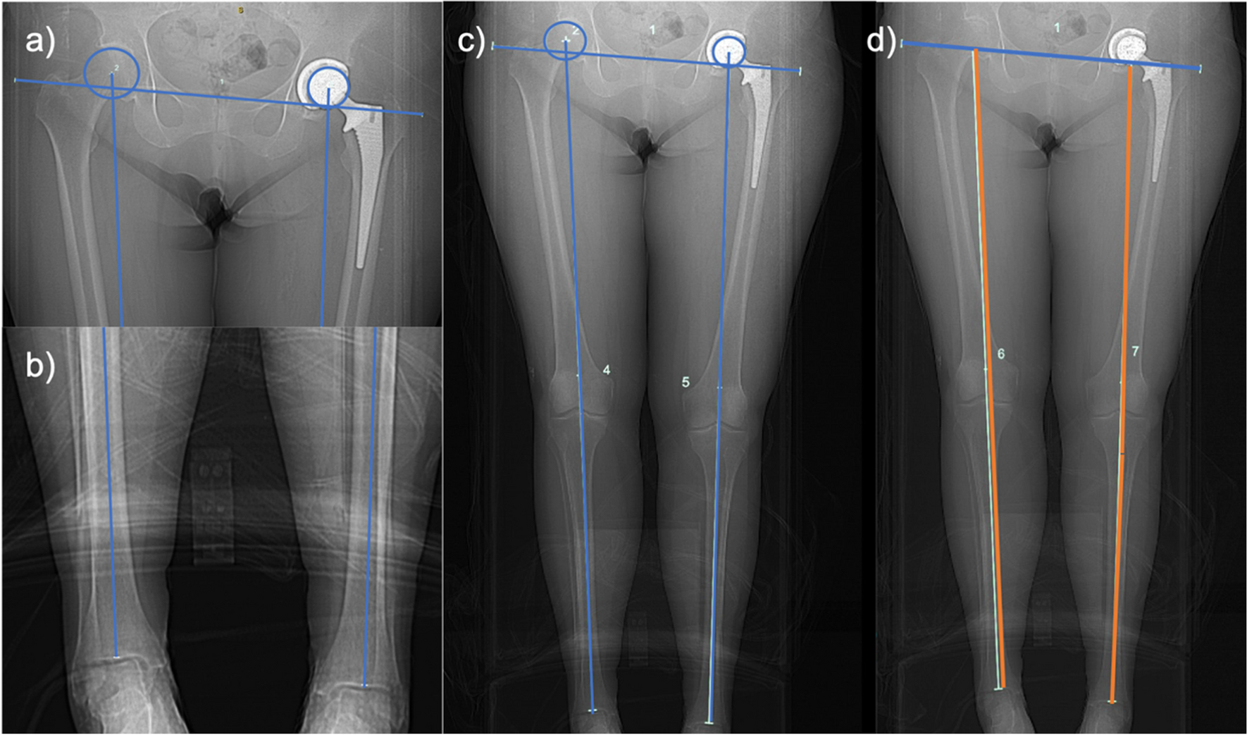
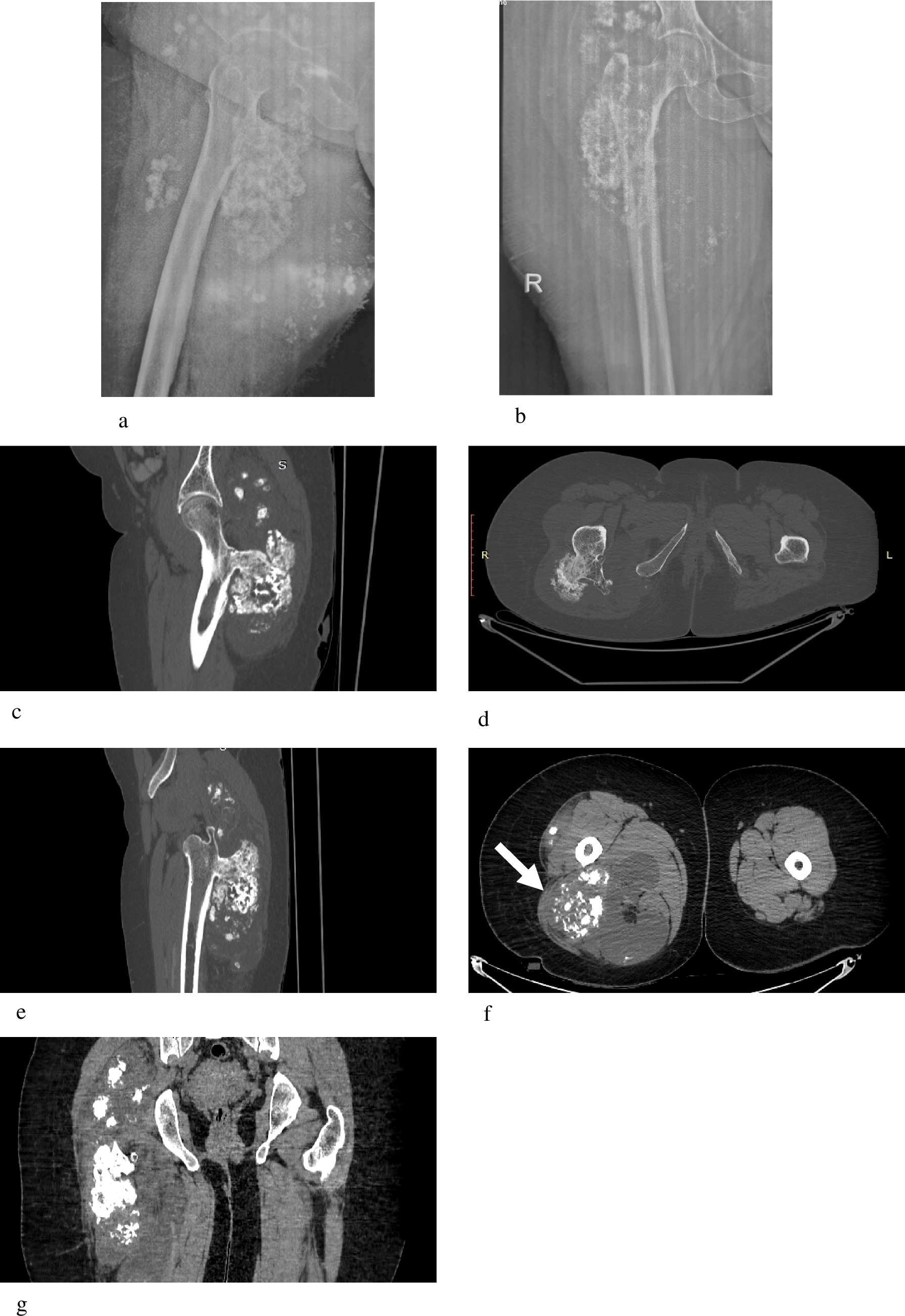
We prospectively compared measurements of the weight-bearing line ratio (WBL), hip-knee-ankle (HKA) angle, and joint convergence angle (JLCA) in 46 sets of supine weight-bearing CT scanograms with those obtained from full-length standing anteroposterior lower extremity radiographs. We achieved the weight-bearing CT scanograms by applying six different levels of axial force: zero, 1/5 of body weight, 2/5 of body weight, 3/5 of body weight, 4/5 of body weight, and full body weight. We assessed the impact of age, body mass index, HKA, and JLCA on the observed mechanical axis deviation differences between the two methods.
Result
The average absolute difference between standing radiographs and supine CT scanograms was 4.32% for the WBL ratio (p < 0.05), 1.25° for HKA (p < 0.05), and 0.46 for JLCA (p < 0.05). The mean absolute difference was minimal when applying full body weight axial pressure during CT scanograms (p > 0.05). Age, body mass index, HKA, and JLCA had no effect on the deviation in the mechanical axis measurements obtained through supine weight-bearing CT scanograms with full body weight.
Conclusion
No significant differences were found in assessing lower limb alignment between standing radiographs and supine weight-bearing CT scanograms with full body weight. Weight-bearing CT scanograms prove to be a valuable method for assessing lower limb alignment while in a supine position.


Comments (0)