
Remember me

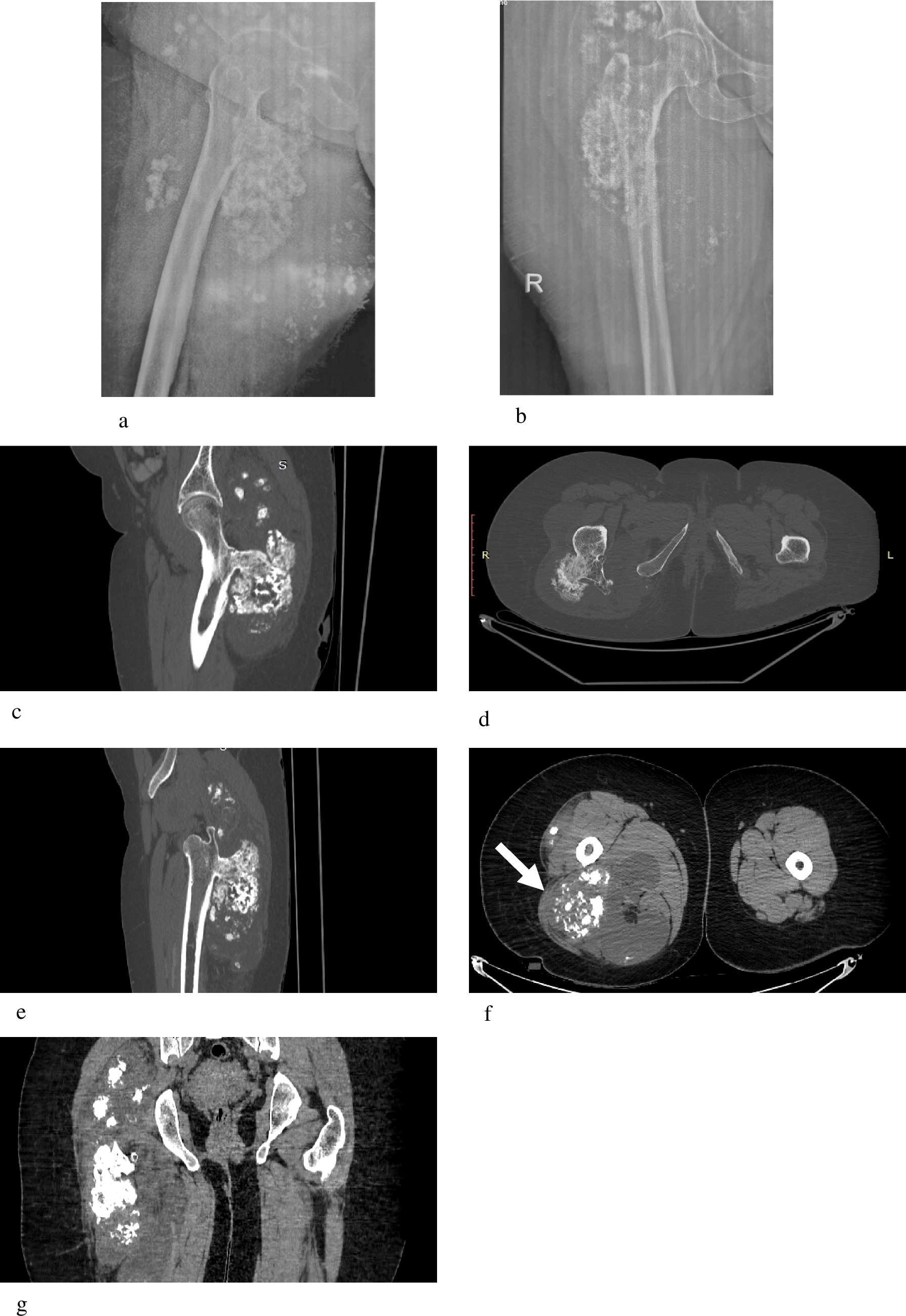
A 78-year-old male presented with a past history of high-grade right-sided pelvic chondrosarcoma for which he had previously undergone right hindquarter amputation. This had subsequently metastasized to the lumbar spine and he required radiotherapy and excision of paravertebral disease.
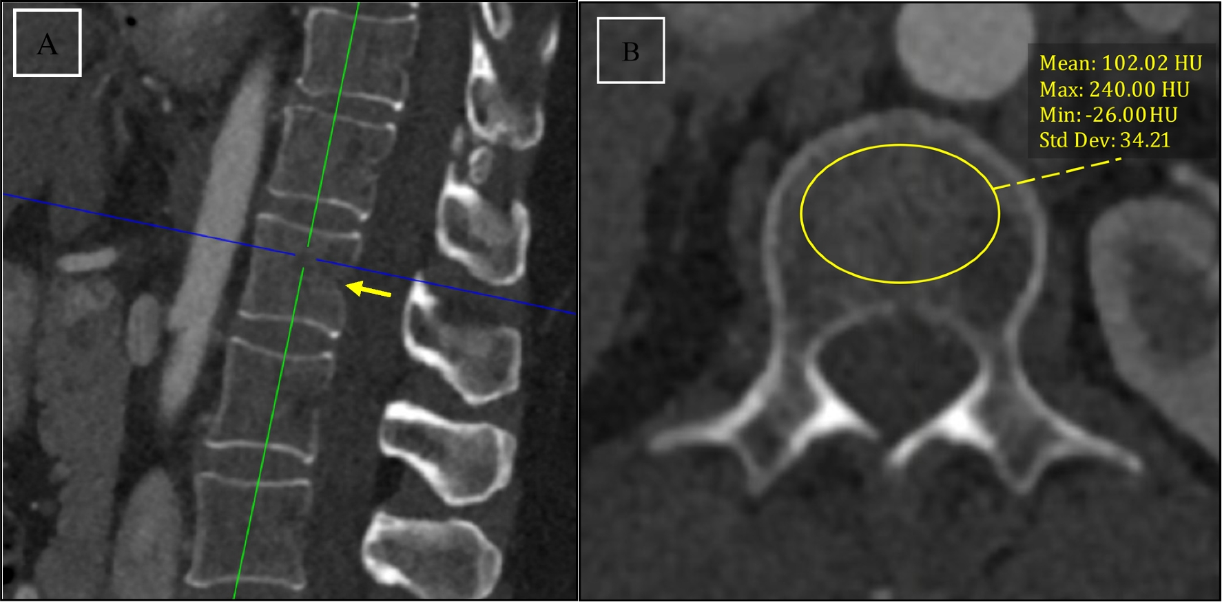
Several years later, he presented with double incontinence and motor deficit of his left lower limb in-keeping with cauda equina syndrome. Magnetic resonance imaging (MRI) of the spine and computed tomography (CT) of the body were performed. The following imaging depicts findings in his spine (Figs. 1, 2, 3 and 4).
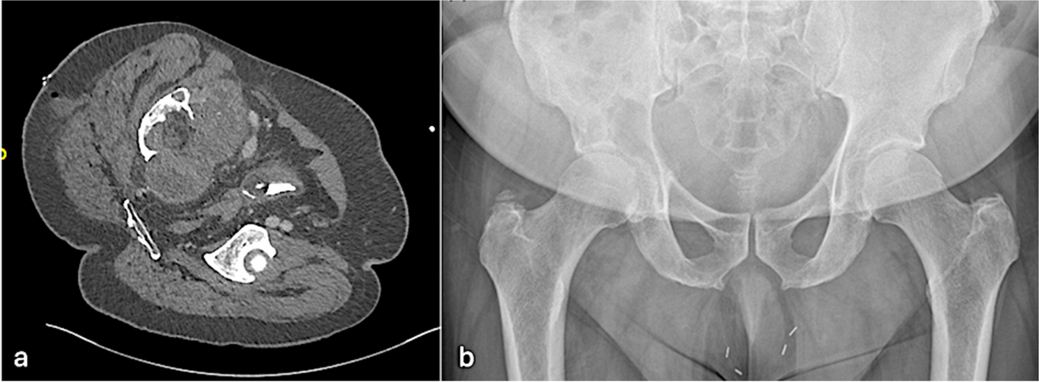
Fig. 1
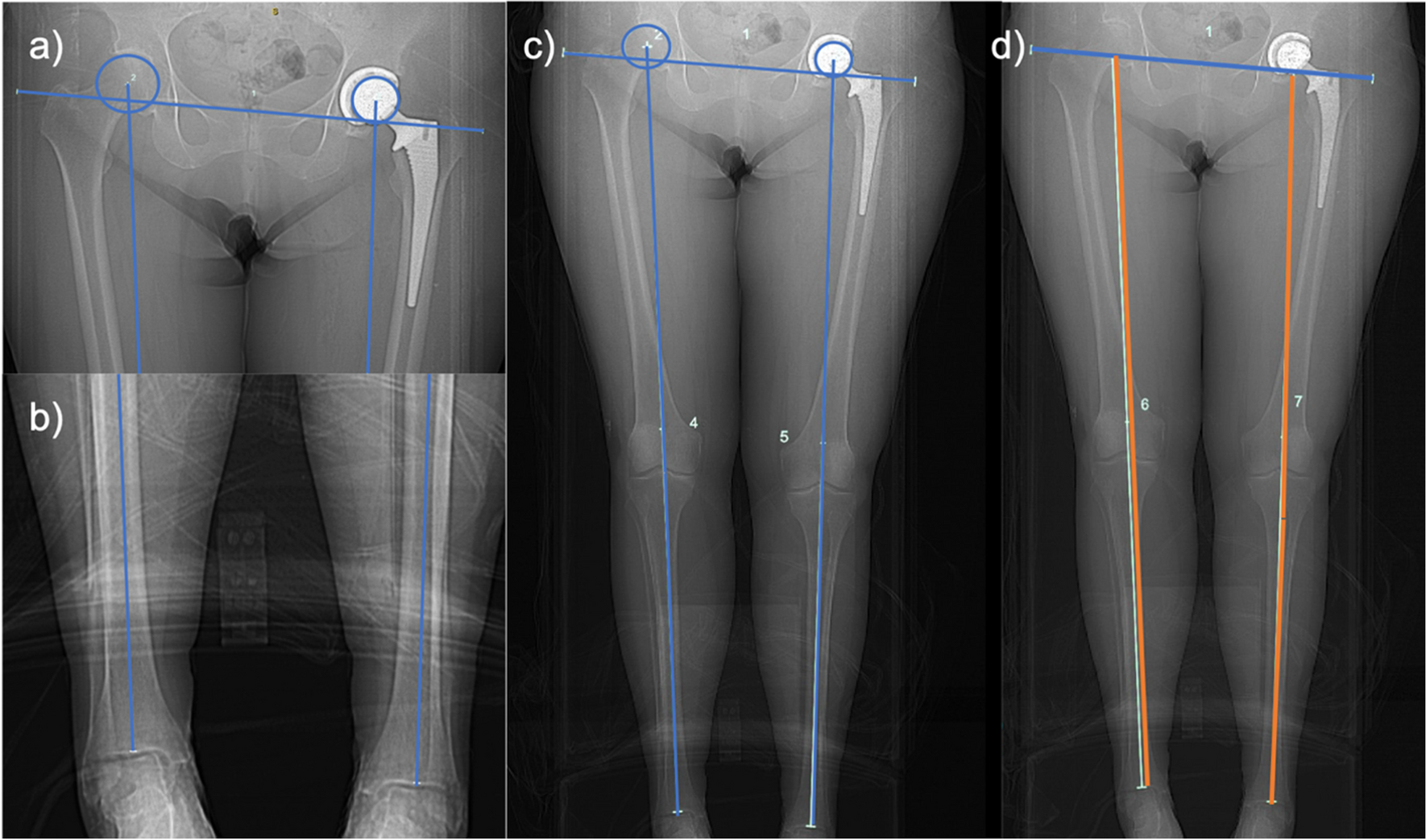
a, b Axial CT image (a) and AP radiograph of pelvis (b)
Fig. 2
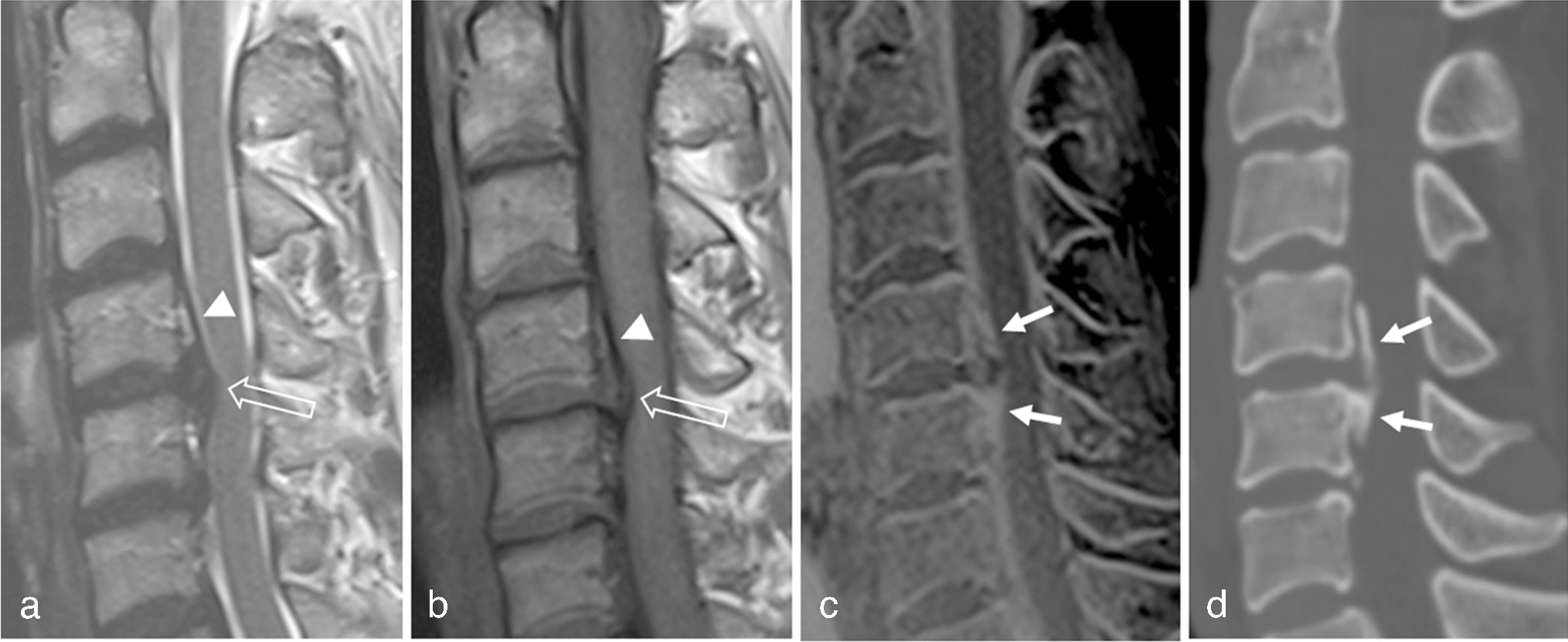
Sagittal T1 (a), T2 (b), and STIR (c) image of lumbosacral spine
Fig. 3
Axial T2 images at L5 (a) and S1 (b) and sagittal T1 (c) and STIR (d) sequence images of the lumbosacral spine acquired 5 years after Fig. 2
Fig. 4
a, b: Low (a) and high magnification (b) hematoxylin and eosin sections
Comments (0)