
Remember me


This study was aimed to demonstrate the outcomes of a novel “trans-facial” approach used to simultaneously access and reconstruct small RMT tumors through an islanded Nasolabial Flap (NLF). Patients with newly diagnosed squamous cell carcinoma of RMT region requiring surgery were included from January 2021 to September 2022. Case selection for this specific approach was done base on the location of the lesion and clinically T1 or T2 status. A total of eight patients were included in this study. The study was approved by the Institutional Ethics Committee (IEC). Radiological assessment was performed to determine skin involvement along with any underlying bone erosion.
Treatment decisions were made as per the disease management group’s joint clinic consisting of surgical, medical, radiation oncologists as well as a radiologist and pathologist. Patients were excluded if other head and neck sites were involved, previously received any treatment or if the disease process warranted a segmental mandibulectomy or skin excision.
Stringent case selection included RMT or posterior buccal mucosa lesions planned for surgical excision of the primary and appropriate for a local or regional flap reconstruction, along with ipsilateral neck dissection was done. A mouth opening of at least 20 mm at presentation was required. A minimum of 30 mm tumor free mucosal margin from the commissure was also needed as shown in Fig. 1, so that the base of the nasolabial flap would not be near the tumor excision area. This was confirmed by examining under anesthesia as well as radiological mapping and estimation of the approximate defect size. All patients underwent surgical resection with a clinical discernable margin of at least 1 cm. This could entail a marginal mandibulectomy with or without partial upper alveolectomy using per oral and trans-facial approach.
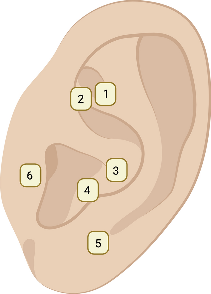
Fig. 1
Case selection for Trans-facial approach- Retromolar trigone lesion
Surgical ProcedureThe surgical plan included the completion of the ipsilateral selective neck dissection before commencing with the primary resection. The extent of neck dissection was based on the status of neck metastasis and the level of involvement. For cN0 patients, clearance of ipsilateral levels I to III/IV was performed; while, level V was included in cN + cases with lower neck involvement or clinical extracapsular spread (ECS).
During the neck dissection, care was taken to preserve the facial artery and vein in continuity across the lower border of the mandible. In case the facial vessels were not preserved, the nasolabial flap was not islanded and instead based on a random pattern blood supply with a broad base. After the neck dissection was complete, the planned nasolabial flap was marked (Fig. 2). We started by confirming the surface marking of the facial artery at the lower border of the flap using Mason’s point or a Doppler probe [5]. Once traced, a broad flap is marked based on the defect size. Figure 3 represents a schematic diagram of the Nasolabial flap and its reach to the retromolar trigone region.
Fig. 2
Marking of Islanded Nasolabial Flap
Fig. 3
Schematic diagram of NLF based on facial vessels and its reach to RMT lesion
The mucosa is incised intraorally, confirming the adequacy of the base of the nasolabial flap. The medial edge of the flap was incised and the facial vessels and its labial branches were identified and preserved by careful dissection. The superior/distal end of the vessel is then ligated and the incision is extended as per the marked flap design. The flap is elevated with the skin, subcutaneous tissue along with the underlying muscle by carefully preserving the artery and veins in the flap. Once the flap is partially raised, we enter the oral cavity by connecting through the anterior mucosal margin.
The anterior mucosal resection margin in the ipsilateral buccal mucosa should correspond to the posterior margin of the modified nasolabial flap. Next, the anterior mucosal cut is deepened to open into nasolabial defect and sufficiently extended to improve access. Further excision of the tumor can be completed through a combined approach using this defect and through the oral cavity (Fig. 4). Due to the wide access obtained transfacially, marginal mandibulectomy with coronoidectomy as well as an upper aleveolectomy can be performed. The specimen is then rotated outward and the posterior tonsillar and soft palate mucosal margins can be accessed intraorally—might be difficult to perform through the defect due to the specimen obstructing vision.
Fig. 4
Resection of the tumor through the Trans-facial approach
Once the specimen is delivered, the margins are assessed. The nasolabial flap is sutured in the defect (Fig. 5). Once the defects is covered adequately, the donor site is carefully closed along the nasolabial crease avoiding and correcting any Burrow’s triangle formation and any deviation of the oral commissure (Fig. 5).
Fig. 5
NLF used for reconstruction and final closure
The histopathology reports were reviewed to record the distance from tumor to mucosal, soft tissue and bone margin, separately, in grossing as well as microscopic examination. Adjuvant radiotherapy (RT) was given when depth of invasion was more than 10 mm, or more than 5 mm with other adverse factors like perineural invasion (PNI) or poor grade of differentiation, or in the presence of positive neck nodes. Concomitant chemotherapy (CCRT) was added in cases of positive margins or presence of extracapsular spread (ECS). All other clinical and pathologic parameters were obtained from the electronic medical records of the hospital.
Comments (0)