This case series provides insights into the use of diode lasers for managing common muco-gingival issues in orthodontics. This paper presented seven cases in four common scenarios that can delay treatment due to lengthy oral surgery/periodontic referrals. The delay may arise from the need to expose an impacted tooth or the delayed provision of an orthodontic retainer, following removal of fixed braces, due to unsightly gingival overgrowth requiring surgical recontouring. Diode lasers are frequently utilized in dental practices due to their compact size and cost-effectiveness [4,5,6,7, 16,17,18]. These lasers, mainly operating in the 800–980 nm range, offer deep soft tissue penetration and effective coagulation with minimal interaction with hard tissues [4,5,6,7, 16,17,18]. The low absorption of diode laser light by dental hard tissues, such as enamel and bone, makes it ideal for soft tissue procedures. This includes exposure of unerupted or impacted teeth, gingivectomy, frenectomy, as well as the excision of fibro-epithelial oral lesions, hyperplastic gingival tissue, or benign and premalignant oral lesions, such as leukoplakia, erythroplakia, or lichen planus [4,5,6,7, 16,17,18,19,20,21].
Diode lasers are part of the deeply penetrating laser category (visible and near-infrared spectrum, 450 nm–1100 nm), which allows transmission through water and results in a lower absorption coefficient in water [6, 16]. This characteristic enables diode lasers to penetrate deeply into healthy soft tissue, with the laser light scattering extensively within the tissue [6, 16]. However, these lasers are selectively absorbed in inflamed areas by blood components and tissue pigments, making them particularly suitable for soft tissue procedures. Compared to Nd:YAG lasers (1064 nm), diode lasers have a shallower penetration depth, reducing the likelihood of causing pulpal damage [6, 18].
Laser-tissue interactions may induce tissue warming, welding, coagulation, protein denaturation, drying, and even vaporization (ablation) and carbonization, where soft tissues are evaporated or incised [6, 11, 17]. This latter process also provides hemostasis, microbial inhibition and destruction [6, 11, 17, 22,23,24]. Increasing evidence suggests that the appropriate use of lasers is associated with reduced intraoperative and post-operative pain, as well as enhanced wound healing or tissue regeneration, compared to conventional scalpel surgery [6, 11, 15, 17, 22,23,24,25]. Diode lasers can be operated in continuous-wave or gated-pulsed mode [5]. Continuous-wave mode produces higher levels of heat which can result in soft tissue damage [26]. Overall, the laser-tissue interaction is determined by interplay between the incident photonic power density and exposure time [26].
Orthodontists often refer patients for the exposure of impacted teeth, removal of frenum, or excision of hyperplastic gingival tissue. These issues can delay orthodontic treatment, increase the risk of complications, and affect patient satisfaction [27, 28]. They can also be detrimental to the cost efficiency and productivity of a practice [27, 28]. The adjunct in-office use of a diode laser can improve the esthetic outcomes of orthodontic treatment, decrease treatment duration for patients requiring surgical exposure of superficially impacted teeth, reduce the number of appointments, and collectively increase patient satisfaction [4].
When performing gingival recontouring, as illustrated in Figs. 5 and 6, it is crucial to leave at least 1 mm of pocket depth and preserve at least 2 mm of keratinized tissue to prevent further soft tissue complications, such as gingival recession or absence of protective keratinized tissue [4, 18]. Gingival hyperplasia is common in orthodontic patients, occurring in about 10% of cases [4, 29, 30]. This condition impedes oral hygiene maintenance leading to esthetic and functional issues [4, 29, 30]. Extensive and fibrotic gingival hyperplasia often does not respond to conventional treatments, such as oral hygiene instructions, scaling, root planing, and prophylaxis, compromising patient self-care [4, 29].
The adjunctive use of diode laser gingivectomy can significantly improve gingival health in patients with gingival enlargement [6]. As shown in Figs. 7a–d, a patient with palatal gingival hyperplasia who did not respond to conventional treatment was successfully managed with diode laser gingivectomy. The purpose of this paper is not to discuss the combined gingivectomy and ostectomy, such as cases with ‘altered passive eruption’ or ‘crown lengthening’ that involves alveolar bone removal with surgical burs or hard tissue lasers such as ER:CR:YSGG laser (2780 nm) [31,32,33]. Instead, this paper focuses on gingivectomy procedures within soft tissue boundaries after exclusion of other causes of excessive gingival display [34]. Various lasers such as ER:CR:YSGG laser (2780 nm) [31,32,33], CO2 [35, 36], and Nd:YAG [36] laser have been used for gingivectomy. However, diode lasers appear to be becoming the popular option. Diode lasers (wavelengths of 808, 810, 940, and 980 nm) were compared to scalpel surgery in the literature [4,5,6,7, 24, 34,35,36,37]. A recent systematic review revealed that diode laser gingivectomy results in lower levels of pain and bleeding compared to conventional scalpel surgery [34].
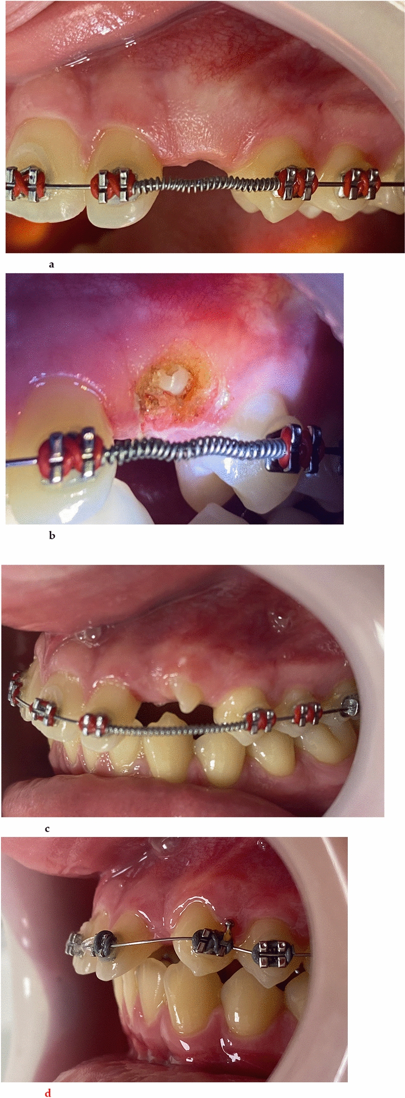
Diode lasers can facilitate the exposure of superficially impacted teeth, either with (Figs. 2, 3 and 4) or without (Fig. 1) simultaneous bonding of the exposed tooth. The bloodless exposure site created by the diode laser enables the bonding of brackets or attachments, which is difficult to achieve when using a punch or scalpel. When using a laser to expose impacted teeth, as depicted in Fig. 3a–d, it is important to keep the exposed crown within the keratinized mucosa and preserve the protective keratinized as much as possible [4,5,6,7, 38,39,40]. This approach helps avoid future complications, such as developing a thin periodontal biotype, the combination of gingival phenotype (three-dimensional gingival volume) and bone morphotype (thickness of the bone plate) [4,5,6,7, 38,39,40].
A recent scoping review revealed that laser exposure of impacted teeth is more efficient, with reduced treatment time, no bleeding, and less need for infiltration anesthetics and analgesics [41]. Diode laser surgical exposure eliminates the need for suturing and allows for immediate bonding of an attachment to the crown of the impacted tooth, compared to only 60% in the conventional scalpel group [41]. This method not only shortens treatment time but also results in less pain during orthodontic treatment, a shorter overall treatment duration, and fewer post-surgical complications [13, 24, 52]. For all reported cases, we initially used topical lignocaine anesthetic gel, applied for 3–5 min, followed by small amount of local infiltration (e.g., 2% lidocaine) approximately 5 min before the procedure to enhance postoperative comfort [41].
As illustrated in Fig. 4a–l, author introduced the concept of using a diode laser in combination with a surgical bur to remove bone and control excessive bleeding at the surgical site. While hard tissue lasers, such as Erbium lasers [Er:YAG (2940 nm) and the Er, Cr:YSGG (2780 nm)] can also perform these tasks, they are costly, require larger equipment setups, and are less effective at hemostasis due to poor absorption in pigmented chromophores [42].
Conventional surgical exposure of impacted teeth covered by bone typically involves making apically positioned flaps, performing releasing incisions apical to the adjacent teeth, cortical bone removal, and managing intra- and post-operative bleeding. This method often requires suturing, which can result in post-operative pain and potential infection. Conventional full-thickness mucoperiosteal flap procedures are relatively aggressive, often leading to minimal alveolar bone loss that can potentially compromise the integrity of the periodontium, particularly with thin alveolar bone thickness (< 2 mm) [43]. A full-thickness mucoperiosteal flap usually necessitates suturing and the placement of a protective dressing (pack) over the surgical site during healing. This process can be costly and stressful for patients [43]. Additionally, sedation or in rare situations general anesthesia may be required, and sutures typically need to be removed 1–2-week postoperatively [44,45,46,47].
Kokich [48] suggested three main methods for exposing labially impacted maxillary canines based on the position of their cusp relative to the muco-gingival junction. These are:
1.
The “closed eruption technique,” where the canine cusp is significantly above the muco-gingival junction and located intra-alveolarly high within the buccal sulcus.
2.
The “excisional uncovering or gingivectomy” when the canine cusp is below the muco-gingival junction, as seen in Case 2. The excisional uncovering can be accompanied with removal of any bone covering the crown of the impacted canine (48).
3.
If the canine cusp is above the muco-gingival junction, an “apically positioned flap” is suggested.
In Case 4, a combination of ostectomy, soft tissue removal, and subsequent gingivectomy was successfully employed, resulting in a healthy periodontal profile with adequate (2 mm) (48) keratinized tissue coverage. Considering that the canine crown was positioned partially below the mucogingival junction, any of the 3 techniques could be used to uncover the tooth (48), which might have involved raising a flap and significant bone removal; therefore, a more conservative approach was chosen to preserve alveolar bone during surgical exposure, given the patient’s age and the uncertain outcome of the procedure. Ectopic maxillary canines that are most favorable for alignment typically exhibit a mesial inclination and are located within 14 mm of the canine site on the occlusal plane, as observed in this patient [49, 50]. However, in adult patients over 30 years of age, the risks associated with surgical exposure and alignment must be carefully evaluated, as treatment outcomes can be compromised due to an increased incidence of ankylosis and slower rates of tooth movement [50, 51].
Previous research has shown that the success rate of orthodontic treatment for impacted maxillary canines in adults is lower than in adolescents [51]. For instance, research indicates a success rate of 69.5% in adults aged 20–47 years, compared to 100% in younger individuals aged 12–16 years [51]. The reduced success in older patients is attributed to factors such as increased bone density and decreased cellular activity, which can impede tooth movement. Consequently, orthodontic traction in adults may require longer treatment durations and carries a higher risk of failure, often leading to the consideration of surgical removal of the impacted teeth.
Nevertheless, surgical removal of the impacted teeth carries the risk of creating a significant bony defect in an esthetically critical area, which may necessitate bone grafting and complicate both orthodontic space management and subsequent dental implant placement [50,51,52]. As illustrated, the combined use of a diode laser and surgical bur to expose the crowns of impacted teeth located below or partically above the muco-gingival junction can be performed in carefully selected cases. Future research can investigate the long-term periodontal status of impacted teeth exposed in this manner compared to conventional exposure using apically repositioned flap or closed eruption technique.
In addition to managing gingival enlargement or hyperplasia, cosmetic gingival contouring, and exposing impacted teeth, diode lasers are used to uncover temporary anchorage devices (TADs) [52,53,54,


Comments (0)