
Remember me


The core microbiome plays a crucial role in the process of crop growth. Because the common microbiome of soil and rhizosphere soil in the three producing areas was the microbiome that existed regardless of region and plant, it was defined as the core microbiome of honeysuckle soil. The Venn diagrams shown in Fig. 1A-C indicated that there were 398 genera of bacteria in rhizosphere soil, 382 genera of bacteria in soil, and 333 genera of core bacteria in rhizosphere soil and soil, which belong to 23 bacterial phyla. The core bacteria mainly included Proteobacteria, Actinobacteriota, Firmicutes, Bacteroidota, Chloroflexi, Myxococcota, and Acidobacteriota. In addition, according to the dominant bacteria abundance heatmap, 22 of the 25 dominant bacteria genera were core bacteria genera (abundance > 2%) (Fig. 1D). The relative content of Bacillus in soil (2.93%) was higher than that in rhizosphere soil (2.39%), whereas the relative content of Streptomyces in rhizosphere soil (2.48%) was higher than that in soil (1.54%). The relative content of Burkholderia-Caballeronia-Paraburkholderia was 2.23% in rhizosphere soil and 0.67% in soil. The contents of Arthrobacter and Sphingomonas in rhizosphere soil were 2.50% and 2.48%, respectively, which were higher than those in soil. The average relative contents of Bradyrhizobium and Allorhizobium-Neorhizobium-Pararhizobium-Rhizobium in rhizosphere soil were 0.85% and 1.89% higher than those in soil, respectively, which indicated that more beneficial bacteria were enriched in rhizosphere soil.
Fig. 1
Abundance map of dominant bacteria and core bacteria genera. A: Venn diagram of Fengqiu, Pingyi, and Julu rhizosphere soils. B: Venn diagram of Fengqiu, Pingyi, and Julu soils. C: Venn diagram of Coreobacteria from Fengqiu, Pingyi, and Julu soils and rhizosphere soils. D: Heatmaps of rhizosphere soil and soil dominant bacteria (abundance > 2%) in Fengqiu, Pingyi and Julu. (RPY: Pingyi rhizosphere soils; RJL: Julu rhizosphere soils; RFQ: Fengqiu rhizosphere soils; SPY: Pingyi soils; SJL: Julu soils; SFQ: Fengqiu soils)
The Venn diagrams in Fig. 2A-C demonstrated that there were 157 genera of core fungi in the rhizosphere soil of the three producing areas, 139 genera of core fungi in the soil, and 115 genera of core fungi in the rhizosphere soil and soil. The genera were classified into the following seven phyla: Ascomycota, Basidiomycota, Chytridiomycota, unclassified Fungi, Mortierellomycota, Rozellomycota, and Glomeromycota. According to the abundance heatmap of dominant fungi, 31 of the 41 genera of dominant fungi were core fungi (abundance > 2%) (Fig. 2D). The average relative content of Mortierella in rhizosphere soil (11.92%) was higher than that in soil (11.72%). Moreover, the average contents of Metarhizium and Beauveria in rhizosphere soil were 5.63% and 2.29%, respectively, which were higher than those in soil (2.64% and 0.14%, respectively). The average relative content of Fusarium in soil (4.75%) was higher than that in rhizosphere soil (2.25%), but the average relative content of pathogens, such as Alternaria, Paraphoma, Cladosporium, and Rhizoctonia, in rhizosphere soil was higher than that in soil. These results demonstrated that rhizosphere soil enriches both beneficial fungi and pathogenic fungi to a greater extent than soil.
Fig. 2
Abundance map of dominant fungi and core fungal genera. A: Venn diagram of Fengqiu, Pingyi, and Julu rhizosphere soils. B: Venn diagram of Fengqiu, Pingyi, and Julu soils. C: Venn diagram of core fungi from Fengqiu, Pingyi, and Julu. D: Heatmap of rhizosphere soil and soil dominant bacteria (abundance > 2%) in Fengqiu, Pingyi, and Julu. (RPY: Pingyi rhizosphere soils; RJL: Julu rhizosphere soils; RFQ: Fengqiu rhizosphere soils; SPY: Pingyi soils; SJL: Julu soils; SFQ: Fengqiu soils)
Characteristics of soil microbial community structure and interaction network analysis in honeysuckle soilThe Shannon index and Simpson index describe the diversity and uniformity of the community. The larger the Shannon index and the smaller the Simpson index, the higher the species diversity of the community. Chao1 and Ace indices describe the number of species in a community, and the larger the value, the greater the number of communities. Alpha diversity analysis (Table 1) showed that there was no significant difference in the Shannon, Simpson, Chao1, and Ace indices of soil and rhizosphere soil in the core bacteria, but the mean values of the Shannon, Chao1, and Ace indices of rhizosphere soil were greater than those of soil. These results indicated that the community diversity and species quantity of bacteria in the rhizosphere soil core were higher. To further clarify the difference in bacterial community structure between rhizosphere soil and soil, the core bacteria of rhizosphere soil and soil at the genus level were evaluated by linear discriminant analysis effect size (LEfSe) analysis. Figure 3A shows that there were 25 different bacteria between the two groups. Among these, 18 rhizosphere soils had significantly higher bacterial abundance than soils, and 7 rhizosphere soils had lower bacterial abundance than soils, which indicated that the differences in core bacteria were attributed to the rhizosphere soils. The differential bacteria belonging to the dominant core bacteria (marked in red) were mainly beneficial bacteria, such as Allorhizobium-Neorhizobium-Pararhizobium-Rhizobium, which have a nitrogen fixation effect.
Table 1 Alpha index of core bacteria communityAlpha diversity analysis (Table 2) indicated that the mean values of the Shannon, Chao1, and Ace indices of core fungi in rhizosphere soil were greater than those of soil, while the mean value of the Simpson index of rhizosphere soil was less than that of soil, which indicated that the community diversity and species number of core fungi in rhizosphere soil were higher. To further clarify the differences between rhizosphere soil and soil fungal community structure, LEfSe was used to analyze the different core fungi in rhizosphere soil and soil at the genus level. Figure 3B shows that there were eight genera with different fungi between the two groups. Among these, three genera had significantly higher fungal abundance in rhizosphere soil than in soil, and five genera had lower fungal abundance in rhizosphere soil than in soil, indicating that the differences in core fungi were mainly attributed to soil, which contrasted the differences in core bacteria. Among these fungi, the dominant core differentiator, Paraphoma, was the pathogenic fungus.
Table 2 Alpha index of core fungi communityFig. 3
A: LEfSe analysis of core bacteria in soil and rhizosphere soil. B: LEfSe analysis of core fungi in soil and rhizosphere soil
To investigate the interaction of microbial communities in soil and rhizosphere soil, bacteria at the classification level were analyzed. Bacteria with correlations greater than 0.6 and P < 0.05 were selected to draw the interaction network among microbial communities (Fig. 4A-B), and the network attribute parameters were calculated (Table S1). As shown in Table S1, the average degree of rhizosphere soil was higher than that of soil, which indicated that one node in the rhizosphere soil bacterial network had more connections with other nodes, suggesting a more complex network. In addition, the average clustering coefficient of rhizosphere soil was higher than that of soil, indicating that the adjacent nodes of rhizosphere soil were more connected, and the nodes were more easily clustered together. These findings suggested that rhizosphere soil bacteria are more densely connected than soil and that their structures are more compact and complex.
The key species were further determined according to the topological characteristics of the nodes in the network. For this experiment, all the network nodes of soil and rhizosphere soil bacteria were peripheral nodes when the average relative abundance of the bacteria at the taxonomy level was greater than 1%, which allowed calculation of the Zi and Pi network attributes. In total, 12 nodes of soil bacteria and 25 nodes of rhizosphere soil bacteria fell in the connector, and neither node fell in the module hub or network hub (Fig. S1A-B). According to the bacteria corresponding to each key node, the distribution and abundance of the critical bacteria in soil and rhizosphere soil at the gate level were analyzed (Table S2). The key bacteria group in the soil belonged to 12 genera and 5 phyla, including Firmicutes (5 genera), Proteobacteria (4 genera), Desulfobacterota (1 genus), Verrucomicrobiota (1 genus), and Myxococcota (1 genus). The key bacteria groups in the rhizosphere soil belonged to 25 genera and 9 phyla, including Proteobacteria (8 genera), Firmicutes (6 genera), Desulfobacterota (3 genera), Actinobacteriota (2 genera), and Bacteroidota (2 genera). All other phyla belonged to 1 genus. In addition, according to the calculation, the abundance of key bacteria groups in soil and rhizosphere soil were almost all low-abundance species without dominant bacteria.
Fungi at the genus classification level were selected, and those with correlations greater than 0.6 and P < 0.05 were used to plot the interaction network among microbial communities (Fig. 4C-D), and the network attribute parameters were calculated (Table S3). As shown in Table S3, the average path length and network diameter were lower in the rhizosphere soil network than in the soil network. The average degree of rhizosphere soil was higher than that of soil, which indicating that one node in the rhizosphere soil fungal network had more connections with other nodes, suggesting a more complex network. In addition, the average clustering coefficient of rhizosphere soil was higher than that of soil, which indicated that the adjacent nodes of rhizosphere soil were more connected, allowing the nodes to be more easily clustered together. Thus, these findings demonstrated that rhizosphere soil fungi have more dense network connections than soil and that their structures are more compact and complex.
According to the topological characteristics of the nodes in the network, the key species were determined. Zi and Pi calculations indicated that 10 nodes of soil fungi fell in the connector, and the other nodes all fell in the peripheral nodes. Five nodes of rhizosphere soil fungi fell within the connector, and one node was located within the module hub. The remaining nodes were located within the peripheral nodes (Fig. S1C-D). According to the fungi corresponding to each key node, the distribution and abundance of the critical fungi in soil and rhizosphere soil at the gate level were analyzed (Table S4). The key fungi groups in soil belonged to 3 phyla in 10 genera, including Ascomycota (7 genera), Basidiomycota (2 genera), and Zoopagomycota (1 genus). The key fungi groups in rhizosphere soil belonged to 6 genera and 3 phyla, including Ascomycota (4 genera), Basidiomycota (1 genus), and Glomeromycota (1 genus). In addition, the abundance of key bacteria groups in soil and rhizosphere soil was comprised of low abundance species without dominant bacteria, and the key genera of soil and rhizosphere soil had no intersection.
Fig. 4
Graph of correlation network analysis of microbial community at genus level. A: soil bacterial network; B: rhizosphere soil bacterial network; C: soil fungal network; D: rhizosphere soil fungal network;
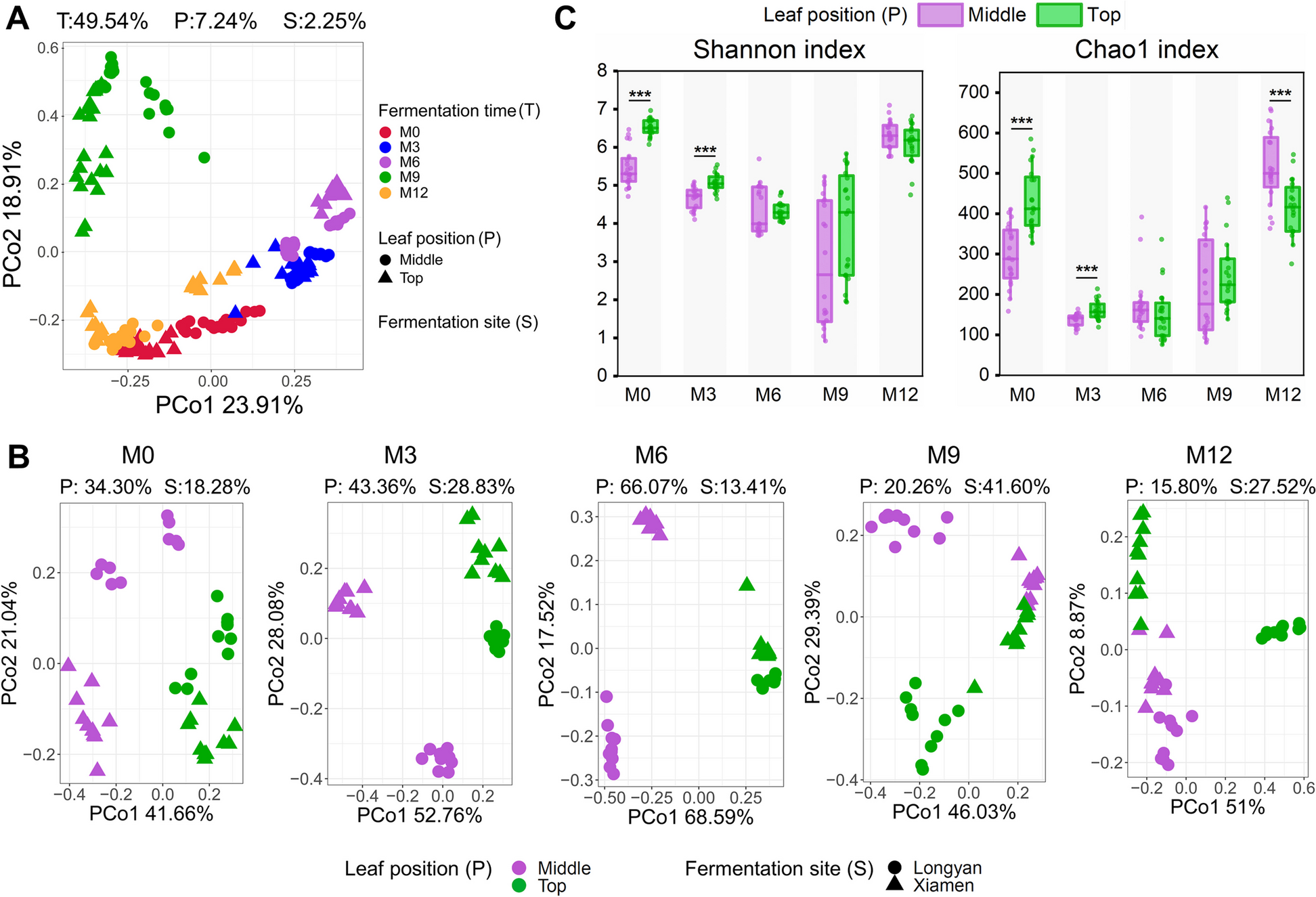
Comparison of microbial composition and analysis of microbial diversity in roots, stems, leaves, and flowers of honeysuckleBased on the OTU level, a Venn diagram was used to show the OTU number of unique endophytes and common endophytes in each tissue of honeysuckle. Endophytic bacteria Venn diagram as in Fig. 5A and endophytic fungi Venn diagram as in Fig. 5B.
For further analysis of species composition, the top 11 species were classified based on the phylum level and genus level (Fig. S2A-D). Endophytic bacteria were identified in 34 phyla and 770 genera. Endophytic fungi were noted in 11 phyla and 581 genera. Among them, the top four endophytic bacteria were Proteobacteria (83.99%), Actinobacteriota (6.83%), Firmicutes (6.16%), and Bacteroidota (2.35%). The abundance of other phyla was less than 1% in the root, stem, leaf, and flower tissues. The top two phyla of endophytic fungi were Ascomycota (54.42%) and Basidiomycota (44.32%), and the abundance of the other phyla was less than 2% in the roots, stems, leaves, and flowers.
At the genera level, the top four genera with the highest abundance of root endophytic bacteria were Pseudomonas (50.13%), g_unclassified_f__Enterobacteriaceae (10.96%), Allorhizobium-Neorhizobium-Pararhizobium -Rhizobium (5.92%), and g_unclassified_f__Rhodocyclaceae (5.27%). The top four genera with high abundance of stem endophytic bacteria were Ensifer (31.09%), g_unclassified_f__Rhodocyclaceae (9.45%), Pseudomonas (7.99%), and Bradyrhizobium (6.21%). The top four genera with high abundance of leaf endophytic bacteria were Ensife (25.25%), g_unclassified_f__Rhodocyclaceae (17.59%), Pseudomonas (13.41%), and Bradyrhizobium (7.04%). The top four genera with the highest abundance of floral endophytes were g_unclassified_f__Rhodocyclaceae (19.23%), Pseudomonas (18.70%), Pantoea (10.20%), and Aquabacterium (4.46%). At the endophytic fungal level, Gibberella and Fusarium were the highest in roots, with abundances of 17.55% and 7.94%, respectively. Phylloporia had the highest abundance in stems (59.13%), followed by roots (27.30%). Ceratobasidium was the highest in stems (33.37%) but less than 1% in roots, leaves, and flowers. Alternaria and Erysiphe were enriched in leaves with an abundance of 20.93% and 29.41%, respectively, followed by flowers with an abundance of 17.41% and 16.25%, respectively. In addition, Filobasidium and Cladosporium had the highest abundance in flowers, with an abundance of 21.13% and 13.77%, respectively.
As shown in Fig. 5C, according to the Shannon index analysis of the roots, stems, leaves, and flowers of honeysuckle, there were significant differences in the Shannon index of the bacterial communities of the roots and flowers, indicating that the endophytic bacteria of the flowers had higher diversity than that of the roots. In addition, the Ace index of stems and flowers was significantly higher than that of leaves, indicating that endophytic bacteria were more abundant in stems and flowers than in leaves. In addition, the Shannon and Ace indices of the fungal communities in roots, leaves, and flowers were significantly higher than those in stems, indicating that the diversity and richness of stems were lower than those in roots, leaves, and flowers. In addition, the Ace indices of endophytic fungi in leaves were significantly lower than those in flowers, indicating that the richness of flowers was higher than that in leaves.
PCoA analysis of endophyte communities based on Bray-curtis distance and testing of PCoA results were shown in Fig. 5D, which indicated that the overall structure of endophytic bacterial and endophytic fungal communities in roots, stems, leaves, and flowers of Honeysuckle was significantly different from that of the other endophytes. Further pairwise comparison showed that there were significant differences in endophytic bacterial community structure between the two pairs of each site in both endophytic bacteria and fungi (Fig. S3, Fig. S4).
Fig. 5
A: The endophytic bacteria in the roots, stems, leaves, and flowers of honeysuckle unique and common OTUs. B: The endophytic fungi in the roots, stems, leaves, and flowers of honeysuckle unique and common OTUs. C: Alpha diversity index of endophytes of roots, stems, leaves and flowers of honeysuckle. D: PCoA analysis of endophytes from roots, stems, leaves and flowers of honeysuckle. (R, root; S, stem; L, leaf; F, flower; P ≤ 0.05:*; P ≤ 0.01:**; P ≤ 0.001:***)
Relationship between endophytes and soil microorganisms of honeysuckle and prediction of endophyte functionsBy screening the common microorganisms of soil core bacteria of honeysuckle and endophytic core bacteria of honeysuckle, 60 genera of common microorganisms were obtained, which were defined as common core bacteria, and their content distribution in rhizosphere soil and soil is shown in Figure S5A. In addition, the common core dominant bacteria in honeysuckle plants were further screened (Fig. S5B), which indicated that the common core dominant bacteria in honeysuckle belonged to the dominant bacteria in soil (marked in red), indicating that the endogenous common core dominant bacteria may come from the soil. Pseudomonas, which was in the top five endogenetic dominant bacteria, is a conditioned pathogen, but it also has an anti-inflammatory effect, indicating that other bacteria may be beneficial bacteria. The results also suggested that honeysuckle flower selected some functional bacteria from the soil to colonize its body.
Through screening the common microorganisms of soil core fungi of honeysuckle and endophytic core fungi of honeysuckle, 30 genera of common core fungi were obtained, and their content distribution in rhizosphere soil and soil is shown in Fig. S6A. In addition, the common core dominant fungi in honeysuckle plants were further screened (Fig. S6B). The common core dominant fungi in honeysuckle belonged to the dominant fungi in soil (marked in red), indicating that the endogenous common core dominant fungi may also come from the soil. Most of the endophytic dominant fungi were common pathogens. The results suggested that honeysuckle selects for certain functional bacteria from the soil, as well as pathogenic fungi, speculating on their potential for certain functions.
Endophytic bacterial communities were predicted based on the PICRUST2 process. According to the abundance distribution of primary functional genes of endophytic bacteria in roots, stems, leaves, and flowers, genes related to metabolism had the highest abundance among the six known biological metabolic pathways (Fig. 6A), indicating that metabolism was the main function. Regarding the abundance of secondary functional genes of endophytic bacteria in roots, stems, leaves, and flowers, endophytic bacteria also had microbial resistance and other functions in addition to numerous metabolic functions (Fig. 6B), indicating that endophytic bacteria in various tissues of honeysuckle may function to resist some pathogenic microorganisms. FUNGuild was used to predict the fungal function of endophytic fungal communities in the roots, stems, leaves, and flowers of honeysuckle, which demonstrated that wood saprophytes, plant pathogens, and other saprophytic functions were more abundant (Fig. 6C).
Fig. 6
A: Abundance of KEGG first-level functional genes of endophytic bacteria in roots, stems, leaves and flowers of honeysuckle. B: Abundance of KEGG second-level functional genes of endophytic bacteria in roots, stems, leaves and flowers of honeysuckle. C: FUNGuild analysis of endophytic fungi in roots, stems, leaves, and flowers of honeysuckle (R, root; S, stem; L, leaf; F, flower)
Comments (0)