Animal ethics and animal feeding
Animal ethics in this paper are by the Institutional Animal Care and Use Committee of the Center for Excellence in Brain Science and Intelligent Technology of the Chinese Academy of Sciences.
Unless indicated otherwise, animals in this paper were subjected to a 12-h light–dark cycle and provided with unrestricted access to both diet and water. The rats obtained from the supplier underwent follow-up testing for a minimum of 2 weeks. Each rearing cage contained three male rats and they were non-aggressive towards each other. Animals were randomly selected for inclusion in the experimental group.
METH injection protocol
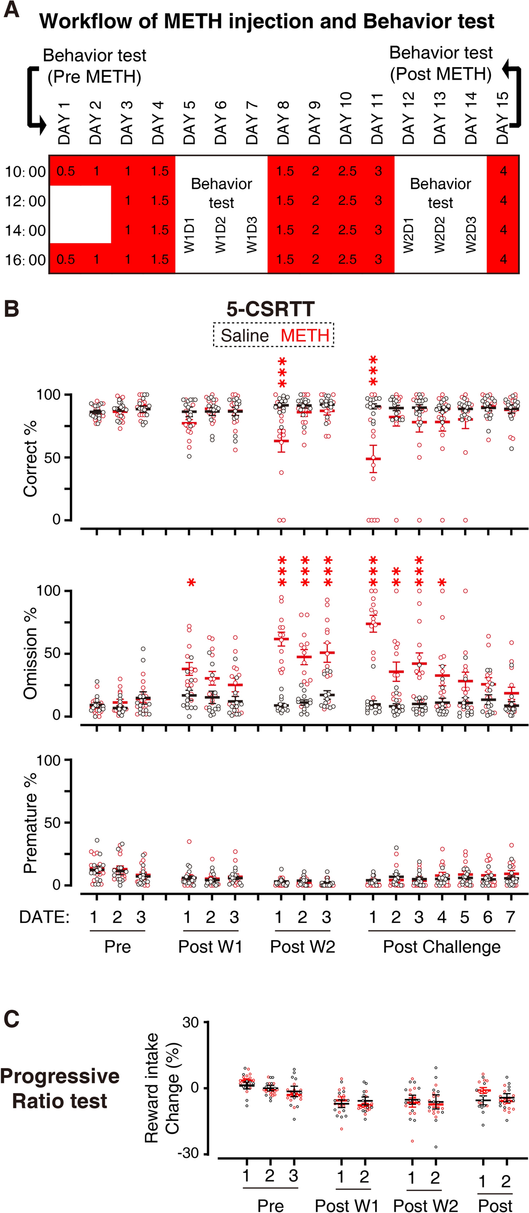
The male Sprague–Dawley rats (400–500 g) were subjected to an intraperitoneal injection of 200uL METH saline solution, following a 4-day injection-3-day rest cycle as the published protocol [21, 22]. The METH crystals were gifted from the Anti-Drug Brigade of the Shanghai Public Security Bureau.
Initially, on the first day, the rats received two injections of 0.5 mg/kg each. On the second day, they were given two injections of 1 mg/kg each. On the third day, four injections of 1 mg/kg each were administered. On day 4, the rats were injected four times with a dosage of 1.5 mg/kg. The same injection frequency and dosage were repeated on day 8. On day 9, four injections of 2 mg/kg each were given, followed by four injections of 2.5 mg/kg each on day 10. On day 11, the rats received four injections of 3 mg/kg each. Finally, on day 15 (challenge injection), they were injected four times with a dosage of 4 mg/kg. All injections were administered at 2-h intervals throughout the day.
Single-nuclei sequencing
Single-nuclei sequencing was conducted following the official protocol provided by the 10 × genomic platform. The rats were anesthetized at 72 h after the final injection of METH using sodium pentobarbital at a dosage of 50 mg/kg. Cardiac perfusion was performed using a solution of cold saline. The brain was then extracted and the specific regions of interest were dissected from bregma − 5 to − 9 in a brain module and frozen in liquid nitrogen. Subsequently, the frozen samples were transferred to a temperature of − 80 °C for preservation (< 1 month) and future utilization. For the isolation of single nuclei, the experiment followed the sample preparation guidelines outlined in the “Isolation of Nuclei for Single Cell RNA Sequencing” provided by 10 × Genomic company. The cDNA library was constructed using the 10 × Chromium Single Cell 3′ Reagent Kits (v.2 Chemistry). Sequencing was conducted on the Illumina HiSeq X10 platform, following the manufacturer's instructions. To annotate the raw *. fastq data for subsequent analysis, CellRanger was utilized.
Single-nuclei sequencing data analysis
We used the Seurat R package to analyze the data after obtaining the single nuclei sequencing data from CellRanger. The analysis process began with data normalization, selecting the top 2000 genes with the highest degree of variation for further investigation. A total of 4 samples, 2 treated with METH and 2 treated with Saline, were included in the workflow. The workflow followed the standard approach. For cell type identification and differential gene expression analysis, we utilized the Seurat package following the guidelines provided by Satijalab. To gain insights into the biological functions, KEGG and Gene Ontology analysis were applied using the KOBAS website [23], utilizing the feature gene list of Transcriptomic cluster-15.
Immunofluorescent
The process began with euthanizing the rats using carbon dioxide and the brain was collected 3-day post METH or saline challenge injection. The brain was then extracted without perfusion [24] and submerged in a 4% PFA-PBS (PBS: 137 mM NaCl, 2.7 mM KCl, 10 mM Na2HPO4·2H2O, 2 mM KH2PO4, pH 7.4) solution. Subsequently, it was stored overnight at 4 °C. The brain tissues underwent gradient dehydration using 15% and 30% sucrose-PBS solutions at 4 °C. Following this, the brains were sectioned into desired blocks using brain molds and embedded in OCT (Sakura 4583) compound.
For the coronal brain sections, the RTTG brain region was sampled at a thickness of 25 μm using a 1:8 ratio (i.e. for 25 µm brain slices, we collected one slice every 200 µm). For the sagittal brain sections, the brain sample was collected at a thickness of 40 μm. A blocking buffer was prepared with 0.1% triton-X, 5% BSA in 1 × TBS (20 mM Tris, 0.137 M NaCl, pH7.6) The brain sections were incubated in the blocking buffer at room temperature for 30 min with shaking. Then, the primary antibody was added and the sections were further incubated at 4 °C for 24 h. After two washes with wash buffer (0.1% Tween20 in 1 × TBS), the secondary antibody was introduced and incubated at room temperature for 1.5 h with continuous shaking. All antibodies were diluted in a blocking buffer. Subsequently, the sections were positioned on top of 1 × TBS in a sterile cell culture dish, mounted onto glass slides, and sealed with nail polish after surplus moisture evaporation. The mounting medium containing DAPI (Beyotime P0131) was applied for nuclear staining.
The primary antibodies used in this experiment and their dilutions were as follows: NFIB (Abcam ab186738) 1:2000, NeuN (Abcam ab104224) 1:2000, CaMKII (Abcam 181052) 1:2000, Gad1/2 (Abcam 183999) 1:2000 and c-fos (Abcam ab190289) at a 1:2000. As for the secondary antibodies, they were Donkey anti-Mouse Alexa Fluor Plus 488 (Thermofisher A32766) diluted at 1:1000 and Donkey anti-Rabbit Alexa Fluor Plus 647 (Thermofisher A31573) diluted at 1:2000.
The sections were then screened using the Olympus VS120 and analyzed with ImageJ software [25]. Integrated intensity from multiple brain slices containing the RtTg (or BPN) region was obtained for each rat and divided by the total area (measured in ImageJ) to calculate the mean intensity per sample. These mean intensities were subsequently normalized by dividing each by the average mean intensity of RtTg from saline-treated rats, yielding the relative fold change. For c-fos quantification, ImageJ’s “Analyze Particles” feature was used to count c-fos and NeuN-positive cells.
In situ hybridization
In order to perform in situ hybridization, the brain sections were first affixed onto slides and then subjected to staining, following the manufacturer's instructions of ACD's RNAscope™ 2.5 HD Duplex Assay (332430) or RNAscope™ Multiplex Fluorescent Reagent Kit (323100). Frozen brain sections were cut into 15 μm and collected at a 1:10 ratio then mounted onto glass slices for the following test. The probes used are RNAscope™ Probe-Rn-Hapln2 (588221) diluted at 1:2 and RNAscope™ Probe-Rn-Slc17a7-C2 (317001-C2) diluted at 1:100. The diluent used is RNAscope™ Probe Diluent (300041). For the fluorescent ISH, the probe utilized was RNAscope™ Probe-Rn-Slc17a6 (317011), RNAscope™ Probe-Rn-Slc32a1 (424541), the RNAscope™ Probe-Rn-Slc17a7-C2 and RNAscope™ Probe- Rn-Fos-C3 (430591-C3) was diluted in accordance with the manufacturer’s instructions. The sections were then screened using the Olympus VS120 and analyzed with ImageJ software as the IF step.
RNA–protein in situ co-detection
The Integrated Co-Detection workflow (ICW) was performed in accordance with the official guidance of RNA–Protein Co-detection kit (RNAscope™ 323180). The combination of the RNA ISH kit and Probe was previously described in the ISH procedure. In the ICW, the NFIB antibody dilution was 1:500 and the Donkey anti-Rabbit Alexa Fluor Plus 647 was 1:1000.
The NFIB antibody was diluted in Co-detection Antibody Diluent and incubated in 4 °C overnight. The secondary antibody was diluted in Co-detection Antibody Diluent and incubated at room temperature for 1 h.
Stereotactic surgery
According to the study conducted by Ali Cetin et al. [26], stereotactic surgery was performed. Anesthesia was administered to the rats using a 2% isoflurane solution, which was then sustained at a concentration of 1% using a stereotaxic apparatus. The injection site for AAV injection was determined based on Bregma coordinates. For RtTg injection, the chosen site was (ML: 0, AP: − 8.5, DV: − 10.5). A control injection sites were used at (ML: 0, AP: − 8.5, DV: − 7) in Fig. 4E. The AAV activity was diluted to a concentration of 2E+12 viral genomes per microliter (V.E./mL), and a total volume of 300 nanoliters (nL) was used.
The AAV was provided by Shanghai Taitool Bioscience Co., Ltd. The catalog of the used AAV in our experiment was:
AAV2/9-mCaMKIIa-hM4D(Gi)-mCherry-ER2-WPRE-pA: S0494.
AAV2/9-mCaMKIIa-hM3D(Gq)-mCherry-ER2-WPRE-pA: S0484.
AAV2/9-mCaMKIIa-mCherry-WPRE-pA: S0242.
For the RTTG lesion, the injection sites for Kainic Acid and PBS were randomly assigned to (ML: 0.5, AP: − 8.5, DV: − 10.5) or (ML: − 0.5, AP: − 8.5, DV: − 10.5). Kainic Acid (MCE HY-N2309), at a concentration of 1 mg/ml in a 1 × PBS solution, was injected at a volume of 250nL. For the Sham surgery group, 250nL of 1 × PBS was injected.
The injection was performed using glass microelectrodes. After the injection, it was standard practice to leave the needle in place for 5 min before removing it. Following hemostasis, disinfection, and suturing procedures, the subject was placed in a cage to recover naturally. No experimental procedures were conducted within 2 weeks after the surgery. Each rat was housed in an individual cage with access to ad libitum food and water. After a 2-week recovery, the rats gradually reintroduced the practice of the 5-CSRTT. This behavior resorting process continued 1 week and then discard the rats unable to restore their 5-CSRTT performance.
5-Choice serial reaction time task
The 5-choice serial reaction time task (5-CSRTT) is conducted as published protocol [17, 27]. Given that attentional impairment has been observed in rodents following METH administration, our 5-CSRTT is a simplified version with a 2-s stimulation duration [18, 27,28,29,30,31,32,33]. In our test, a food and water-restricted rat is placed in the 5-CSRTT apparatus, where one of five poke lights is illuminated 5 s after the rat triggers the food tray, and the rats are rewarded for correctly touching the illuminated light in the next 5 s (Fig. S1A, B). The 5-CSRTT was performed between 11:00 AM and 11:00 PM.
The training process involves gradually shortening the poke lights’ duration. In this experiment, the gradient configuration includes time intervals of 30 s, 10 s, 5 s, 3 s, and 2 s. After achieving stable performance in the 2-s 5-CSRTT task, rats may proceed to subsequent experiments if their average Omission% was below 30% for three consecutive days. Following each training session, rats have 1 h of free access to food and water. The reward employed in the 5-CSRTT task consists of a single droplet of sucrose solution with a concentration of 20%.
In the data collection step, rats enter the behavior training box at similar times daily and perform a 30-min 5-CSRTT test, with data automatically recorded by the software provided by Anilab (AES-130). To ensure consistency, all experimental groups were evenly distributed across the time slots through a cross-arrangement. The rats were also food and water restricted and rats have 1 h of free access to food and water after they complete daily task. The software records the Correct action, Error action, Omission action, and Premature action. The calculation method for Correct%, Omission%, and Premature% as follows:
$$}\% = }\;}/\left( }\;} + }\;}} \right)*00\%$$
$$}\% = }\;}/\left( }\;} + }\;} + }\;}} \right)*00\%$$
$$}\% = }\;}/\left( }\;} + }\;} + }\;} + }\;}} \right)*00\% .$$
For rats that have received AAV injections for neuronal activation, a 10 μg/ml deschloroclozapine (abbreviation: DCZ, catalog: MCE HY42110) solution is administered intraperitoneally at a dosage of 4 μg/kg, 40 min before the behavioral experiment begins. For the purpose of inhibiting neuronal activity and prevent RtTg activation in subsequent c-fos staining, we prolonged the inactivation duration by the DCZ was i.p. inject-ed at a dosage of 4 μg/kg, 2 h and 40 min prior to the 5-CSRTT and an additional DCZ injection is administered 1d prior to the first day to balance the subsequent daily DCZ intake.
The compound DCZ is initially dissolved in dimethyl sulfoxide (DMSO) at a concentration of 10 mg/ml and then combined with saline in a ratio of 1:1000. The control Vehicle (Veh) is a 0.1% DMSO-Saline solution.
The data was collected in 2–4 continuous days in the 5-CSRTT experiment without METH administration. The injection schedule for DCZ or Veh was as presented in the previous figure. The data presented in Fig. 3C, G were collected over the course of two consecutive days (1-day and 2-day post-weekly METH injection, respectively) and subsequently averaged for analysis. Challenge 5-CSRTT is provided in the 3-day and 4-day post-METH challenge injection. On the 1st and 2nd-day post-METH challenge injection, the rat undergoes a 2-s pokelight duration 5-CSRTT test. On the 3rd day, the pokelight duration is 1 s. On the 4th day, the pokelight duration is 3 s.
Shuttle test
The Shuttle test was post-completion of the 5-CSRTT test. The content of the Shuttle test involves the utilization of the behavior paradigm previously described, wherein all five pokelights are illuminated simultaneously and remain lit until they are triggered. Touching any pokelight at any given moment will yield a reward (Fig. S5B). If the data remains stable, the test proceeds to the data collection phase. This experiment is also conducted under food restriction, and rats are provided with 1 h of free feeding time after completing their daily tasks. The data is automatically collected by the 5-CSRTT software as previously mentioned. The DCZ or Veh injection protocol was as the previous 5-CSRTT described. The data were collected over the course of three consecutive days, after which the mean values were calculated for subsequent analysis.
Progressive ratio test
The Progressive ratio test, which is based on the fundamental principle that rats receive food as a result of repeated activation of the Foodtray, determines the ratio between the number of times the rat triggers the Foodtray and the reward it receives. The ratio is determined based on the reward amount, with a ratio of 1 for rewards between 0 and 20, a ratio of 2 for rewards between 20 and 30, a ratio of 5 for rewards between 30 and 35, and a ratio of (5*e^0.2(Reward-35) − 1) for rewards greater than 35.
For rats injected with METH, the Reward Desire test is conducted on the same day as the 5-CSRTT tests. The rats in the METH injection and 5-CSRTT group differ from the rats in the METH injection and Reward Desire test group. Both groups of rats are exposed to the same experimental conditions in terms of the 5-CSRTT testing environment and the environment for administering METH injections.
Data analysis
For all behavioral and optical microscopy data in this experiment, we employed ANOVA or 2-way ANOVA analysis. Subsequently, statistical corrections were applied using methods such as Bonferroni. In the case of a comparison between two groups, t-tests were utilized. All calculations were performed using GraphPad Prism 9.0.0. All error bars in this study are presented as SEM (standard error of the mean).
The optical density of the imaging data was measured using ImageJ, and the normalized integrated optical density was calculated by dividing it by the average value of the control group's integrated optical density.
For the analysis of single-nuclei sequencing data, we followed the methods implemented in the Seurat [34].


Comments (0)