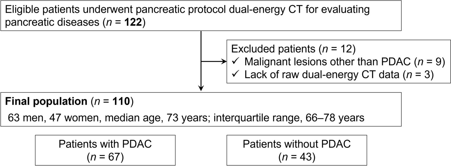
Our institutional review board approved this retrospective, single-center study, and the need for written informed consent was waived owing to the retrospective nature of the study.
Patient data
Patient data were collected exclusively at our institution. The study cohort comprised individuals who met the following criteria: (a) previously underwent FLAIR MRI revealing multiple white matter hyperintensities, (b) underwent follow-up MRI between July 2022 and October 2022, (c) were imaged using a specific MRI device capable of storing raw data used for this investigation, and (d) had successful acquisition of raw FLAIR data.
MRI image acquisition
All patients were scanned using a 3-T MRI scanner with a 32-channel head coil (FUJIFILM Healthcare Corporation, Tokyo, Japan).
The acquisition parameters for two-dimensional (2D) axial FLAIR were as follows: repetition time 12,000 ms, echo time 118.8 ms, inversion time 2770 ms, flip angle 90 \(^\circ\), field of view 240 × 240 mm, reconstructed matrix 512 × 512 (acquired matrix 320 × 376), slice thickness 5 mm, and slice gap 1.5 mm. The scan time of fully sampled data was 4 min 37 s. The image obtained using this fully sampled method was referred to as the standard FLAIR (std-FLAIR) image.
We retrospectively reconstructed 3 \(\times\) accelerated images (i.e., images obtained by applying AF = 3) using one-third of the fully sampled raw k-space data. MATLAB (version 2022a, MathWorks Inc., Natick, MA, USA) was used for the reconstruction. Intel Core i9-7900X CPU (Intel Inc., CA, USA) was used, with a memory of 32 GB. Images with a 3 \(\times\) AF were generated by first performing a Fourier transform on the fully sampled complex image of each channel to generate a k-space signal. This signal was then reduced by performing equidistant under-sampling in the phase encoding direction, sampling every third line. The 3 \(\times\) accelerated image was generated from the reduced k-space signal using a conventional reconstruction method (sensitivity encoding) or iterative reconstruction. An image equivalent to that obtained via 3 \(\times\) acceleration was designated as an accelerated FLAIR (acc-FLAIR) image.
To estimate the anticipated scan time for 3 \(\times\) acc-FLAIR, the time required for full sampling was divided by the AF (AF = 3).
Deep learning-based reconstruction
PI produces spatially non-uniform noise when the AF is high. To reduce non-uniform noise, we developed a method that combines iterative reconstruction with soft-thresholding and deep-learning-based denoising, with details published in the literature [22]. Our iterative reconstruction method was based on projections onto convex sets (POCS) [23]. In the POCS method, noise amplification depended on the sensitivity distribution, and spatial noise non-uniformity was generated. Wavelet-based soft-thresholding was added to the POCS method to reduce noise amplification. After spatial noise nonuniformity was reduced via wavelet-based soft-thresholding, spatial noise uniformity was reduced via deep learning-based denoising. Deep learning-based denoising combines a super-resolution convolutional neural network [24] with a residual network [25]. Super-resolution convolutional neural network comprises three layers, and each layer performs convolution, bias, and rectified linear unit operations. The kernel size was 9 × 9 for the first layer and 5 × 5 for the second and third layers. The first, second, and third layers had 64, 32, and 1 channels, respectively. Training was performed using reference image/noisy image pairs [22].
We applied deep learning-based reconstruction to the 3 \(\times\) acc-FLAIR images and designated the resulting images as DLR–FLAIR images.
Qualitative analysis (reader study)Image quality evaluation
As part of the qualitative assessment, three board-certified neuroradiologists with 14, 10, and 9 years of experience in radiology, respectively, visually evaluated the std-FLAIR, acc-FLAIR, and DLR–FLAIR images of all patients. All FLAIR images were anonymized and randomly displayed. The images were independently and blindly evaluated by the three neuroradiologists using a dedicated viewer (VOX-BASE Browser/View; J-Mac System, Sapporo, Japan).
To assess the image quality, two aspects were emphasized: (i) the amount of noise and (ii) the visibility of the gray/white matter contrast in the frontal lobes. Each aspect was rated on a 4-point scale. The amount of noise was evaluated as follows: the image contained excessive noise, and thus was unsuitable for diagnosis (1 point); noise was present, potentially impacting the diagnostic interpretation (2 points); noise was minimal, posing little to no effect on diagnosis (3 points); and almost no noise was evident, providing minimal diagnostic limitations (4 points). The visibility of gray/white matter contrast in the frontal lobes was assessed as follows: obscure (1 point), partially obscure (2 points), mostly visible (3 points), and fully visible (4 points).
White matter hyperintensity identification
Two radiologists with 15 and 17 years of experience, distinct from the three evaluators, selected 100 white matter hyperintensities, each measuring 3–10 mm in diameter. These hyperintensities were identified from the std-FLAIR images of 30 patients. Each patient had at least one hyperintensity, with a preference for those situated at the level of the centrum semiovale. The 100 hyperintensities were randomly displayed using combinations of std-FLAIR and either acc-FLAIR or DLR–FLAIR images. The same three board-certified neuroradiologists who assessed the image quality evaluated hyperintensity visibility in acc-FLAIR and DLR–FLAIR images compared with that in std-FLAIR images.
The boundaries of hyperintensities were rated on a 4-point scale as follows: unclear and different from std-FLAIR (1 point), partially different (2 points), nearly identical (3 points), and equivalent (4 points).
Quantitative analysisImage quality
The structural similarity index measure (SSIM) and normalized root mean square error (NRMSE) were calculated as pixel-based quantitative measures across the full image set, encompassing the entire brain. SSIM values (ranging from 0 to 1) indicate the percentage deviation of the acc-FLAIR and DLR–FLAIR images from the std-FLAIR image, with higher values denoting greater similarity to std-FLAIR image [26]. The NRMSE, a normalized variant of the root mean square error, offers a scale where lower values suggest less error, and thus closer resemblance between the acc-FLAIR or DLR–FLAIR and the std-FLAIR baseline image.
White matter hyperintensity identification
In addition, regions of 25 × 25 pixels containing 100 extracted white matter hyperintensities were harvested from each image at precisely the same coordinate axis. For the 100 white matter hyperintensities, the SSIM and NRMSE values in the region of 25 × 25 pixels (regional SSIM and regional NRMSE) for acc-FLAIR and DLR–FLAIR images were computed and compared with those for the std-FLAIR image.
MATLAB (The MathWorks Inc.) was used for the quantitative analysis.
Statistical analysis
The neuroradiologist-assigned image quality assessment scores for std-FLAIR, acc-FLAIR, and DLR–FLAIR were evaluated for statistical significance using the Wilcoxon signed-rank test. Bonferroni-corrected p values were employed, with statistical significance defined as a p value < 0.05. Results are discussed based on these corrected p values. The neuroradiologist-assigned white matter hyperintensity identification and quantitative (SSIM, NRMSE, regional SSIM, and regional NRMSE) scores for acc-FLAIR and DLR–FLAIR images were evaluated for statistical significance using the Wilcoxon signed-rank test. Statistical significance was defined as a p value < 0.05.
For qualitative assessment, the intraclass correlation coefficient (ICC) (2,1) was employed to evaluate inter-rater reliability among the evaluators (neuroradiologists) with values falling into the following categories: < 0.50 (poor), 0.50–0.75 (moderate), 0.75–0.90 (good), > 0.90 (excellent) [27].
The Wilcoxon signed-rank test was performed using JMP Software (version 16.1.0; SAS Institute Inc., Cary, NC, USA), and the ICC (2,1) was calculated using the Bell Curve for Excel (Social Survey Research Information Co., Tokyo).


Comments (0)