Reagents and antibodies
Cisplatin and paclitaxel were procured from MACKLIN (Shanghai, China). The CDH17 small interfering RNA (siRNA) and riboFECT™ CP Transfection Kit were purchased from RIBOBIO (Guangzhou, China). The CDH17 overexpression plasmid and the control plasmid were purchased from GeneChem (Shanghai, China). The Lipofectamine™ 3000 transfection kit was purchased from Invitrogen (Carlsbad, CA, USA). Antibodies against ABCG2, ALDH1, CD44, SOX2, OTC-4, α-catenin, p-α-catenin, YAP/TAZ, p-YAP, and Ki-67 were obtained from Abcam (Cambridge, UK). Antibodies against GAPDH, CDH17, 14-3-3, caspase-3, cleaved-caspase-3, survivin, goat anti-mouse IgG-HRP, and donkey anti-rabbit IgG-HRP were obtained from Affinity Biosciences. Antibodies specific for CD133 were purchased from Miltenyi Biotec.
Cell culture
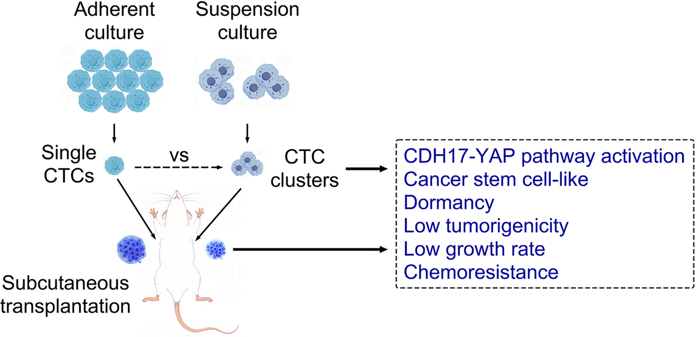
Lung cancer patient-derived CTCs (CTC-TJH-01) were established by our laboratory as we previously reported [22, 23]. CTC-TJH-01 cells were cultured in F12K medium (Gibco, CA, USA). An ultralow adsorption cell culture plate (Corning, no. 3473) was used to establish the CTC-TJH-01 cell suspension culture system.
Animals
We used 6-week-old male nude mice and NOD/SCID mice, which were obtained from GemPharmatech (Nanjing, Jiangsu), as the mouse models. The animals were housed under pathogen-free conditions in accordance with the Guide for the Care and Use of Laboratory Animals. All the animal experiments were approved by the Animal Ethical and Welfare Committee of Shanghai Municipal Hospital of Traditional Chinese Medicine, Shanghai University of Traditional Chinese Medicine (approval no. 2020-0014), in compliance with the guidelines of the Basel Declaration.
Morphological observation
CTC-TJH-01 cells in suspension and adherent cultures were observed and photographed under an inverted microscope with a digital camera (Leica, Wetzlar, Germany).
In vitro cell growth assays
CTC-TJH-01 cells (3000 cells/200 μl) were seeded in precoated or ultralow adsorption 96-well plates. Cell proliferation was assessed every 24 h using a Cell Counting Kit-8 (Dojindo, Shanghai, China).
Cell cycle analysis
For the cell cycle analysis, CTC-TJH-01 cells were cultured in adherent media and suspensions for 48 h. Then the cells were digested, collected, stained with PI, and analyzed with a FACSVerse™ flow cytometer (BD Biosciences, CA, USA).
Quiescent cancer cell analysis
CTC-TJH-01 cells were cultured in adherent media and suspensions for 48 h. Then the cells were digested and collected, and stained with PI and Ki-67 separately. Flow cytometry was subsequently used to analyze the proportion of Ki-67 negative cells in the G0/G1 phase, which were considered cancer cells in the quiescent stage.
Drug sensitivity assays
CTC-TJH-01 cells (5000 cells/100 μl/well) were seeded in ordinary or ultralow adsorption 96-well plates. After overnight incubation, the cells were treated with cisplatin or paclitaxel for 24, 48, or 72 h, and cell viability was assessed via a CCK-8 assay.
Apoptosis analysis
The annexin V-FITC/PI apoptosis assay was performed as previously described [24]. CTC-TJH-01 cells were seeded in ordinary or ultralow adsorption six-well plates. After overnight incubation, the cells were treated with cisplatin or paclitaxel for 48 h, and cell apoptosis was assessed via an annexin V-FITC/PI apoptosis assay (BD Biosciences, CA, USA).
Western blot analysis
Western blotting was conducted as described previously [25]. Briefly, the cells were lysed, and the proteins were extracted. Then, 40 µg of protein were used for western blot analysis.
Transfection
RNA interference assays were performed as described previously [26]. In brief, CTC-TJH-01 cells were seeded in ultralow adsorption 24-well plates. After 4 h, the cells were transfected with CDH17 siRNA, via a riboFECT™ CP Transfection Kit (RiboBio, China). An unrelated, scrambled siRNA was used as a negative control. For the CDH17 plasmid, 2 μg of the CDH17 plasmid was transfected via a Lipofectamine™ 3000 transfection kit, as instructed by the manufacturer. An empty vector plasmid was used as a negative control.
Immunofluorescence staining assays
In brief, CTC-TJH-01 cells were seeded in ultralow adsorption 6-well plates or laser confocal small dishes. After 48 h, the cells were first stained with CDH17, and then red fluorescent secondary antibody and DAPI. The expression of CDH17 was captured via a Leica TCS-SP8 laser confocal microscope.
Transcriptomics, proteomics, and bioinformatics analysis
Briefly, CTC-TJH-01 cells were collected after adherent and suspension culture, after which the cells were sent to Sinotech Genomics (Shanghai, China) for RNA sequencing and Hangzhou Jingjie Biotechnology Co., Ltd (Zhejiang, China) for protein mass spectrometry. The overlapping differential genes and proteins were analyzed.
Tumor growth assays
CTC-TJH-01 cells in suspension or adherent culture were injected subcutaneously into the right or left armpit of 6-week-old male nude mice with a density of 5 × 10⁵ cells (100 μl). The development and growth of the tumors were measured twice a week with a Vernier caliper. The tumor volumes were calculated using the formula: [sagittal dimension (mm) × cross dimension (mm)2]/2 and are expressed in mm3. The animals were euthanized once the volume of the tumor exceeded 2000 mm3. Approximately 9 weeks after inoculation, the mice were sacrificed, and the tumors were sectioned, stained with hematoxylin and eosin (HE) and subjected to immunohistochemistry.
Immunohistochemistry assays
Subcutaneous tumors were fixed with 4% paraformaldehyde, embedded in paraffin, and sectioned. The tumor sections were stained with HE and antibodies against human CDH17 and Ki-67. The slides were scanned with an automatic digital pathological section scanner (KFBIO, Zhejiang, China).
Statistical analysis
The significance of the differences was determined via Student’s t-test or one-way ANOVA. Kaplan–Meier analysis was employed for survival analysis and the differences in the survival probabilities were estimated using the log-rank test. All the statistical analyses were performed via GraphPad Prism 8.0 (GraphPad, San Diego, CA, USA). All experiments were performed at least in triplicate (n = 3). The data are expressed as the mean ± standard deviation (SD) or the mean ± standard error of the mean (SEM). The levels of statistical significance were set at *P < 0.05, **P < 0.01, and ***P < 0.001.


Comments (0)