
Remember me
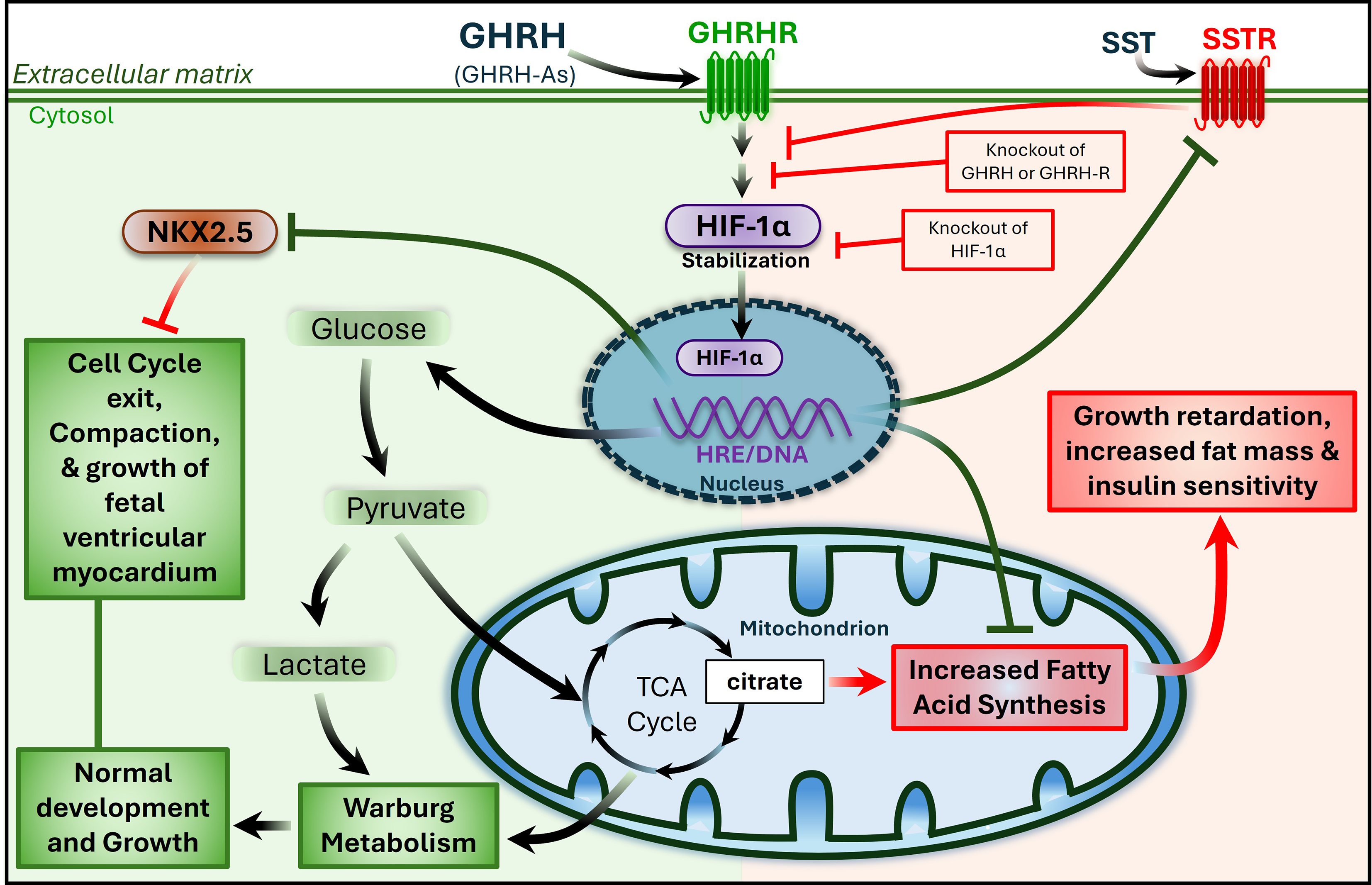


Since the beginning of the last century, investigations have suggested that raw BIA-derived parameters, particularly PhA, are determined by several factors directly influencing the overall characteristics of cells and the organization of structural tissues. By adopting a biophysical point of view, this review will describe how relevant atomic-molecular, cellular, and tissue-level determinants can influence the conductive and capacitive properties of the human body, which will further modulate PhA (Fig. 1).
Fig. 1
Iceberg model of bioelectrical impedance analysis: surface biophysical indicators and underlying biological properties
2.1 Atomic and molecular-level determinantsWhen discussing micro-level determinants of raw BIA variables, we typically refer to distinct constituents implied in atomic-molecular processes that can influence the cell’s biophysical properties. Within this research field, relevant cell constituents include water, as well as other elements such as inorganic ions and organic molecules (Fig. 2, Atomic - Molecular).
2.1.1 Inorganic ionsAccounting for 70 to 80% of BCM, water is the most abundant molecule in cells [20]. In the human body, cellular water is not inherently pure. Instead, it contains multiple dissolved ions that are electrically charged and sustain electrical conductivity [20, 21]. This fundamental concept was first demonstrated by Rudolf Höber in 1910 through in vitro experiments showing that the electrolytic cytoplasm of compacted blood and muscle cells, which contained free ions within a plausible physiological range, is a relevant determinant of electric conductivity [21]. While hypothesizing that conductivity directly correlates with the concentration of ions dissolved in an aqueous solution, Meguid and colleagues demonstrated a direct association between Na+ concentration and conductivity using dialysis tubing [22]. Similar trends were documented in other research areas exploring water conductivity properties from various sources [23]. Higher conductivity rates were found in highly enriched water with ions but not in relatively pure types of water (e.g., distilled solutions), independently of the measurement methods (i.e., number of electrodes and frequency levels) [23].
While certain cellular processes supporting growth and bone mineralization (e.g., signal transduction and gene expression) [24], could suggest increased intracellular levels of specific electrolytes, particularly those directly involved in these processes (e.g., Ca²⁺ and PO₄³⁻), potentially favoring intracellular conductivity, there is still limited evidence to confirm this hypothesis. Instead, the available literature points towards similar concentrations of the most prevalent inorganic ions (e.g., Na⁺, K⁺, Mg²⁺) across all ages, particularly in healthy populations. Nevertheless, direct verification is still required, especially through experiments using in vivo methods (e.g., neutron activation analysis), to determine the real cytoplasmatic concentrations of inorganic ions and assess their relationship with BIA-derived parameters. Moreover, further evidence is required in youth to investigate the frequency-dependent nature of conductivity and its role in how atomic processes (e.g., drifting ions) influence BIA measurements. A major challenge relies on the fact that most available techniques provide only a systemic overview of inorganic ions without distinguishing between intracellular and extracellular compartments, whereas other physiological factors, such as the cell structure and internal temperature [25], may also influence the electrical path within the cell environment.
2.1.2 Organic molecules2.1.2.1 LipidsLipids, such as fatty acids (FA), are the fundamental building blocks of the cell membrane and other cellular organelles, operating structural, metabolic, and signal transduction functions. While looking at the implications of lipids on electrical conductivity, Hodkin and Huxley [26] have early documented the bioelectrical phenomena when the electric current is carried through lipidic structures, such as the cell membrane. The organization of the cell membrane as a bilayer lipid membrane (BLM) enables the formation of an insulating barrier between the electrically conductive solutions of the intra- and extracellular mediums, with the potential to retain energy through the capacitive properties of the FA chains (i.e., the hydrophobic surface of each monolayer; capacitance of ~ 1 microfarad µF/cm2) [27].
Despite the rapid turnover of membrane lipids, the dietary FA composition can significantly influence membrane lipid structure and function. Previous evidence has shown that membrane-linked cellular processes contributing to energy metabolism remain relatively constant in response to saturated and monounsaturated FA diets but are significantly affected by polyunsaturated FA, such as n-6 and n-3 polyunsaturated FA [28]. A similar pattern has been observed in electrical capacitance, with this biophysical property being particularly sensitive to changes in membrane lipids. For instance, studies on NG108-15 cells demonstrated that dietary supplementation with polyunsaturated FA induced significant changes in membrane capacitance, reflecting alterations in membrane lipid dynamics [29]. More recently, in vivo experiments using whole-cell patch clamping to record cardiomyocyte electrical activity demonstrated that treatment with multiple FA enhances membrane capacitance, likely reflecting an increase in cell surface area [30].
When looking at lipidic structural characteristics in youth, although phospholipids composition assumes adult levels of most FA before the second year of life, some FA are expected to be higher in youth [31], potentially enhancing the capacitive properties of the cell membrane. For example, monounsaturated FA, such as oleic acid (i.e., 18:1 n-9 FA; most prevalent monounsaturated FA), are expected to peak in the erythrocyte membrane lipids during childhood and then gradually decrease until adulthood [31]. Considering the unique characteristics of this monounsaturated FA (i.e., kink structure), previous evidence has shown a close relationship between the content of this unsaturated FA in the membrane phospholipids and overall membrane integrity and cell growth [32]. In this context, cell growth refers to an increase in the cell surface area-to-volume ratio, a factor with significant implications for membrane capacitive properties. The relationship linking capacitance to cell membrane area was previously demonstrated in experimental research employing a biophysical membrane model approach, specifically focusing on artificial lipid bilayers composed of egg phosphatidylcholine, cholesterol, and n-decane [33].
All membrane-bound organelles (e.g., endoplasmic reticulum, Golgi apparatus, mitochondria, nuclear envelope) display similarities with the cell membrane regarding their lipidic composition and organization as BLM, potentially functioning as dielectric materials capable of storing electrical charge [27]. However, further evidence is needed to prove that these cellular components can act as true capacitors. If proven, higher levels of capacitance might be found in the membranes of organelles that have a relatively pure lipid composition, which is often observed in the early stages of life [34]. However, as age progresses, genetic mutations and environmental stressors lead to the aggregation of abnormal proteins within the internal cellular membranes, disrupting membrane integrity and signaling pathways [35] and potentially diminishing its capacitive properties.
2.1.2.2 ProteinsIn the human body, the key location of proteins includes the cell membrane, cytoplasm, nucleus, and organelles, where a diverse range of structural and signaling processes occur. Despite these molecules not inherently exhibiting capacitive characteristics in the same way as others (e.g., lipids), their constitution and functional roles contribute to characterize them as facilitators of intracellular conductivity. From a biophysical perspective, great attention has been given to proteins coupled in the cell membrane – i.e., voltage-gated ion channels. When facing voltage, mechanic, or binding-related stimulus, these particular proteins act as channels that selectively or non-selectively allow inorganic ions with electrical charge to cross the membrane, thereby representing the primary mechanism to enhance conductance across the membrane [20]. This process follows the principle of ion selectivity and can be found in various protein complexes responsive to external electric current – i.e., Na+, K+, and Ca2+ ion channels [20]. When an external electrical stimulation, operating within the optimal frequency range (e.g., kHz), reaches the outside of the cell membrane, two sequential events favoring cell membrane depolarization occur. While there is an initial low current flow through leak channels in a passive manner, as the electrical stimulus builds up across the cell membrane, distinct ion-selective channels actively open, allowing positively charged ions to migrate according to the electrochemical gradient.
Although this physiological process is not expected to impact intracellular electrical conductivity, as it only represents the primary mechanism to allow current flow within the cell, previous evidence has shown that it slightly enhances membrane capacitance. This is relevant when using frequencies close to the low-frequency end of the surface membrane (i.e., 500 Hz). This was demonstrated in animal experiments, where the voltage-dependent capacitance of isolated muscle fiber membranes from post-mortem Rana pipiens was shown to increase by approximately 5% in response to 500 Hz frequency without the prepulse [36]. According to the author, it is likely that the origin of charge displacements in muscle membranes relies on the multiple ion channels (e.g., Na+ and K+) and non-linear processes (e.g., Ca2+ and Cl− fluxes) [36]. When translating these findings to humans, given the high abundance of ion-channel proteins, efficient cellular signaling processes, and limited exposure to oxidative stress in youth [37], it is plausible that mechanisms enhancing capacitance in response to similar frequencies (e.g., multifrequency devices) are refined in early ages. However, an accompanying exposure to reactive oxygen species with aging can cause protein misfolding and aggregation [37, 38], further impacting overall cells’ electrophysiology. This also applies to other cytoskeleton proteins, including tubulin, which have also been highlighted as important electrical resistors [39]. Recent in vitro experiments have highlighted that elevated concentrations of tubulin, i.e., polymerized microtubules accompanied by increased counter-ion concentrations, directly contribute to enhancing intracellular conductivity [40].
2.1.2.3 OthersDespite the evidence highlighting lipids and proteins as the most relevant organic elements of electrical capacitance and conductivity, the biophysical implications of other organic molecules deserve consideration. Already in 1906, Zuntz et al. [41] demonstrated that glycogen, the most common molecular form of carbohydrates in humans, exhibits water-binding properties by changing the surrounding ionic environment. Considering the strong affinity of glycogen with high molecular weight for water molecules at a proportion of 1:3 g (i.e., water binding) [42, 43], one can expect glycogen to have a positive relationship with electrical conductivity. Particularly in children and adolescents, who typically require higher demands for energy storage in the form of intracellular glycogen to support growth and metabolic processes [44], it is reasonable to anticipate that increased glycogen levels will yield a rise in ICW. This physiological adaptation alone can, in turn, enhance ions’ mobility within the cell, favoring intracellular electrical conductivity [45]. On the other hand, the amplified presence of intracellular glycogen in younger individuals also contributes to arranging water molecules as “hydration shells” (i.e., water molecules with the negatively charged oxygen atoms binding to the carbohydrate molecule and the positively charged hydrogen atoms facing away). From a biophysical perspective, this intracellular compound reduces the ability of the cell environment to insulate or polarize [35], making the intracellular medium less effective in resisting the flow of electric current.
Similar to glycogen, the presence of creatine, as an osmotically active substance, is expected to increase ICW retention in muscle cells by drawing water into the intracellular space [46]. Although this effect is particularly evident after creatine loading, resulting in temporarily decreased intracellular conductivity and increased muscle volume across all ages, it is not clear whether creatine supplementation alters TBW relative to muscle mass over the long term [47].
2.1.3 WaterDespite ICW representing the central water compartment in the body (i.e., ~ 60% of TBW), great attention has been placed on quantifying and qualifying ECW, which compromises approximately 92% of all extracellular fluids (i.e., cellular-level) [48], thus representing around 40% of TBW [49]. Often regarded as the combined volume of plasma and interstitial fluids - i.e., two fluids with high conductive properties owing to their abundant electrolyte composition [50], ECW has been highlighted as the primary determinant of conductivity in the human body. Contrary to ICW, whose intracellular pathway primarily depends on the capacitive effects of BLM, the ECW pathway is known to be purely resistive. As a result, the resistivity of ECW is expected to differ from the ICW by a factor of 3 to 4, regardless of the frequency range employed [51]. Notwithstanding that increased levels of ECW per se may contribute to increase conductivity, evidence suggests that the rise in ECW proportion relative to ICW (ECW: ICW) may also reflect a state of decreased cellular health (i.e., loss of BLM capacitive properties) [52]. In line with this, 50 kHz BIA-derived PhA has been found to be inversely associated with several protein markers such as adrenomedullin and N-terminal prohormone brain natriuretic peptide, which are implicated in the proteomic profile of heart failure – i.e., a condition often characterized by overhydration and altered fluid distribution [53]. On the other hand, the decreased absolute ECW levels may suggest systemic dehydration and fluid loss [52], which impacts PhA mostly due to loss of conductivity (i.e., dominating mechanism). This is particularly true in conditions where ECW decreases independently of changes in ICW (e.g., acute dehydration; suggesting maintenance of capacitance properties). Interestingly, this event can also reflect improved cellular health (e.g., improved cellular metabolism and protein synthesis), as well as better hydration balance (e.g., water retention through glycogen and creatine). Particularly in this last scenario, the shift towards a higher proportion of ICW relative to ECW (i.e., cell swelling phenomena) is expected to be positively associated with PhA, as this association is primarily driven by the capacitive properties resulting from the integrity of cell membranes [54].
While ECW accounts for most of TBW in fetuses, the ratio between ECW and ICW progressively falls during childhood to the point where it stabilizes until young adulthood (i.e., a ratio of 3:2) and ICW becomes the significant component of TBW (i.e., ~ 60% of TBW) [55, 56]. Due to the physiological changes between sexes, favoring SMM and BCM accumulation in males [19, 57], the ECW content decreases from around 43–40% in males from 3 to 18y of age [58]. Conversely, females maintain their proportion of ECW (i.e., ~ 42%) relatively stable during the same development stages [58]. Until puberty, when the absolute volume and relative portion of ECW in females match the numbers of males, similar levels of electrical conductivity within the body would be expected. However, recent evidence using a tetrapolar 50 kHz frequency device demonstrated that males exhibit higher levels of conductivity than females [59], eventually opening the discussion to potential sex-specific qualitative factors impacting conductivity. As age progresses, males, in particular, shift towards greater ICW, slightly contributing to decreased ECW: ICW ratio [58]. Despite the decrease in ECW: ICW ratio initially suggesting a decrease in conductivity, the available evidence clearly shows that it is not merely the ratio but the absolute of ECW that strongly influences conductivity [60]. Furthermore, this rise in ICW is expected to reflect an increasing number of cells and, consequently, a larger amount of cell membranes. This expansion enhances capacitance, further contributing to the observed PhA increases with age.
Fig. 2
Potential implications of multilevel body composition components on dielectric properties in humans
2.2 Cellular-level determinantsWhile moving up the scale of biological complexity, attention must be given to major biological components sustaining cellular levels with implications on how the electric current flows through the body. Amongst the various interrelated factors influencing raw BIA parameters, particular attention will be given to the biophysical significance of BCM (Fig. 2, Cellular), which includes the full spectrum of cells exclusively containing intracellular fluids and solids [61, 62]. BCM has been proposed as all the cells of the body capable of oxidizing substrates to obtain or convert energy - i.e., oxygen-requiring, carbon-dioxide-producing, glucose-burning mass of tissue - and reproducing through mitosis processes [61]. Despite being true that BCM will be predominantly influenced by the cell content of larger organs, such as the muscle tissue (i.e., myocytes account for up to 80% of BCM), the characteristics of other cells (e.g., adipocytes) may also play a role on the relationship of BCM and raw BIA variables [62, 63].
Due to the inherent characteristics of BCM in the human body (i.e., 20–55% of body mass), which compromises approximately 85% of ICW and 15% of cell solid content [62], two direct implications of this component can be seen in the electric current flow. First, due to the high proportion of ICW content, previous evidence has consistently shown that increased BCM can enhance overall electrical conductivity [23]. When looking at additional underpinnings of BCM, the available evidence stands out that increased portions of cell solids (e.g., lipid and protein structures localized at the BLM and within the cell) may also improve bioelectrical properties of the body by favoring overall cell capacitance and electrical conductivity [36, 39], potentially suggesting an increase in PhA. While the positive relationship between BCM and PhA has been well established over time [60], recent findings offer a novel perspective, demonstrating that protein markers, including myoglobin, matrix metalloproteinase-9, and protein-glutamine gamma-glutamyltransferase, which are strongly linked to muscle mass and, consequently, BCM, also exhibit a positive association with PhA (50 kHz frequency BIA device) [53]. Expanding on this relationship, other investigations using a tetrapolar 50 kHz frequency BIA device highlighted that BCM may have a more pronounced impact on other raw BIA variables, such as Xc in parallel (i.e., an indicator of ICW) and capacitance (i.e., an indicator of cell mass) [64]. The authors demonstrated that both variables are expected to display strong relationships with ICW [64] and, hence, BCM, potentially even more so than PhA. These findings underscore the need for further research to identify raw BIA variables that should be targeted when predicting specific components of the body.
2.2.1 Skeletal muscle cells – myocytesGreat interest has been placed on skeletal muscle fibers, not only for representing the highest portion of muscle cells [63] - i.e., at the age of 17y, more than two-thirds of BCM is composed of muscle fibers (i.e., 69%) [57, 65], but also because of their high plasticity and metabolic activity. With these factors potentially interfering with the dielectric properties of the human body, several investigations examined the biophysical impact of specific characteristics of these cells (e.g., muscle fiber length, girth, and type). Previous evidence has demonstrated that muscles have most of their fibers arranged in parallel arrays along the muscle’s longitudinal axis, potentially representing a source of enhanced conductivity compared with other muscle type arrangements. While longer muscle fibers usually observed in parallel muscles may provide more surface area for current flow with influence on the capacitive properties of this tissue, the increased length typically results in lower conductivity [66]. This knowledge follows the principle that in a uniform conductor with constant geometry and biological composition (e.g., resistivity and CSA), the conductivity is directly associated with the length of the segment [1, 67, 68]. When looking at other muscle shapes with shorter muscle fibers, such as the pennate muscles, an opposite trend can be expected in markers of conductivity. This fact, together with the anisotropic characteristics of distinct muscle shapes previously described, suggests a “conductivity paradox”. On the one hand, muscles with a parallel arrangement of fibers are more aligned but have generally less and longer muscle fibers compared to pennate muscles, and on the other hand, pennate muscles have an oblique arrangement of fibers but show a more significant amount of muscle fibers, in particular muscle fibers of reduced length.
To minimize these confounding effects of muscle fiber size and shape on whole-body BIA assessments, several authors have proposed using multiple voltage-sensing electrodes positioned along the muscle, allowing for the extraction of spatial dependencies in localized BIA [69, 70]. This geometric approach facilitates the development of mathematical models that connect impedance data to underlying muscle anatomy, improving the accuracy of muscle composition assessments. In this context, there has been particular interest in current flow transverse to muscle fibers, as it e
Comments (0)