This study provides several important insights into the use of nerve US in patients with GBS. The key findings include: (1) significant differences in nerve conduction study (NCS) parameters between GBS patients and controls; (2) increased CSA of peripheral nerves in GBS patients as measured by US; and (3) limited correlation between US and NCS findings, suggesting these methods offer complementary diagnostic value. In addition, we propose CSA cut-off values at specific anatomical landmarks of the median, ulnar, and PTN nerves to aid in the early diagnosis of GBS.
Sociodemographic characteristics, including age, gender, weight, height, and body mass index, did not differ significantly between GBS patients and controls, ensuring valid group comparisons. Similarly, no significant differences were observed among GBS subtypes in terms of age, gender, symptom onset, comorbidities, or presenting symptoms. These findings are consistent with Liu et al. (2021), who reported similar demographic and severity profiles across AIDP and AMAN/AMSAN subtypes [27]. However, they contrast with Mori et al. (2016), who found that older age and lower MRC sum scores were present in the AIDP group [29]. Such inconsistencies highlight the limitations of clinical features alone in distinguishing GBS subtypes and reinforce the importance of electrophysiological studies.
As expected, NCS revealed significant abnormalities in GBS patients, including prolonged DML, reduced CMAP, slowed MCV, and prolonged F-wave latencies. These findings align with the underlying pathophysiology of GBS, encompassing both demyelination and axonal injury, and emphasize the utility of NCS in diagnosis and subtype differentiation [4].
One of the most notable findings of this study is the consistent nerve CSA enlargement in GBS patients, regardless of subtype. This enlargement, detected at multiple sites, suggests nerve swelling as a common feature of GBS and supports previous studies [14, 26, 27, 31, 46, 47]. The observed enlargement likely reflects inflammatory edema, demyelination, and/or axonal degeneration [12, 26, 48], supporting the potential use of nerve US as an early diagnostic tool for these conditions.
Interestingly, CSA values did not significantly differ among electrophysiological GBS subtypes. This finding aligns with other studies [18, 30, 46], suggesting that nerve enlargement in GBS may not be dependent on the specific pathophysiological subtype. However, Liu et al. (2021) reported intermediate nerve CSA values in axonal subtypes compared to demyelinating and control groups [27], while Mori et al. (2016) observed that AIDP patients had predominantly proximal nerve enlargement, contrasting with more distal enlargement in AMAN/AMSAN subtypes [29]. These discrepancies underscore the need for larger, standardized studies.
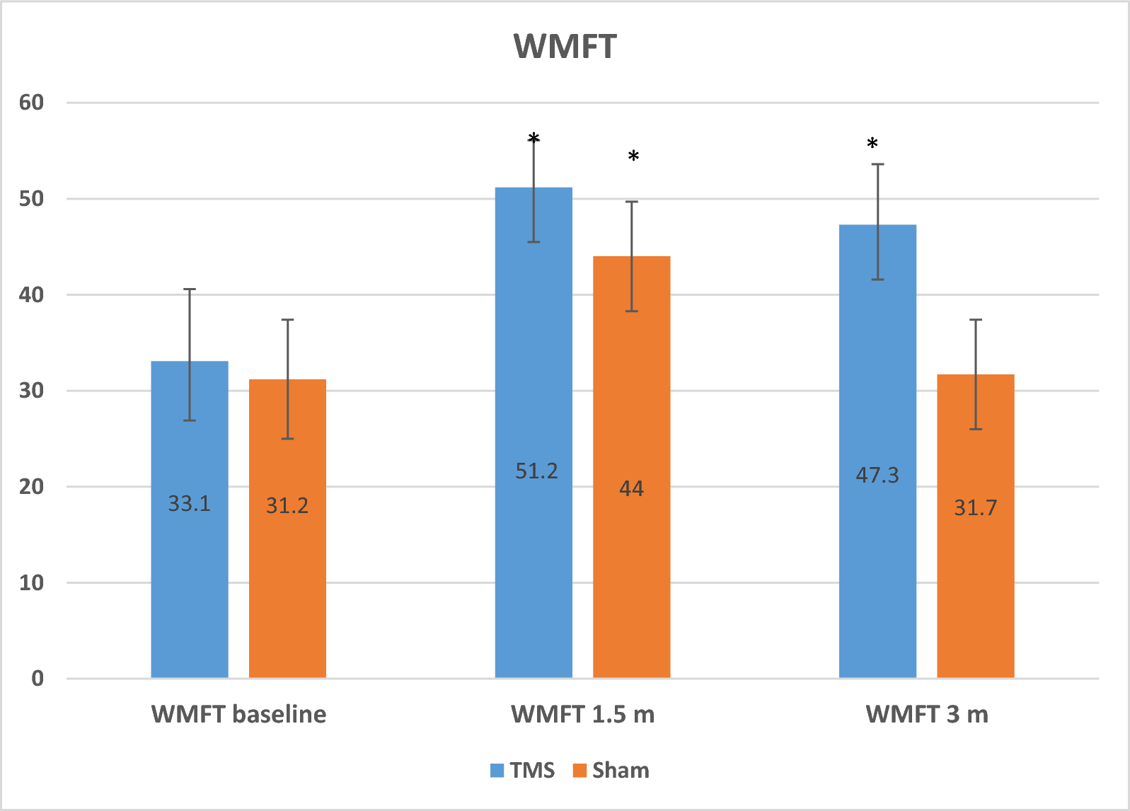
Our ROC analysis highlights the diagnostic potential of nerve US in GBS. In this study, the proposed CSA cut-offs demonstrated high sensitivity, with abnormalities detected in all patients at the median mid-forearm, pronator quadratus, and posterior tibial nerve levels, and in the vast majority at the median pronator teres (97.6%) and ulnar mid-forearm (90.3%) sites. These findings support the potential role of nerve ultrasound in the early diagnosis of GBS, particularly when electrophysiological studies are inconclusive [19].
Among the examined nerves, the median nerve showed the highest diagnostic accuracy at the mid-forearm, pronator quadratus, and pronator teres levels, while the ulnar nerve was most reliable at the mid-forearm. Although data on the PTN remain limited, the initial results are encouraging. Notably, our findings contrast with those of Liu et al. (2021), who reported diagnostic relevance primarily in spinal and vagus nerves, rather than peripheral nerves [27]. However, our study did not include assessments of proximal nerve segments, cervical roots, or the vagus nerve, which may account for this discrepancy. These differences underscore the need for larger, multicenter studies with standardized ultrasound protocols further to validate both peripheral and central nerve assessments in GBS.
Correlation analysis between US and NCS findings revealed only weak to moderate associations, limited to specific nerves and parameters. Notably, the median nerve CSA did not correlate with its corresponding NCS values, whereas some associations were observed for the ulnar and posterior tibial nerves. These findings partially align with earlier work [31], but diverge from studies reporting no correlation at all [14, 26, 31, 46]. The inconsistent relationships suggest that US and NCS assess different aspects of nerve pathology—US detects structural changes, while NCS evaluates functional integrity, supporting their complementary roles in assessing GBS, as highlighted in prior research [19].
These findings contrast with those in chronic neuropathies such as CIDP and diabetic neuropathy, where stronger correlations between CSA and NCS are typically observed [49, 50]. The discrepancy may be explained by the patchy and dynamic nerve involvement seen in early-stage GBS [8, 37], reinforcing the notion that US and NCS provide distinct yet complementary insights: NCS characterizes nerve dysfunction and helps classify subtypes, while US offers data on nerve size, vascularity, echogenicity, and mobility [27].
To our knowledge, this is one of the few studies to propose early diagnostic ultrasound cut-off values for multiple peripheral nerves in GBS, supported by correlation with both clinical and electrophysiological findings. The proposed CSA cut-offs—particularly for the median nerve—demonstrate strong potential for screening and early diagnosis, which is crucial during the evolving phase of the disease. If externally validated in larger, multicenter cohorts, these ultrasound parameters could serve as a rapid, non-invasive adjunct to standard diagnostic tools, enhancing clinical decision-making when electrophysiological studies are inconclusive or delayed.
While our study offers valuable insights into the role of nerve US in GBS, several limitations must be acknowledged. The relatively small sample size, mainly when subdivided by GBS subtype, may limit the statistical power and generalizability of our findings. The cross-sectional design, with all assessments conducted at baseline before treatment, precludes evaluation of longitudinal changes in ultrasound parameters during disease progression or in response to therapy. Additionally, our ultrasound protocol focused on accessible nerve sites, such as the PTN at the medial malleolus, and did not include more proximal segments, cervical roots, or the vagus nerve—regions that may yield important diagnostic information, as highlighted in previous studies [17, 19, 27]. Parameters such as vascularity, echogenicity, and fascicular patterns were also not assessed, though they may offer complementary value in ultrasound-based evaluation of GBS.
Future research should prioritize large-scale, prospective, multicenter studies employing standardized protocols that include proximal nerve segments, cervical roots, and the vagus nerve. Incorporating longitudinal assessments and a broader range of ultrasound parameters, including vascularity, echogenicity, and fascicular patterns, may enhance our understanding of disease dynamics and improve diagnostic accuracy across diverse patient populations.


Comments (0)