The most important finding of this study is that primary ACLR in EDS patients leads to good midterm survival rates and acceptable short term survival rates in patients with revision ACLR. Survival probabilities for primary ACLR were significantly higher compared to revision ACLR in patients with EDS. These survival rates are similar to those reported in patients with generalized joint hyperlaxity (GJH) but without hEDS [36,37,38]. However, GJH patients have been shown to carry a higher risk of graft rerupture compared to individuals without hyperlaxity, and clinical outcomes may also be inferior [36, 37]. Whether these findings apply similarly to patients with hEDS remains unclear and should be further investigated in future studies. Regarding anatomic risk factors, only the lateral femoral condyle ratio (LFCR) and tibiofemoral rotation (TFR) showed significant differences compared to non-EDS and intact ACL controls (p < 0.01). These findings suggest that, despite the distinct connective tissue characteristics of EDS patients, successful ACLR is achievable, especially in primary reconstructions.
The results align with the broader orthopedic literature, which shows that revision ACLR patients generally have a higher risk of re-injury and lower patient-reported outcome measures (PROMs) compared to primary ACLR patients [15,16,17, 39]. However, this study uniquely establishes that EDS patients undergoing primary ACLR can achieve comparable survivorship to non-EDS populations [40, 41].
With regard to possible risk factors for ACLR failure, no factors were identified in this study. However, the literature highlights numerous risk factors in non-EDS populations such as age, sex, training regimen, early return to sport and biomechanical deficits [15, 16]. The use of allografts has also been associated with higher ACL graft rerupture rates [17]. This was also not observed in this current study.
Another factor that is emerging as a key contributor to ACLR failure is insufficiently restored rotational stability. Numerous studies have shown that the use of lateral extra-articular tenodesis leads to a significant reduction in the rerupture rate, particularly in hypermobile patients [15, 42, 43]. Only a small number of patients in our cohort underwent LET, and accordingly, no statistically significant differences in outcomes were observed. However, the low frequency of LET procedures limits the ability to draw definitive conclusions.
In addition, several osseous morphologic characteristics have been associated with an increased risk of ACL injury or failed ACLR [29]. One of the most investigated factors is the posterior tibial slope [21, 26, 27, 29, 34]. A PTS of > 12° has been established as a risk factor for failure after ACLR [34, 44]. This association was not be demonstrated in this study, suggesting that the hypermobility in these patients is primarily due to soft tissue imbalance rather than bone-related factors.
Awareness of the bone morphology of the distal femur is increasing in recent research [23, 29]. Specifically, the morphological and morphometric characteristics of the LFC, such as an increased posterior femoral condylar depth, are thought to play a significant role in the occurrence of ACL injuries [23, 29]. This may be due to an increased posterior depth of the LFC, which leads the femur to a more oval shape. This alteration can cause greater anisometry and elongation of the lateral and anterolateral knee structures, potentially reducing the contact surface between the femur and tibia [29]. As a result, there may be an increase in rotational knee laxity, particularly near full extension [29]. The data from this current study suggests that this relationship may also exist in patients with EDS.
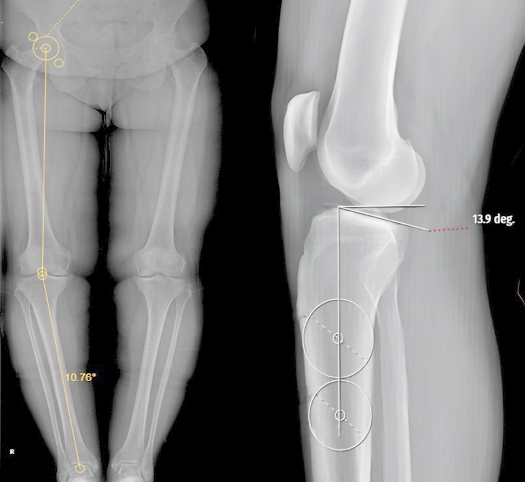
While factors such as internal tibial rotation and TT-TG distance are routinely considered in the evaluation of patellofemoral instability, they are not commonly assessed in the context of ACL injuries [25, 30, 45]. However, radiographic measurements of axial rotation may be relevant to reinjuries following ACLR, especially in a patient population that is massively affected by patellofemoral instability [7, 18, 20, 30]. There is evidence that the TT-TG distance can be an additional tool for detecting increased rotational laxity after ACL injury, especially in combination with ALL injury [30, 45]. This finding does not appear to be relevant in our patient population, as the values were lower than in the study by Leite et al. and also lower than the threshold for healthy non-EDS patients [30, 45].
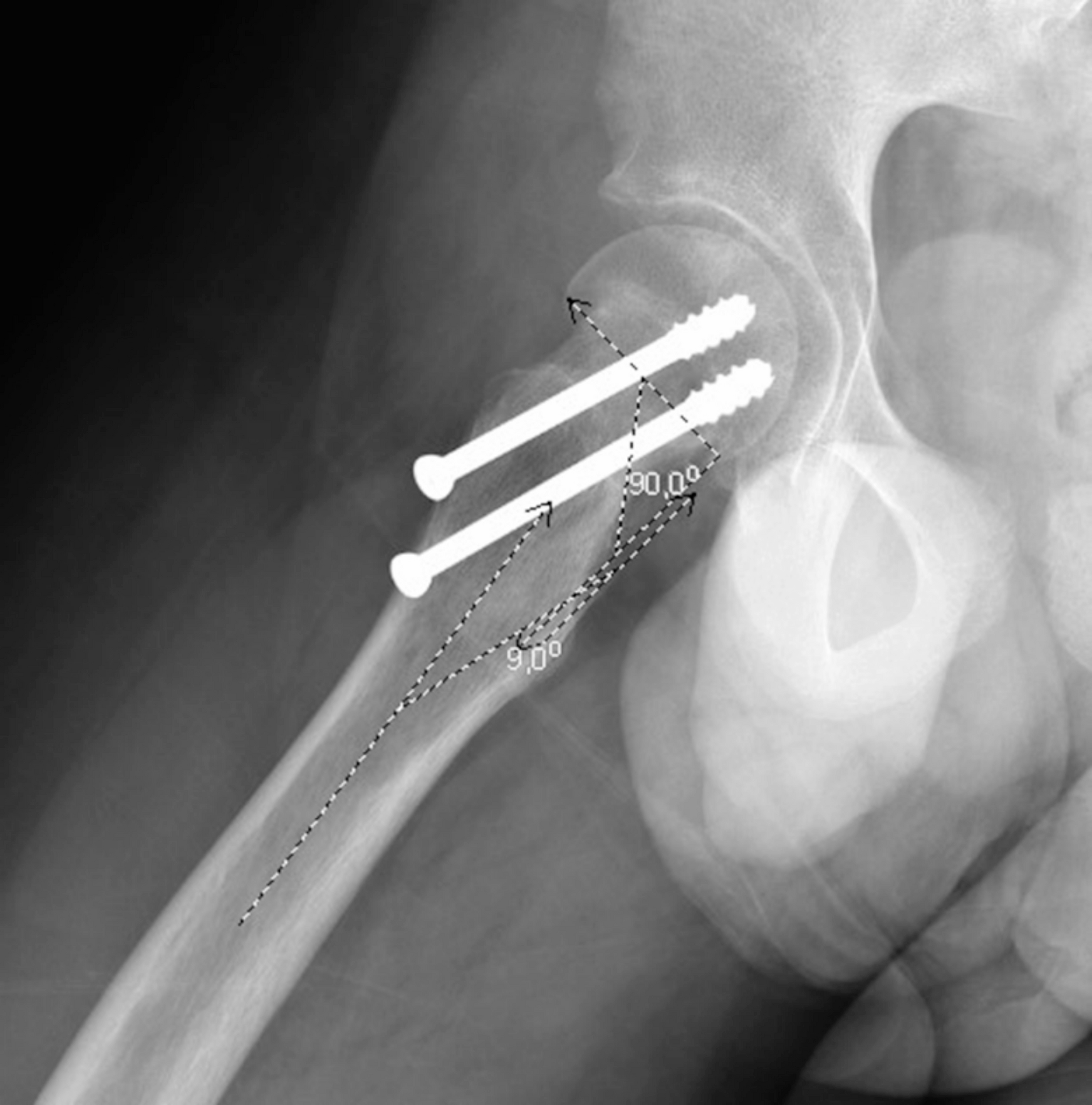
Regarding the TFR measured by the TFA, patients with intact ACLR presented similar TFAs to healthy intact controls [25, 30]. Our study found that the TFA was significantly higher in EDS patients compared to healthy controls and ACLR patients, underlining that the proposed threshold of 4.9° or greater is a sign for the diagnosis of ACL tears [25].
Leite et al. suggests that patients with ACLR failure exhibit greater internal tibial rotation compared to those with intact ACLRs [30]. Increased tibiofemoral rotation can place more reliance on secondary stabilizers, potentially leading to their earlier failure. This excess laxity may contribute to a higher risk of ACLR failure. In patients with EDS, these challenges are magnified due to the connective tissue abnormalities. To address this, adjunctive stabilizing procedures, such as anterolateral ligament (ALL) reconstruction or lateral extra-articular tenodesis, have been proposed to provide better rotational control of the knee [42, 43, 46].
The availability of outcome data for this patient group in the literature is very poor. A recent review looked at the outcomes after ACLR in EDS patients. In total, there were only six case reports with a maximum follow-up of 36 months, all of them reporting good outcomes, including no further knee instability and return to normal activity. These results are in line with the findings of our study. After successful ACLR, the patients showed no increased instability at the follow-up examinations.
Several studies have raised concerns about using autograft tissue in patients with EDS due to intrinsic changes in peri-articular connective tissues that compromise strength and stiffness [18, 46, 47]. Therefore, the use of allografts and lateral extra-articular tenodesis has been recommended for this patient population [42, 43]. However, in our cohort, both graft types were used and no significant differences in outcomes could be detected, likely due to the small sample size. However, in our cohort, both graft types were used and no significant differences in outcomes could be detected, likely due to the small sample size.
Currently, no evidence-based postoperative protocols exist that are specifically tailored to patients with EDS undergoing ACL reconstruction. All patients in our study followed the institutional standard rehabilitation regimen. However, given the characteristic delayed wound healing, joint instability, and risk of graft elongation in this population, protocol modifications, such as extended bracing duration, delayed initiation of sport-specific drills, or prolonged restriction from high-impact activities, may be warranted.
Importantly, this study contributes to the limited evidence available on ACLR in EDS patients. The data challenges traditional assumptions that connective tissue fragility precludes successful surgical intervention, providing a foundation for further research into graft selection and adjunctive procedures tailored to the unique needs of this population.
Limitations
This study has several limitations. Its retrospective design and reliance on electronic medical records may have introduced selection bias and incomplete data. In addition, a minimum follow-up period of only six months was used, which may not be sufficient to capture long-term failures or complications. The small sample size (25 knees) reflects the rarity of EDS but substantially limits the statistical power to detect significant associations, especially in survival analysis and multivariable modeling. Therefore, nonsignificant findings in this study should be interpreted with caution, as the lack of statistical significance does not necessarily imply the absence of an effect. Furthermore, the study spans a period of 30 years (1993–2023), during which surgical techniques, graft choices, fixation methods, and rehabilitation protocols have evolved considerably. This temporal variability could have influenced outcomes by introducing heterogeneity in surgical management and postoperative care. For example, changes in graft preference from bone–patellar tendon–bone to hamstring autografts, advances in fixation devices, or shifts towards accelerated rehabilitation protocols may have affected graft survival and complication rates. However, to address this, we reported the smallest common denominators across all procedures, showing that despite differences in details, all surgeries shared the same base technique (arthroscopic reconstruction with medial portal femoral tunnel drilling) and a standardized rehabilitation protocol involving initial brace-protected weight bearing and return to full activities after functional testing at approximately six months. Due to the limited sample size, further subgroup analyses to adjust for temporal factors were not feasible. Additionally, the study lacks a matched control group for direct comparison of outcomes, but published results of ACLR in the general population allow comparison with existing literature as in other studies of this specific patient population [48].
To our knowledge, this is the first study to investigate the survival of ACLR and examine anatomical risk factors for ACLR failure in a cohort of patients with EDS. This research provides valuable insights into graft selection, risk factors, and injury mechanisms, particularly in rare populations such as those with EDS.


Comments (0)