
Remember me


A 31-year-old-man presented to our institution with chest pain and 6 months of worsening palpitations. He had no prior clinical diagnoses, took no regular medication and was generally fit and active. However, in the past 6 months he had noticed high heart rates on his smartwatch. Family history was notable on the maternal side for a history of heart failure and septal defects in 3 separate relatives.
Initial 12-lead electrocardiogram (ECG) demonstrated narrow complex tachycardia at 140 beats/min with p wave inversion in lead I and aVL and positive deflection in the inferior leads. P wave morphology suggested atrial tachycardia (AT) with focus from the left atrial appendage (LAA) (Fig. 1A). Initial bedside echocardiography demonstrated severe global left ventricular (LV) systolic dysfunction. Initial treatment with adenosine resulted in a brief slowing of the atrial rate but no termination, while Verapamil bolus temporarily restored sinus rhythm followed by prompt recurrence. At this stage the patient was transferred to our cardiac centre.
Fig. 1
(A) ECG of atrial tachycardia. The negative p-waves in leads 1 and aVL can be appreciated, consisted with a LAA origin. (B) 6 months follow-up after successful final ablation and LAAO. The ECG p wave morphology is no longer abnormal
On arrival he was persistently tachycardic with heart rate of 142 beats/min, blood pressure of 111/70 mmHg and oxygen saturations of 99% on room air. His lung fields were clear to auscultation, and no heart murmurs were heard. His laboratory workup demonstrated unremarkable full blood count and biochemistry including a normal troponin, C-reactive protein and thyroid function tests. His NT-proBNP was normal at 166 ng/L. Transthoracic echocardiography (TTE) confirmed a dilated LV cavity with severely impaired systolic function, LV ejection fraction (LVEF) of 20–25% and structurally normal valves. The right ventricle (RV) was also impaired with fractional area change of 26%. This was corroborated by cardiac magnetic resonance imaging which revealed biventricular impairment (LVEF 20%, RVEF 46%) without regional wall motion abnormalities. Importantly there was no evidence to suggest a concomitant myocardial disorder since tissue characterisation demonstrated homogonous T1 and T2 mapping signals and no late gadolinium enhancement was seen. Management included guideline directed optimal heart failure therapy.
Despite treatment with verapamil he remained in persistent tachycardia and therefore on day 4, underwent endocardial catheter ablation. The left atrium (LA) electro-anatomical mapping showed the earliest site of activation to originate from the distal region (mid-tip) of the LAA. Careful ablation initially terminated the tachycardia but early recurrence was seen after 15 s. The LA was remapped and a focus identified within the anterior aspect of the LAA base. Further ablation here terminated the tachycardia with no recurrence after 30 min.
However, a few hours after the ablation the tachycardia returned. Ivabridine was introduced and titrated to 7.5 mg twice daily with the aim of controlling the rate to allow LV recovery. Tachycardia persisted albeit at a slowed rate of 100/min and his LVEF remained impaired at LVEF 27%. On day 10, repeat ablation of the distal LAA tip was performed with TEE and CARTO (Biosense Webster, Johnson & Johnson, Irvine CA, US) guidance. Consolidate lesions with eventual bump termination were delivered with various permutations. Following the final lesion, sinus rhythm was acutely restored. Unfortunately, post-procedure atrial tachycardia returned. Amiodarone was then introduced, and the patient was referred for surgical LAA exclusion.
An Atriclip device (AtriCure, Inc, West Chester OH, US) was placed thoracoscopically which resulted in immediate cessation of the AT. Disappointingly, the tachycardia returned in the early post-operative period.
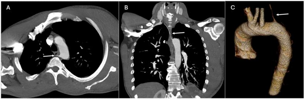
Following further multidisciplinary team discussions a detailed assessment of the LAA and surrounding anatomy was performed with delayed phase contrast computed tomography (CT). This demonstrated suboptimal positioning of the Atriclip (Fig. 2A). Contrast opacification of the residual LAA lobe was noted all the way to the distal tip. CT analysis to define the dimensions of LAA orifice, the device landing zone and available depth, along with computational modelling for optimal sizing for a virtual LAA occluder (LAAO) device, in this case the Amplatzer™ Amulet™ LAAO (Abbott, Santa Clara CA, US) was performed using FEops Heartguide Platform workflow (Fig. 2B). Therefore, it was felt a further attempt at percutaneous LAA ablation with concomitant LAA occlusion was potentially feasible and should be attempted.
Fig. 2
(A) delayed phase contrast CT 3D multiplane reconstruction of the LAA following surgical AtriClip placement (white arrow). The AtriClip device can be seen to be displaced and contrast opacifies the whole residual appendage. The yellow dotted line depicts the optimal position for the AtriClip for correct coaxial alignment with the LAA orifice (base of LAA). PA pulmonary artery, LSPV left superior pulmonary vein. (B) FEops CT 3D reconstruction showing the relationship of the AtriClip to the LAA and LA, the green circles show the orifice and landing zone measurements for the Amulet LAAO device. The yellow circle depicts the position of the fossa ovalis. Computational modelling depicts optimal LAAO device size and positioning. (C) shows the 3D TEE multiplane reconstruction of the residual LAA with measurements of the LAA orifice, landing zone and depth. These measurements corresponding to the same device choice as the predicted FEops simulation, 18 mm Amulet. (D) X-plane image of the 18 mm Amulet LAAO device after deployment at the time of the final procedure. (E) Fluoroscopic image of the LAAO device and its relationship to the AtriClip
The patient then underwent combined pulsed field ablation (PFA) of the LAA followed by percutaneous LAA occlusion. Using 3D TEE the ostium measured 23 × 13 mm, the landing zone 13 × 12 mm and available LAA lobe depth of 8 mm (Fig. 2C). The 3D transoesophageal echocardiography (TEE) measurements were consistent with the predicted FEops CT derived measurements. An 18 mm Amulet device was selected. The device disc diameter is 24 mm and requires a minimum depth of approximately 10 mm for the compressed lobe length. Therefore the LAAO device lobe might sit a little proud of the orifice. The intention was to fully seal the ostium, and avoid risk of peri-device leak, particularly important in this case as there was high risk of thrombus formation following LAA electrical isolation. Sphere-9™ (Medtronic, Minneapolis MN, US) was used to map the LAA and confirmed distal to proximal activation. After the pulsed field lesions were delivered, sinus rhythm and a heart rate drop was observed. Next, the 18 mm Amulet™ device was delivered via a 12Fr sheath. On the first deployment the device sat well with 22% lobe compression (14 mm compressed diameter), good separation and complete occlusion of the LAA orifice. Using the standard post deployment checks, no peri-device leak was seen. The device stability was confirmed using tug-testing and remained in the satisfactory position following release (Fig. 2D, E).
Six months later, repeat cardiac testing confirmed maintenance of sinus rhythm (also confirmed by no tachycardias recorded on his smartwatch) and normalization of LV systolic function with TTE LVEF 52% (Fig. 1B shows ECG at 6 months). The patient’s symptoms had resolved, and he had returned to regular exercise.
Comments (0)