In vivo experimentsAnimal model establishment
Experimental SD rats (Pengyue, Jinan, China) were housed individually with ad libitum access to water and food, and acclimatized for 1 week prior to procedures. All animal experiments were conducted in compliance with ethical regulations of the Affiliated Hospital of Qingdao University, China, under approval from the Animal Ethics Committee (Approval No. AHQ-MAL20230407DYQ).
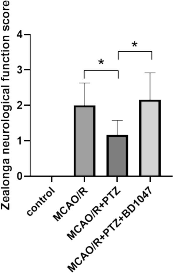
Rats weighing between 220 and 250 g were randomly assigned to four groups (n = 6): a control group, an Middle Cerebral Artery Occlusion/Reperfusion (MCAO/R) group, an MCAO/R + PTZ (China Resources Double-Crane Pharmaceutical) group, and an MCAO/R + PTZ + BD1047 (a sigma-1R inhibitor MCE, USA) group. The processing is based on relevant references [17], all groups except the control received treatment prior to modeling; specifically, the MCAO/R + PTZ group was received intraperitoneal injection 3 mg/kg of PTZ daily for three consecutive days. The MCAO/R + PTZ + BD1047 group received both 3 mg/kg of PTZ and 3 mg/kg of BD1047. The MCAO/R group was given an equivalent volume normal saline.
MCAO was induced using the common carotid artery (CCA) filament insertion method. Rats were anesthetized with 5% isoflurane and positioned supine after cervical hair removal. A 2-cm midline neck incision was made, followed by layered blunt dissection to expose the left carotid bifurcation. The CCA was isolated rostrally while performing concurrent blunt vagal nerve dissection. After nerve separation, two sutures were placed beneath the CCA: a permanent proximal ligation and an adjustable slipknot near the bifurcation. The external carotid artery (ECA) was permanently ligated along its length, while the internal carotid artery (ICA) received a proximal adjustable ligature and vascular clip. A 45° V-shaped micro-incision was created on the anterior CCA wall using ophthalmic scissors. The filament was introduced through this incision, and the bifurcation slipknot tension was adjusted to achieve hemostasis while permitting filament mobility. Upon ICA clip removal, the filament was advanced continuously into the middle cerebral artery through the ICA, with trajectory adjustments following vascular anatomy. Following successful insertion, the bifurcation slipknot was tightened for primary fixation, and the ICA ligature secured for secondary fixation. Proper occlusion was confirmed by vascular collapse and darkening, with distal traction verifying absence of leakage. All CCA and ECA ligation have been validated to prevent bleeding and ensure survival after modeling. Successful MCAO surgery was confirmed by the presence of ipsilateral Horner's syndrome and contralateral hemiparesis predominantly affecting the forelimbs upon rat awakening [18].
Zea-Longa neurological behavioral scoring
The grouping of rats was consistent with that in “2.1.1” (Control group, MCAO/R group, MCAO/R + PTZ group, and MCAO/R + PTZ + BD1047 group).Neurological impairment in rats was assessed using the Zea-Longa scoring system [19]: 0 (no deficit), 1 (failure to fully extend left forepaw), 2 (circling to the left), 3 (falling to the left), and 4 (no spontaneous ambulation with loss of consciousness). The Zea-Longa neurological assessment was performed 3 days after ischemia–reperfusion.
Immunohistochemical analysis of sigma-1R expression in hippocampal tissue of MCAO/R-induced rats
The grouping of rats was consistent with that in “2.1.1” (Control group, MCAO/R group, MCAO/R + PTZ group, and MCAO/R + PTZ + BD1047 group). Hippocampal tissues were harvested 3 days post-modeling and immediately fixed in 4% neutral buffered formalin for 24 h at room temperature (avoiding over-fixation to prevent epitope masking). Samples underwent graded ethanol dehydration, xylene clearing, and paraffin embedding before sectioning. Deparaffinization was achieved through xylene immersion followed by ethanol rehydration. Antigen retrieval was performed using citrate buffer (pH 6.0) with heat-induced epitope retrieval (92 °C, 20 min). Endogenous peroxidase activity was quenched with 3% H₂O₂, and non-specific binding was blocked with 5% normal goat serum (Solarbio, China). Primary antibody incubation: anti-sigma-1R (Santa Cruz, USA) 1:200 in 1% Bovine Serum Albumin/ Phosphate Buffered Saline at 4 °C for 16 h. Secondary detection: Horseradish Peroxidase (HRP)-goat anti-rabbit IgG (1:500, Elabscience, China) at 37 °C for 30 min protected from light. Target proteins were visualized via HRP-catalyzed oxidation of 3,3'-diaminobenzidine (DAB Solarbio, China) generating insoluble brown precipitates, with reactions terminated by running water rinsing. Nuclei were counterstained with hematoxylin, followed by dehydration, clearing, and permanent mounting.
Tissue sections were imaged at 400 × magnification using Case Viewer 2.4 software. Following image acquisition, analysis was performed using Image-Pro Plus 6.0 software. Within each section, the integrated optical density (IOD) and corresponding tissue area (expressed in pixels) were quantified in three randomly selected fields of view. Areal density was calculated using the formula: Areal Density = IOD / Area.
Western blot detection of sigma-1R, IRE1, XBP-1s and CHOP proteins in hippocampal tissue from MCAO/R rats
The grouping of rats was consistent with that in “2.1.1” (Control group, MCAO/R group, MCAO/R + PTZ group, and MCAO/R + PTZ + BD1047 group). Under deep anesthesia with inhaled isoflurane, rats were euthanized and fresh hippocampal tissues were immediately dissected due to their high sensitivity to ischemic-hypoxic injury. Total protein was extracted using RIPA lysis buffer (Elabscience, China) supplemented with protease/phosphatase inhibitors. Proteins were separated by sodium dodecyl sulfate–polyacrylamide gel electrophoresis (SDS-PAGE) (Solarbio, China) and electrophoretically transferred to polyvinylidene fluoride (PVDF) membranes (Millipore Sigma, USA). After 2 h room temperature blocking, membranes were incubated overnight at 4 °C with primary antibodies against: sigma-1R (1:500, Santa Cruz, USA), IRE1 (1:2000, ABclonal, China), XBP-1s (1:1000, CST, USA), and CHOP (1:1000, Wanleibio, China). This was followed by 1 h incubation at room temperature with HRP-conjugated goat anti-rabbit or anti-mouse secondary antibodies (1:10000, Elabscience, China). Protein bands were visualized using a chemiluminescence imaging system (Thermo Fisher,USA), and band intensities were quantified with ImageJ software. Target protein expression levels were normalized to β-actin (1:5000, Elabscience, China) and expressed as relative band intensity ratios.
In vitro experimentsCell culture and model development
Compared to primary neurons, human neuroblastoma cells (SH-SY5Y, Shanghai Cell Bank, China) exhibit similar morphological and physicochemical characteristics and can be stably passaged. These properties make them widely utilized in the study of neurological diseases. According to reference [20], SH-SY5Y cells were cultured in DMEM/F12 (Procell, China) complete culture medium supplemented with 10% fetal bovine serum and maintained in a 5% CO2 incubator at 37 °C. Approximately 70–80% of the cells underwent passage. The cells were randomly divided into four groups: control group, OGD/R group, OGD/R + PTZ group, and OGD/R + PTZ + BD1047 group (n = 6). The control group received no treatment following passage. After passage, when the cell density of the other three groups reached 70–80%, EBSS medium (Solarbio, China) was applied; hypoxia and glucose deficiency conditions were induced at 37 °C under an atmosphere of 5% CO2 and 95% N2 for a duration of 4 h. Subsequently, fresh complete medium was introduced for reoxygenation in a standard incubator for 18 h. During reoxygenation: OGD/R group received no intervention, OGD/R + PTZ group received 10 μM PTZ, OGD/R + PTZ + BD1047 group received 10 μM PTZ plus 20 μM BD1047.
Cell survival rate
The groups and processing of cells were performed as described in Sect. 2.1.1 above (control group, OGD/R group, OGD/R + PTZ group, and OGD/R + PTZ + BD1047 group). Cell survival rate was detected using the Cell Counting Kit-8 (CCK-8) (Solarbio, China) method. Cell suspension was added into 96-well plates (100 μL/well, 5 × 10³/well), the cells were randomly divided into three groups: control group, OGD/R group, OGD/R + PTZ group, each group had 6 samples and treated as described above. 10 μL of CCK-8 reagent was added to each well, and the 96-well plates were incubated in a CO2 incubator for 2.5 h. The absorbance (A) value of the cells was quantified at a 450-nm wavelength using an enzyme-linked immunosorbent assay reader (Shanghai Meigu, China). The percentage of cell survival rate was calculated using the following formula: [(treatment group A–blank group A) / (control group A–blank group A) × 100].
Immunofluorescence analysis of sigma-1R expression in SH-SY5Y cells subjected to OGD/R
The expression of sigma-1R was detected through immunofluorescence staining. The groups (n = 6) and processing were conducted as described above. After model establishment, cells were fixed with 4% paraformaldehyde at room temperature for 30 min, permeabilized with 0.5% Triton X-100 (Solarbio, China) for 10 min at room temperature, and blocked with 5% goat serum for 1 h. Specimens were then incubated with primary antibody against sigma-1R (1:100, Santa Cruz, USA) at 4 °C overnight, followed by species-appropriate fluorescent secondary antibody (1:500, Elabscience, China) incubation for 2 h at room temperature in the dark. Nuclei were counterstained with DAPI (0.5 μg/mL) for 5 min. After mounting with anti-fade medium, fluorescence images were acquired using an inverted fluorescence microscope (Nikon, Japan). Qualitative analysis of fluorescence intensity was conducted using ImageJ software.
Flow cytometric analysis of cell apoptosis
Group assignment (n = 6 per group) and experimental procedures followed the aforementioned protocol. Apoptosis was quantified by flow cytometry using Annexin V-FITC/propidium iodide (PI) (Yeasen, China) dual staining. Briefly, cells were resuspended in binding buffer (1 × 10⁶ cells/mL) and incubated with 5 μL Annexin V-FITC and 10 μL PI for 15 min at 25 °C in the dark. Stained cells were analyzed within 1 h using a CytoFLEX LX flow cytometer (BECKMAN COULTER,USA). The apoptotic rate was calculated as the percentage of cells in early apoptosis (Annexin V⁺PI⁻) plus late apoptosis (Annexin V⁺PI⁺) populations.
Western blot detection of sigma-1R, P-IRE1, XBP-1s, CHOP protein and Cleaved-Caspase-3 levels in SH-SY5Y cells from OGD/R model
Cells from each group (n = 6) were collected and lysed for total protein extraction. Protein concentrations were determined using the bicinchoninic acid assay. Equal amounts of protein were separated by SDS-PAGE and transferred to PVDF membranes. After 2 h blocking at room temperature in 5% non-fat milk/Tris Buffered Saline with Tween 20, membranes were incubated overnight at 4 °C with the following primary antibodies: sigma-1R(1: 500, Santa Cruz, USA) 、p-IRE1(1: 1000, ABclonal, China) 、XBP-1s(1:1000, CST, USA) 、CHOP(1: 1000, Santa Cruz, USA) 、cleaved Caspase-3 (C-Cas-3)(1: 1000, CST, USA). Subsequently, membranes were incubated with species-matched HRP-conjugated secondary antibodies (goat anti-rabbit or goat anti-mouse, 1:10000) for 1 h at room temperature. The subsequent processing was the same as the Western blot detection (Thermo Fisher, USA) of the above-mentioned rat MCAO/R model.
Statistical analysis
Statistical analysis was performed using GraphPad Prism (GraphPad software 9.0, San Diego, CA). Normally distributed continuous data are expressed as mean ± standard deviation. Multiple groups comparisons were conducted using one-way analysis of variance (ANOVA) with a post-hoc Dunnett’s test. P values of < 0.05 were considered statistically significant.


Comments (0)