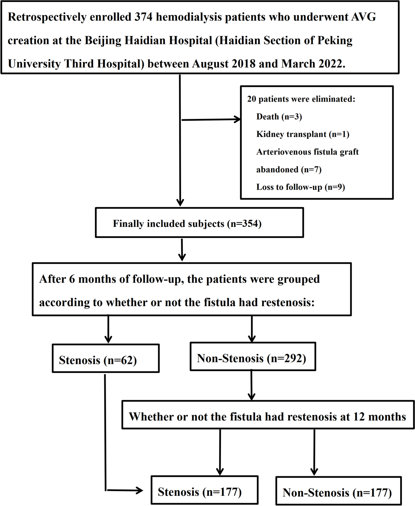
Arteriovenous graft (AVG) remains the second-line option for hemodialysis access, particularly for elderly and diabetic patients with compromised vascular resources. At our center, we perform approximately 200 AVG creations annually to accommodate this growing patient population with challenging vascular conditions. However, it has a limitation of lower long-term patency and thus requires more frequent treatments for maintaining its patency, which results in a higher incidence of stenosis and thrombosis. Several studies have revealed that the primary patency for AVG at 6 months, 1 year, and 2 years is 58%, 33.9–90%, and 25.3–85%, respectively. Halbert et al. performed a systematic literature review and meta-analysis including 3381 AVG accesses (38 study arms) analyzing a timeframe from 2004 to 2019; the mean primary, primary-assisted, and secondary patency rates at 1 year were 41%, 46%, and 70% [15, 16]. The primary patency rates of the present study were 82.49%, 50%, and 16.95% at 6 months, 1 year, and 2 years, respectively, which were higher than previous reports.
Based on our findings, we propose that the long-term patency of arteriovenous grafts is significantly influenced by the functional capacity of the venous outflow tract, which can be assessed through early postoperative hemodynamic and morphological parameters. This concept is strongly supported by our data showing that brachial artery blood flow and draining venous diameter at 1 month post-operatively serve as independent predictors for AVG patency at both 6 and 12 months. Previous literature lacked the analysis of specific clinical or demographic features linked with AVG survival. Notably, no clinical factors—including age, body mass index, race, diabetes, cardiovascular disease, sex, or access location—have been reliably associated with early AVG failure in multicenter studies [16]. This contrasts with the well-established pathophysiology of AVG failure; experimental models and human studies have demonstrated neointimal hyperplasia at graft-vein or graft-artery anastomoses as the primary contributor to AVG stenosis. Research by Michael Allon and colleagues further confirmed that pre-existing vascular abnormalities (arterial medial fibrosis, intimal hyperplasia, and arterial calcification) may paradoxically reduce AVG failure rates. Our results provide substantial evidence for this concept through several key findings. Here, based on the multivariable risk adjustment of possible confounding factors, it was found that brachial artery blood flow (HR 0.998 (0.997, 0.999), p < 0.001) and draining vein diameter (HR 0.731 (0.573, 0.934), p = 0.012) at 1 month post-operatively were independent risk factors for patency at 6 and 12 months. These findings raise an important question: Does the preoperative venous status determine the fate of AVG by influencing these indicators? Although this study did not directly evaluate the preoperative venous features, the existing data strongly suggest this possibility.
The pathophysiological mechanism underlying our findings involves a complex interplay between venous anatomy and hemodynamic factors. Specifically, a smaller diameter of the drainage vein may reflect preoperative venous dysplasia or pre-existing lesions; reduced brachial artery blood flow may be a secondary manifestation of poor venous outflow tract. These morphological and hemodynamic alterations likely create a vicious cycle that exacerbates AVG dysfunction through two synergistic pathways. First, the presence of pre-existing vascular pathologies in the draining vein to which an AVG is anastomosed can accelerate neointimal hyperplasia and restrictive vascular remodeling, leading to early AVG failure. Second, the implantation of an AVG generates non-physiological oscillatory wall shear stress in the vicinity of the graft-vein anastomosis. This condition may contribute to the development of intimal hyperplasia and progressive luminal narrowing, ultimately leading to thrombosis and loss of graft patency. Additionally, high blood flow can maintain normal endothelial cell function through laminar shear stress (where the direction of blood flow aligns with the direction of shear stress), reducing endothelial injury and intimal hyperplasia. The diameter of reflux veins may also influence reflux resistance, generating oscillatory shear stress, which can damage endothelial cells and lead to intimal hyperplasia.
Stenosis most commonly occurs at the venous anastomosis, but it can also be detected in the central vein, draining vein, feeding artery, or within the AVG itself [17,18,19]. The present study revealed that the most frequent locations of stenosis were graft-vein anastomosis (51.72%) and draining vein (48.28%), consistent with the above studies. The observed phenomenon may be attributed to the pronounced severity of neointimal hyperplasia (NIH) at the graft-vein anastomosis, which is exacerbated by aberrant blood flow and non-physiological wall shear stresses in the venous region [20,21,22].
These findings collectively substantiate our central hypothesis that venous outflow tract characteristics fundamentally determine AVG patency outcomes. The observed hemodynamic disturbances appear to be significantly amplified by pre-existing venous wall abnormalities, revealing a crucial interplay between intrinsic venous properties and acquired hemodynamic stressors. This pathophysiological understanding not only validates our hypothesis but also establishes a mechanistic framework for interpreting AVG failure patterns.
The critical importance of venous outflow capacity is further evidenced by the strong predictive value of postoperative surveillance. Given that most thrombosed grafts develop from underlying stenosis, our findings align with previous studies emphasizing the value of clinical monitoring [23,24,25]. We demonstrate that systematic ultrasonography surveillance following surgery provides essential data for predicting AVG patency, corroborating the reports by Krivitski, Gantela et al. [26]. The predominance of duplex ultrasonography as the preferred imaging modality stems from its unique ability to provide both anatomical and hemodynamic assessment. Clear ultrasound criteria enable precise differentiation between evolving and borderline stenoses, allowing early detection of lesions that could progress to AVG thrombosis.
Our hypothesis gains additional support from computational fluid dynamics research [27,28,29,30], which demonstrates that strategic modifications to graft geometry or the graft–vein interface can generate favorable flow patterns and hemodynamic conditions. These engineering approaches, potentially reducing NIH incidence, are further validated by meta-analytical evidence suggesting that geometric optimization of the graft–vein interface enhances AVG patency [21, 31, 32]. This convergence of clinical observations and engineering principles strongly reinforces our proposition that venous outflow characteristics dictate AVG fate.
The detrimental impact of central venous catheterization provides compelling ancillary evidence for our hypothesis. Central venous stenosis, well-established as a factor reducing vascular access lifespan [33, 34], creates upstream resistance that compromises venous outflow capacity. Our multivariate analysis confirms that a history of central venous catheterization (HR 1.939, 95% CI 1.137–3.304, p = 0.015) independently predicts reduced 12 month patency, consistent with Shingarev et al.'s conclusions [35]. The association between ipsilateral central catheters and diminished cumulative access survival further underscores how pre-existing venous compromise adversely affects AVG outcomes.
While pharmacological interventions represent a potential avenue for improving AVG patency, current evidence remains mixed. Although conventional anticoagulants or dual antiplatelet therapy have failed to demonstrate efficacy while increasing bleeding risk, some regimens like adenosine diphosphate plus aspirin show modest benefits in reducing stenosis or thrombosis [35, 36]. The promising results with fish oil supplementation suggest potential avenues for future pharmacological strategies. However, the absence of systematic drug intervention data in our study highlights the need for prospective investigations with standardized medication recording and long-term follow-up to establish evidence-based pharmacological management.
The clinical application of our hypothesis is embodied in the developed nomogram, which translates theoretical concepts into practical tools for personalized risk assessment. By integrating key predictors—brachial arterial blood flow, draining vein diameter at 1 month, and central venous catheter history—this model enables clinicians to generate individualized patency probabilities for 6 month, 1 year, and 2 year intervals. Beyond its clinical utility for risk stratification, the nomogram serves as an effective educational instrument, helping patients visualize their personal risk profiles and make informed decisions regarding treatment options and follow-up adherence. The model's demonstrated capacity to discriminate between different risk strata validates its practical implementation for personalized AVG management.
This study has several important limitations that should be acknowledged. First, its single-center, retrospective design introduces potential selection bias and limits the generalizability of our findings. The lack of randomization and relatively small cohort size may also affect the robustness of our predictive model. While our results demonstrate good discriminative performance, external validation in larger, multicenter populations is essential to confirm their applicability across diverse clinical settings. To address these limitations, we are actively collecting prospective data from multiple centers, which will allow for rigorous external validation in future studies. Additionally, our analysis did not include “preoperative vessel diameter” as a potential predictor of AVG patency, which warrants further investigation. Ultimately, prospective, multicenter studies with standardized protocols are needed to strengthen the clinical validity of our findings and enhance their broader applicability.


Comments (0)