Viruses
The PRCV strain (PRCV/USA/Minnesota/2016, GenBank: KY406735.1) was kindly provided by Professor Song Zhenhui of the Department of Veterinary Medicine at Southwest University in Chongqing, People’s Republic of China. The viral titers of PRCV were determined using swine testis cells through a TCID50 assay (50% tissue culture infective dose).
Establishment of PCLSs culture system
PCLSs were generated as previously reported (Fu et al. 2018). Briefly, lungs from 3-month-old healthy pigs were harvested post-euthanasia, and the upper, middle, and caudal and accessory lobes were isolated. All lobes were infused with 1.5% low-melting-temperature agarose (Agarose, low-melting point, Promega, USA) through the lobar bronchus to maintain the lung’s delicate honeycomb structure. After agarose solidification on ice, cylinder tissues containing a bronchial airway were drilled out using an 8-mm hollow rotating tissue coring tool and sliced to 250 μm thickness using a Krumdieck slicer (Alabama Research & Development, USA). All the generated PCLSs were collected in dishes and incubated at 37 °C and 5% CO2 for 1 h to get out the agarose. Next, the highly quality tissue sections were selected and transferred to 24-well plates. PCLSs with the same thickness, round shape, ciliary-beating, smooth edges were identified as highly quality tissue sections. Next, PCLSs were cultured in RPMI1640 (Merck, Germany), DMEM-F12 (Merck, Germany) and DMEM high glucose media (Merck, Germany). Finally, the adenosine triphosphate (ATP) levels of the PCLSs were measured to determine the optimal culture medium.
Viability analysis of PCLSs
The prepared PCLSs were cultured with different culture media for 72 h. Slices were then collected at the indicated intervals and subjected to the ATP Bioluminescence Assay Kit CLS II kit (Roche Diagnostics, Germany) to measure ATP content according to the manufacturer’s guide. In addition, the slices cultured with RPMI 1640 medium for 72 h were collected and stained using the calcein acetoxymethyl/ethidium homodimer-1 (calceinAM/EthD-1) staining kit (ThermoFisher, USA) to assess PCLS viability, following the manufacturer’s protocol.
Histological analysis
To determine the intact structure of PCLSs, an H&E (Hematoxylin and eosin) assay was employed and performed as previously reported (He et al. 2022). Briefly, PCLSs were maintained in RPMI 1640 medium without fetal bovine serum (FBS) for 72 h, followed by fixed with tissue fixative solution (4% paraformaldehyde). PCLSs were dehydrated with a gradient of alcohol, and then treated with xylene, followed by embedded in paraffin. Then 4 μm-sections were generated using a Leica RM2245 semi-automatic microtome (Leica Biosystems, Germany). Following, sections were stained with hematoxylin and eosin. Finally, sections were observed under an optical microscope (Lecia, Germany) to analyze the integrity of the slices.
PRCV infection in PCLSs
PCLSs exhibiting 100% ciliary activity were selected for viral infection. After aspirating the culture medium, the PCLSs were inoculated with PRCV (300 μL RPMI1640/well at 105 TCID50/mL) for 2 h at 37 °C and 5% CO2. Subsequently, the slices were washed using PBS to remove unattached virions, the culture supernatants and slices were collected at 24 h, 48 h, 72 h and 96 h post-infection to determine viral loads using qPCR and TCID50 assays.
RNA extraction and quantitative real-time PCR
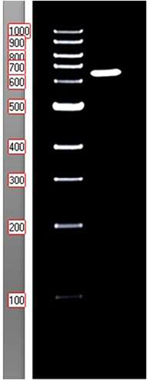
Total RNA of supernatant and PCLSs were extracted using RNAiso Plus (TaKaRa, Japan) according to the manufacturer’s guide. Next, the extracted RNA was reverse transcribed into cDNA for detection of target genes using HiScript III RT SuperMix for qPCR (+gDNA wiper) (Vazyme, China). The copies of PRCV in the infected PCLSs were determined using RT-qPCR assay established in our group (Huang et al. 2019). To validate the RNA-seq results with the RT-qPCR analysis, specific genes expression levels using 2× SYBR-Green Q-PCR Mix (Vazyme, China) and then conducted with the manufacturer’s protocol and normalized to the β-actin expression. The primers used in this study are listed in Table 1.
Cryosection
The PCLSs were embedded in optimal cutting temperature compound (SAKURA, Japan) in a suitable tissue cryomold and then quickly frozen in liquid nitrogen. The embedded PCLSs are stored at − 80 °C until use. Cryosections of 8-μm thickness were prepared using a Leica CM1950 Cryostat (Leica, Germany), and dried at room temperature (RT) overnight, and stored at − 20 °C until assay.
Indirect immunofluorescence assay (IFA)
The cryosections were fixed with tissue fixative solution (4% paraformaldehyde) for 30 min at RT and then treated with 100 mM glycine for 10 min. Following washes with PBS, the cryosections were permeabilized with 0.2% TritonX-100 for 20 min. Subsequently, different primary antibodies were diluted with 1% BSA (bovine serum albumin) and then treated with the cryosections for 16 h at 4 °C. For viral particle detection, a mouse anti-TGEV-N protein monoclonal antibody at a 1:500 dilution with 1%BSA was used and then incubated with an anti-mouse IgG (Alexa Fluor 488 conjugate) (CST, USA). To visualize ciliated cells, basal cells and mucus-producing cells, a rabbit anti-Cy3 labeled monoclonal antibody against β-tubulin at a 1:500 dilution with 1%BSA (Sigma-Aldrich, USA), a rabbit anti-cytokeratin 5 (CK5) monoclonal antibody (Abcam, UK) at a 1:500 dilution and a rabbit anti-mucin-5AC antibody at a 1:250 dilution was used, respectively. After PBS washed three times, the secondary antibodies, goat anti-rabbit IgG (Alexa Fluor 594 conjugate) (CST, USA) were diluted at 1:500 with 1%BSA and incubated with the cryosections for 1 h at RT. Finally, nuclei were stained using DAPI (Solarbio, China) for 3–5 min at RT. All sections were embedded in Mounting Medium and subjected to a TCS SP8 confocal microscope for analysis (Leica, USA).
RNA-seq analysis
PCLSs were infected with or without PRCV and collected at 24 h post-infection. The PCLSs were rapidly frozen in liquid nitrogen and preserved at − 80 °C. The prepared PCLSs were sent to Suzhou GENEWIZ for RNA-seq analysis. The RNA sequencing raw data were processed and mapped based on the Sus Scrofa reference genome. The differentially expressed genes (DEGs) between the PRCV-infected group and the mock group were screened using the expression of DEGs more than two times and FDR (qvalue) ≤ 0.05. If the log2Foldchange of a gene was ≥ 1, the differential gene was considered to be up-regulated; otherwise, if the log2Foldchange was ≤ − 1, the differential gene was considered to be down-regulated.
Statistical analysis
All statistical analyses were conducted using the Student’s t-tests, diagram of statistics was made by GraphPad Prism (Version 9.5). All results in the figures are presented as the means±standard errors of the mean (SEMs) from three independent experiments. The significance level (P value) is indicated as follows: *, P < 0.005; **, P < 0.01; ***, P < 0.001; ****, P < 0.0001.


Comments (0)