To the best of our knowledge, this study is the first to show a strong correlation between the SUVmax in the hottest non-malignant cervical lymph node and the SUVmax in the ipsilateral palatine tonsil. This result is not surprising. Increased uptake in the palatine tonsils may be the result of an infectious process involving the Waldeyer ring, in which satellite adenopathy is common. Our study was done in the pediatric population, since we assumed that the prevalence of benign increased uptake neck lymph nodes would be higher. However, the data for the 16–20 years old show the same correlation with slightly higher values of uptake in the cervical nodes, suggesting that this result may remain true for adults. Of note, we have found no study showing a correlation between the size of non-malignant cervical lymph nodes and the size of the ipsilateral palatine tonsil.
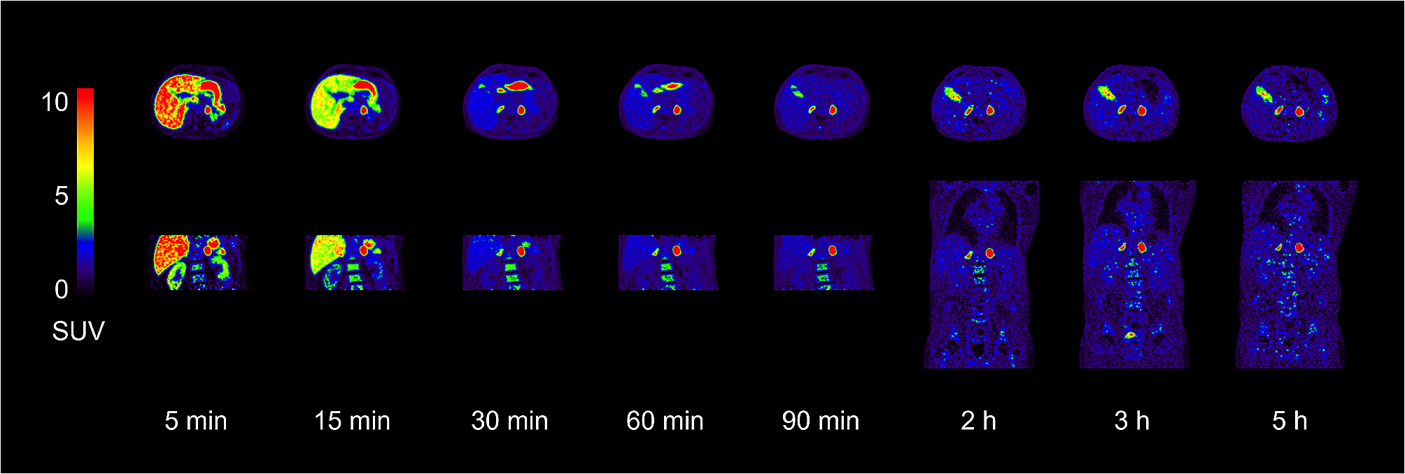
The formula given for the prediction of SUVmax in benign cervical lymph nodes may be useful for the staging of pediatric malignancies that may involve cervical lymph nodes, such as rhabdomyosarcoma and Hodgkin lymphoma when tonsils are not involved (see an example in Fig. 4).
For pediatric Hodgkin lymphoma, neither the EuroNet-PHL protocol [10] nor the NCCN guidelines (Version 1.2022) [11] provide objective criteria for positivity of lymph nodes at staging. (The NCCN guidelines misleadingly show the Deauville criteria in the “Principles of staging” section page 31, although these criteria are validated for early response assessment only after two courses of chemotherapy). Considering cervical lymph nodes at staging as PET-positive is consequently a subjective decision of the interpreting physician, with potential inter-observer variability. Using the results presented here, the interpreting physician can compare the observed SUVmax to the predicted expected value of SUVmax of a benign/reactive node, computed from the uptake value in the non-involved tonsil, in order to decide whether the lymph node is positive or not.
The results presented here may also be apply to solid tumors. For instance, in childhood rhabdomyosarcomas, the rare lymph node involvement at diagnosis classifies the patient as high-risk, with consequences for the treatment plan [12]. By comparing cervical lymph node uptake to tonsillar uptake, the need for lymph node biopsy may be waived in certain cases.
At the end of treatment too the results presented here may alleviate the need for biopsy and justify a more watchful management in the case of low-grade residual uptake in cervical lymph nodes, when the uptake value in the tonsil makes it consistent with a benign etiology.
Moreover, the fact that after adjusting for tonsillar uptake, no correlation is seen between liver uptake and non-malignant cervical lymph nodes uptake suggests that tonsillar uptake may replace liver uptake as a reference value for response assessment of Hodgkin lymphoma involving cervical lymph nodes.
The use of tonsillar uptake as a reference depends upon the low incidence of tonsillar involvement by pediatric malignancies. For Hodgkin lymphoma, Waldeyer ring involvement is extremely rare in the pediatric population with only a few case reports published [13]. For non-Hodgkin lymphomas, Burkitt lymphoma and diffuse large B-cell lymphoma may involve the Waldeyer ring generally with clear clinical symptoms [14]. In these rare cases tonsillar uptake cannot be used as reference. Symmetry of the uptake in both palatine tonsils has been proposed as a predictor of non-malignant uptake in the adult population [15].
Previous published works have reported the occurrence of increased uptake in non-malignant cervical lymph nodes, including in the pediatric population [16]. included 27 pediatric patients after treatment for lymphoma with increased uptake in cervical lymph nodes, all of them with disease-free survival during follow-up, and 3 of them with histologic evidence of benign follicular hyperplasia. An adult study [17] included 87 patients after treatment for diffuse large B-cell lymphoma with increased uptake in cervical lymph nodes, 9% of whom ultimately had malignancy. In the latter study, the authors found no correlation between the unilateral pattern of cervical lymph.
node uptake and the risk of malignancy. The mean SUVmax in these nodes was not reported in these two papers, so a direct comparison with our population is not feasible. In [18], the authors studied in a systematic fashion the frequency of increased uptake in cervical lymph nodes in children without head and neck cancer. For the subgroup of 38 patients that were proved to be free of disease, the authors reported a mean SUVmax of 2.1, as compared to 2.9 in our study when limiting the analysis to the 69 studies performed on PET/CT scanners with similar technology (non-PSF and non-TOF), but their PET/CT system (a Gemini Philips hybrid scanner installed in 2007) was different from those used in our study and their population size was smaller.
The challenges posed by “hot” cervical lymph nodes of unknown significance have been addressed in other ways. Texture analysis [19] and artificial intelligence [20] have been shown to improve the diagnostic accuracy of FDG PET/CT in the lymphatic staging of oral squamous cell carcinoma in adult patients. Other tracers such as Gallium 68 (68Ga)–labeled fibroblast-activation protein inhibitor (FAPI) have been sporadically reported to distinguish reactive lymph nodes from tumor metastatic lymph nodes in a patient with nasopharyngeal carcinoma [21]. Further studies that would include the results presented here as input to train artificial neural networks for the adequate classification of cervical lymph nodes on PET/CT studies would be of interest.
On a technical level, we did not observe any influence of the use of TOF and PSF technologies on the values of uptake of benign cervical lymph nodes. This result differs from the conclusion of several studies that have highlighted the influence of TPF and PSF technologies on the values of SUV for small lesions [22, 23]. A possible explanation for this finding is the relatively low values of uptake in the cervical lymph nodes in our study. For example, in reference [22] the authors studied the influence of TOF and PSF on the value of uptake of lung tumors and observed an increase of SUVmax value of 26%. However, the lesions they studied had an average SUVmax of 7.2 without TOF and PSF, as compared to 2.9 in our study.
Present study also showed that in the pediatric population the uptake value of the hottest non-malignant cervical lymph node is frequently above the liver SUVmean. To the best of our knowledge this observation as well has not been previously reported. This ratio is used for early response assessment of pediatric Hodgkin lymphoma both and in the Euronet-2 protocol [10], and in the American guidelines published by the National Comprehensive Cancer Network (NCCN) [11]. In these protocols, PET/CT is performed after two cycles of chemotherapy. Patients with low risk classical Hodgkin disease are considered to have an inadequate response (IR) to treatment if the uptake in residual lymphoma is higher than 1.3 times the average liver uptake, corresponding to a Deauville score 4 in previous works that were based on visual assessment. These patients only are referred to radiotherapy. No particular attention is given to the age of the patients or to the body area involved by the lymphoma. Since Deauville score of 4 is a common finding in benign cervical lymph nodes, the proportion of patients with IR is expected to be higher in Hodgkin lymphoma involving the neck area than in other body areas, although it is not known to be a pejorative predictive factor [24]. Moreover, physiologic liver uptake increases with age, as observed in our cohort. This result has already been reported [25] although its implications on the Deauville score has not been studied. It implies that younger patients are at higher risk of being classified as IR, despite having a better prognosis [26].
Limitations of the study
This study has several limitations. It is a single center study, and although we pooled the studies of four different PET/CT machines and did not find differences between them, the applicability of these results to other departments is not known.
An intrinsic limitation stems from the methodology of this study. Formally, our methodology of describing the distribution of non-malignant cervical lymph nodes only allows an interpreting physician confronted with a “hot” cervical lymph node to know whether a benign condition may explain such an uptake value. This is a different question than whether such an uptake value is suspicious for malignancy. However, it allows to have a large study population. For the latter purpose, a large cohort of studies with involved cervical nodes is necessary, which is not readily available in clinical practice. Moreover, biopsy is done in general in a single site of disease, chosen for being accessible and unambiguous. As a consequence, uptake values in the sites of disease chosen for biopsy are higher than other potential sites of disease. This may lead to overestimate the uptake values of true sites of disease.
Another limitation is the applicability of this work to cervical lymph nodes stations that were involved with Hodgkin lymphoma at presentation. Present study has assessed the uptake in non-malignant cervical lymph nodes, showing relatively high level of uptake in these areas in a large number of patients and correlation to tonsillar uptake. Whether these results are true for lymph nodes that were previously involved with Hodgkin disease is not known and should be assessed in a suitable patient cohort.
For comparing the uptake in cervical lymph nodes to the liver uptake, we used the definition of liver uptake as Hasenclever et al. [9] but a different definition of the uptake in the target lesions. The authors used SUV peak in the hottest voxel and the three hottest adjacent ones, while we used SUVmax, which by definition is higher than SUVpeak. However, their definition of SUVpeak depends on the size of the voxel, which depends on the PET/CT scanner. Moreover, their definition of SUVpeak requires a dedicated software and is not measurable with standard workstations. It is not clear why the authors did not use SUVmax as a measure of residual uptake, as is common in other studies [27].


Comments (0)