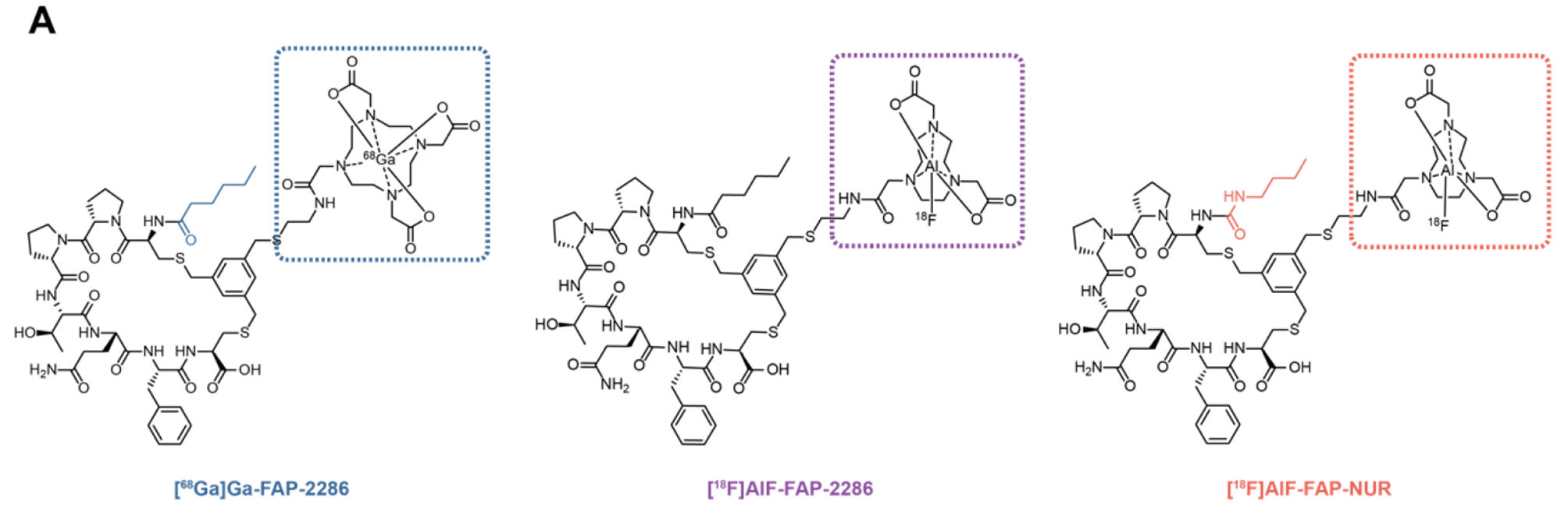
Radiosynthesis
NOTA-FAP-NUR (Nanchang Tanzhen Biotechnology Co., Ltd., Jiangxi, China) (Fig. 1) was radiolabeled with [18F]F. For [18F]AlF-labelings, the tracer was synthesized on a modified GE TRACERlab FxFN platform according to the steps shown in Supplementary Fig. S1. The irradiated [18O]H2O was bypassed through the Sep-Pak QMA Plus Light Cartridge (preconditioned with 10 mL ethanol and 10 mL water) into the vessel for [18O]H2O recovery in vacuo. Subsequently, the [18F]F− (22–29 GBq) trapped on the QMA cartridge was eluted with 0.5 mL of 0.9% NaCl aqueous from Vial 1 into the reaction vessel. Drying was accomplished by azeotropic distillation using 1 mL of acetonitrile from Vial 2. After completing the drying process, a mixture solution containing 400 µL acetonitrile, 250 µL 0.2 M NaOAc buffer (pH 4.0), 7.0 µL 10.0 mM AlCl3 in 0.2 M NaOAc buffer (pH 4.0), and 150 µL precursor (1 mg/mL) in 0.125 M ascorbic acid was added to the reaction vessel from Vial 3. After being heated for 15 min at 100 °C, the mixture was cooled to 70 °C and diluted with 10 mL of water from Vial 5. Next, the dilute solution was transferred over a C18 Light Cartridge (preconditioned with 10 mL ethanol and 10 mL water). Afterward, the cartridge was washed sequentially with 10 mL of water from Vial 6 and 5 mL of water from Vial 8. The product was eluted with 1.5 mL 50% EtOH solution from Vial 7 into the pre-loaded collection vial (6.5 mL 0.9% sterile NaCl for injection containing 10 mg/mL ascorbic acid). The final product solution of 8 mL was transferred into the sterile vial by sterilizing filtration using a Millex GV 33 mm 0.22 μm filter. In addition, a [68Ge]Ge/[68Ga]Ga generator obtained from iThemba LABS was employed for the labeling of [68Ga]Ga. The radiosynthesis of [68Ga]Ga-FAP-2286 followed the previously described method [10].
Stability and partition coefficient
[18F]AlF-FAP-NUR (100 µCi, 10 µL) was added to saline (90 µL) and the mixture was incubated at 37 ℃ for 4 h. The mixture was injected directly into the radio-HPLC (Thermo Fisher Scientific, USA) for analysis. The radio-chemical purity was automatically analyzed by the radio-HPLC. Detailed information is provided in the Supplementary data.
The tracers were added to equal volumes of n-octanol and phosphate-buffered saline (PBS) buffer (0.01 M, pH = 7.4), and centrifuged at 12,000 × rpm for 5 min (n = 3). Then, the mixture was centrifuged, and equal volumes of n-octanol and PBS solutions were taken separately to be counted by an automatic γ-counter (Hidex AMG, Hidex Oy, Finland), and the average LogP value was calculated.
Cell culture and preparation of xenograft models
293T, A549, HT1080 cell lines and the cells transfected with human FAP genes, including 293T-FAP, A549-FAP, and HT1080-FAP were cultured in Dulbecco modified Eagle medium supplemented with 10% fetal bovine serum (Bovogen, Australia) and 1% penicillin-streptomycin solution (Cytiva, USA) at 37 ℃/5% carbon dioxide.
All animal experiments were conducted in compliance with a protocol approved by the Guangzhou Medical University Institutional Animal Care and Use Committee (No. 20230330). Normal male Kunming (KM) mice (6 weeks old with body weight of 25–34 g) were purchased from Zhuhai BesTest Bio-Tech Co., Ltd., and male BALB/c-nude mice (4–5 weeks old with body weight of 16–22 g) were purchased from GemPharmatech Co., Ltd. Mice were housed under standard conditions with temperature and light control (12-h-light/12-h-dark cycle) and had free access to water and food. For the tumor models, 293T-FAP or 293T cells (5 × 10⁶/mouse, n = 8 mice/group) were subcutaneously injected into the right flank of male BALB/c-nude mice, respectively.
Cell assays
For cell uptake experiments, HT1080-FAP cells (5 × 10⁴/well) or HT1080 (5 × 10⁴/well) were seeded in 24-well plates for 24 h and incubated at 37 °C with radiolabeled tracers (74 kBq/well) in 1 mL of serum-free medium for indicated times. Non-specific binding was determined by co-incubating with DOTA-FAP-2286 (3 µM/well). For internalization experiments, the radiotracer-incubated cells were washed twice with PBS (pH 7.4), followed by glycine-HCl (0.05 M, pH = 2.8) solution to distinguish between cell surface-bound (acid-releasable) and internalized (acid-resistant) radioligand. For efflux experiments, cells were preincubated with [18F]AlF-FAP-NUR or [68Ga]Ga-FAP-2286 for 1 h, and subsequently incubated at 37 °C in a medium free of radiotracers and serum for different time points. At the same time, a group without adding medium was kept measuring the total amount bound to the cells. Then the medium was removed, and the cells were washed twice with cold PBS and subsequently lysed with 0.2 mL of NaOH (1 M). Cell lysates were collected, and the radioactivity was determined using a γ-counter. The efflux rate was calculated by using the radioactivity of the total bound to the cells (0 min) as the denominator and the radioactivity at different time points as the numerator.
For competitive FAP binding assays, 293T-FAP cells were incubated with [177Lu]Lu-FAP-2286 (74 kBq/well) in the presence of different concentrations (10− 5–10− 11 M) of competing non-radioactive ligands for 60 min. The cell incubation process was performed by Brandel M-48T Cell Harvester (Biomedical Research & Development Laboratories, Inc., Gaithersburg, MD, USA). Then the cells were collected, and the radioactivity was measured with a γ-counter.
Micro PET/CT imaging and ex vivo biodistribution in mice
Micro PET/CT imaging and biodistribution studies were conducted when the tumor diameter reached 5–8 mm. PET/CT imaging was performed using a PET scanner (MadicLab PSA071, Shandong Madic Technology Co., Ltd., China). Micro-PET scanning was performed after intravenous injection of [68Ga]Ga-FAP-2286 (0.1 mL, 7.4 MBq) or [18F]AlF-FAP-NUR (0.1 mL, 7.4 MBq). The acquisition time of PET scanning for FAP-positive tumor-bearing mice was 120 min, then it was framed at a rate of 5 min per frame. The FAP-negative tumor-bearing mice had a 10-min static scan after receiving the equivalent medication injection for 60 min. Before static scan, mice were able to move freely. The CT scan parameters were tube voltage of 80 kV, tube current of 0.7 mA, and voxel spacing of 200 μm. The PET image was used to determine the radiotracer uptake, and the CT image was used for the attenuation correction of PET images and localization of radiotracer uptake sites. The images and volumes of interest (VOI) were produced using PMOD software (version 4.4, PMOD Technologies LLC, Switzerland).
To characterize the normal biodistribution of [18F]AlF-FAP-NUR in normal male KM mice, mice were divided into 5 groups (n = 4/group) and injected intravenously with 100 µL of [18F]AlF-FAP-NUR (2.22–3.70 MBq/mouse). At different time points after injection, the mice were sacrificed, and blood samples and organs of interest were collected, weighed and counted with a γ-counter. Meanwhile, to further verify the biodistribution in xenograft mice, male BALB/c-nude mice bearing 293T-FAP tumors were divided into two groups (n = 4/group) and injected intravenously with 100 µL of [68Ga]Ga-FAP-2286 or [18F]AlF-FAP-NUR (2.22–3.70 MBq/mouse), respectively. At 1 h after injection, they were subjected to the same operation as described above.
Patient enrolment
The clinical translational study of [18F]AlF-FAP-NUR was approved by the institutional review board of the First Affiliated Hospital of Guangzhou Medical University (ES-2023-083-01) and was registered on the www.Chictr.org.cn under the identification number ChiCTR2300076976. Oral and written informed consent was obtained from all participants. The inclusion criteria were as follows: (1) adult participants (aged 18 years or older), (2) patients with suspected tumors who couldn’t be clearly diagnosed as tumors using other imaging methods such as ultrasound, CT and MRI, and (3) patient’s general condition was acceptable and could tolerate PET examination. Exclusion criteria were as follows: (1) non-tumor disease, and (2) patients who were pregnant.
Clinical PET/CT imaging
For the clinical translational study of [18F]AlF-FAP-NUR, we enrolled two patients, including a 65-year-old woman with a preliminary diagnosis of breast cancer through CT scan, and a 70-year-old woman with a right upper lung lesion. Both patients received an intravenous injection of [18F]AlF-FAP-NUR at a radioactivity dose of 5.55 MBq/kg (0.15 mCi/kg). Approximately 50 min after intravenous administration, static PET/CT scans were acquired using uMI Panorama system (United Imaging Healthcare, Shanghai, China) from the top of the skull to the middle of the femur. The PET scan parameters were: 1 min/bed position, 4 positions in total, 256 × 256 matrix. The CT scan parameters were: 120 kV, auto-mAs, 512 × 512 matrix, 0.5 s per tube rotation, slice thickness of 3.75 mm, and pitch of 0.9625. All data were reconstructed on the workstation (uWS-MI R002.5.0). Reconstruction for all imaging data was performed with an ordered subset expectation maximization algorithm (OSEM) with three iterations per ten subsets. For standardized uptake value (SUV) calculations, a sphere with proper diameter was placed inside the organ parenchyma for evaluating the maximum SUV (SUVmax) of tumors and mean SUV (SUVmean) of normal organs, then the TBRs were calculated as the ratio tumor-SUVmax/normal-organ-SUVmean. Two experienced nuclear medicine physicians independently reviewed all images, and any discordant results were resolved by consensus.
Data analysis and statistics
Statistical analysis was performed using the SPSS 27.0 software (IBM Corp., Armonk, NY, USA) and GraphPad Prism 9.0 (GraphPad Software, Boston, MA, USA). Data were presented as mean ± SD. An unpaired t-test was used to evaluate the differences in each organ uptake between [18F]AlF-FAP-NUR and [68Ga]Ga-FAP-2286 PET/CT. Statistical significance was defined as P < 0.05.


Comments (0)