
Remember me

EM simulations were performed in CST Microwave Studio (CST GmbH, Darmstadt, Germany). Two different RF coil geometries were simulated, each operating in transmit/receive mode. The first is a dome-helix [14] and the second is an elliptical solenoid coil, consisting of 15 and 20 turns of copper wire of 1.5 mm diameter, respectively, with one capacitive segmentation halfway along the wire length. The coils had dimensions of 180 mm width, 240 mm height, and 10 mm gap between wire turns (Fig. 1A). The coils were loaded with the head model, Duke, from the IT IS Virtual Family [29], with an isotropic resolution of 1 mm × 1 mm × 1 mm. Its material properties were categorized into 18 different tissues to simplify the number of dielectric properties in the model; for example, the eye, sclera, cornea, and vitreous were considered to have the same conductivity/permittivity/proton density. Two different geometries, cylindrical and elliptical, of the RF shield were simulated as a continuous copper structure with a length of 350 mm, a thickness of 0.07 mm, and a conductivity of \(5.96\times ^ S/m\). The time domain solver with open boundary conditions in all directions was used, 1-W input power was considered for all simulations, and the computations were ended at an accuracy of − 40 dB. Figure 1B shows the three different simulation setups considered:
First setup: dome-helix coil inside a cylindrical shield with various diameters from 260 to 320 mm with a 5 mm step size.
Second setup: an elliptical solenoid coil inside a cylindrical shield with various diameters from 260 to 320 mm with a 5 mm step size.
Third setup: a dome-helix coil inside an elliptical shield with a symmetric gap between the coil and shield where the major axis changes from 260 to 320 mm with a 5 mm step size.
Fig. 1
A Schematics of the simulated dome-helix (left) and elliptical solenoid (right) coils with the Duke model, from the IT IS Virtual Family. B On the left, three different simulation setups are shown, and on the right, a schematic of the coil and the shield (depicted as an orange circle) is presented
In each case, the RF coils were impedance matched to 50 Ω at 1.965 MHz using variable capacitors in an L-network. The \(_^\) efficiency (\(\mu T/\sqrt\)) map was calculated for each setup.
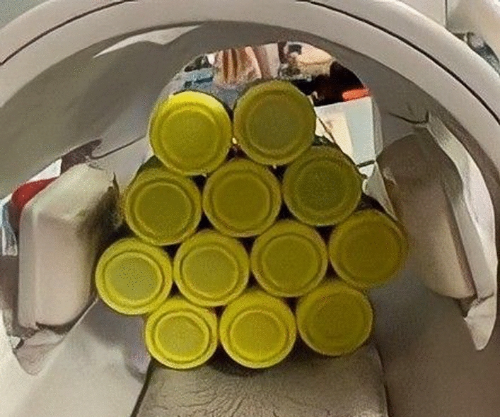
Experimental measurementsTo validate the simulated data, two turbo spin echo (TSE) images and \(_^\) maps were acquired on a Halbach POC low field system [6, 15] using a dome-helix coil placed within a cylindrical shield of different diameters, 300 mm and 260 mm, each 0.07 mm thick, (Fig. 2). \(_^\) maps were acquired from a head model phantom filled with copper sulphate doped water (T1 ≈ 100 ms) the phantom dimension is 240 mm in the head/foot direction, 190 mm anterior/posterior, and 160 mm left/right. \(_^\) mapping used a 3D double angle (60° and 120°) method (DAM) [16], 600 ms/8 ms repetition time/echo time (TR/TE), field-of-view of 220 × 210 × 210 mm3, spatial resolution 4 × 4 × 4 mm3, 20 kHz acquisition bandwidth, and a 200 microsecond RF pulse duration. K-space data were filtered using a sine-bell function. \(_^\) maps were produced from the tip angle (α) maps:
$$\alpha = }^\left(\frac_}_}\right)=\tau \gamma _^\stackrel}} _^=\frac}^\left(\frac_}_}\right)$$
(4)
where \(_\) and \(_\) represents the signal intensities from the 60° and 120° image acquisitions, τ indicates the RF pulse duration and \(\gamma\) is the gyromagnetic ratio. Background noise outside the phantom was masked.
Fig. 2
An illustration of the imaging setups for the TSE and B_1^ + maps on the Halbach POC low field system. A two shields 300 mm and 260 mm in diameter. B Constructed coil with slab phantom inside. C Head model phantom filled with copper sulphate doped water (T1 ≈ 100 ms) used for B_1^ + mapping. D Two-dimensional brain phantom corresponding to axial brain slabs with a thickness of 20 mm and relaxation parameters of the compartments approximately equal to in vivo data at 50 mT [17]
To validate simulated SNR results, two turbo spin echo (TSE) images from a brain slab phantom (Fig. 2) with dimensions of 200 × 150 × 20 mm were obtained. 2D TSE images were acquired with 1000 ms/20 ms TR/TE, field-of-view 220 × 200 mm2, spatial resolution 1 × 1 mm2, 20 kHz acquisition bandwidth, 10 averages, and a 200 microsecond RF pulse duration.
Static magnetic field simulationsMagnetostatic simulations were conducted using Python, building upon the framework established by O’Reilly [18], to obtain the magnetic field strength and homogeneity within a 200 mm diameter spherical volume (DSV). The magnet is based on an existing Halbach-based neuroimaging POC system with a length of 496 mm [15]. Based on the range of shield diameters simulated from 260 to 320 mm, and assuming a 10 mm thickness to account for the presence of the three gradient coils, the magnet inner diameter was changed in eight steps from the smallest possible 270–330 mm. Within the Halbach array, each ring comprised 12 × 12 × 12 \(}^\) N48 neodymium boron iron (NdBFe) magnets, arranged in a dipolar (k = 2) Halbach configuration. In this design each ring contains two layers of magnets and the outer layer has seven more magnets than the inner layer. In all cases, the outer diameter is 81 mm larger than the inner diameter and the number of cubic magnets increases as the diameter of each ring is increased. Finally, by combining the transmit efficiency and magnetic field strength, the SNR of the coil was calculated.
Comments (0)