
Remember me


A total of 22 CSCR patients (16 male and 6 female) aged 46.0 ± 8.4 years were recruited. Twenty patients had uniocular CSCR and 2 patients had binocular CSCR; thus, 24 eyes were included. The clinical details of a typical subject are shown in Fig. 1.
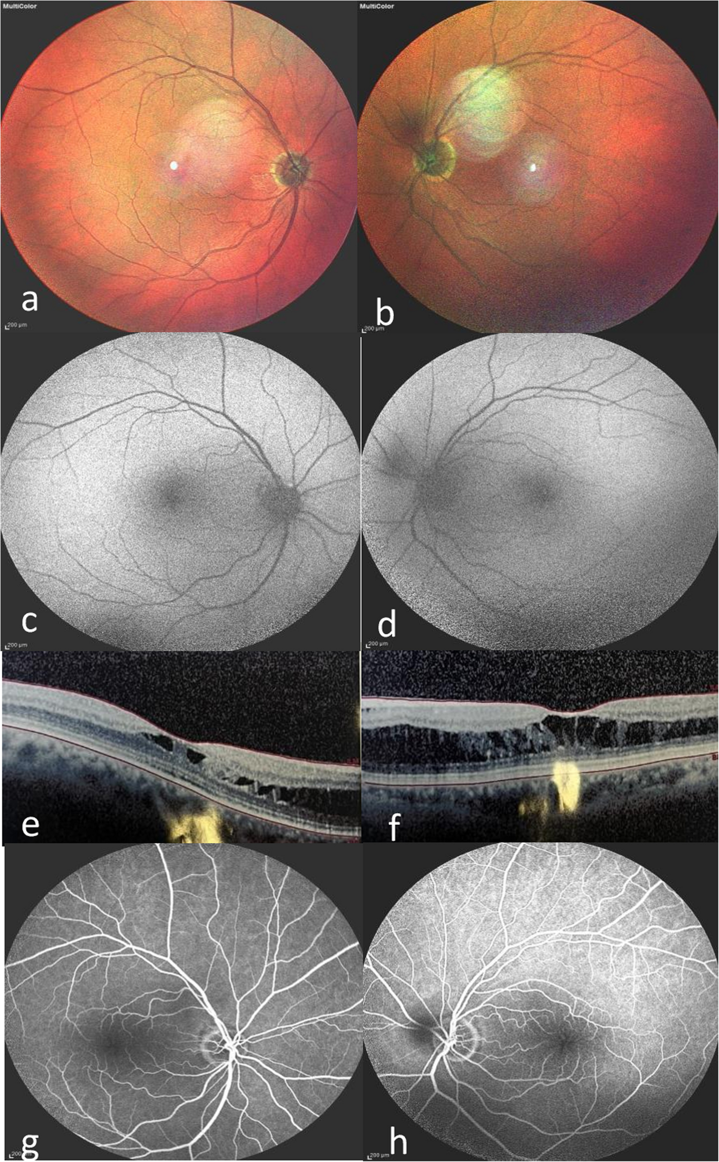
Fig. 1
Clinical examinations of a typical subject. A Early phase (30 s) of FFA and ICGA. B Medium phase (5 min) of FFA and ICGA. C Late phase (20 min) of FFA and ICGA. D Fundus color photography. E Macular OCT
A typical ink-blot leakage site beside macula in the early phase of FFA is shown in Fig. 1A, and it expanded as a “smokestack” pattern in the middle and late phases in Fig. 1B and C. In Fig. 1D and E, the SRF is shown under the macular neural retina; thus, it is a typical CSCR patient.
The therapeutic effects of 577 nm MLP on macular function and structure in early-stage CSCR patientsAll the CSCR patients received 577 nm MLP treatment and were reviewed in 30 days. In review, BCVA, OCT and microperimetry examinations were performed. Figure 2 shows the comparisons of BCVA, the SRF height of the macula on OCT and macular neural function level on microperimetry before and after MLP treatment. In microperimetry, the macular area is divided into 9 parts, including the central part of the macula (C), inferior part of the macula (I), superior part of the macula (S), nasal part of the macula (N), temporary part of the macula (T), inferior inferior part of the macula (II), superior superior part of the macula (SS), nasal nasal part of the macula (NN) and temporary temporary part of the macula (TT).
Fig. 2
Comparisons of BCVA, SRF macular height, and macular neural function level on microperimetry before and after 577 nm MLP treatment, and correlation analyses of SRF and BCVA, and the neural function level of the central part of the macula were performed. A Comparison of macular neural function level on microperimetry before and after MLP treatment in different macular regions. B Macular region division diagram in microperimetry. C Comparison of BCVA before and after MLP treatment. D Comparison of the SRF height of macula before and after MLP treatment. E Correlation analysis of SRF height and BCVA before and after MLP treatment. F Correlation analysis of the SRF of macula and neural function level of central part macula (C) on microperimetry before and after MLP treatment. The hollow circles represent the values before MLP treatment, and the gray circles represent the values after MLP treatment. *: P < 0.05, **: P < 0.01, ***: P < 0.001
As shown in Fig. 2B, the macula was divided into 9 parts. The diameter of the central part of macula (C) is 1 mm, the diameter of the I, S, N and T parts of the circle is 2 mm, and the diameter of the II, SS, NN and TT parts of the circle is 3 mm. As shown in Fig. 2A, the macular neural function levels of all 9 parts on microperimetry significantly increased after MLP treatment, including the C part (t = -4.025, P < 0.001), I part (t = -3.062, P = 0.006), S part (t = -4.685, P < 0.001), N part (t = -4.335, P < 0.001), T part (t = -3.835, P < 0.001), II part (t = -2.790, P = 0.010), SS part (t = -2.924, P = 0.008), NN part (t = -3.143, P = 0.005) and TT part (t = -3.122, P = 0.005). These findings indicate that the MLP treatment improved neural function in all the parts of macula.
As shown in Fig. 2C, the BCVA significantly increased after MLP treatment (t = 3.768, P < 0.001), and it indicating that MLP treatment significantly increased subjective visual function in CSCR patients. Figure 2D shows that the SRF height of the macular area significantly decreased after MLP treatment (t = 8.884, P < 0.001), which also confirmed that the SRF was absorbed and that the CSCR condition improved. Therefore, MLP treatment improved both visual function and retinal anatomy in patients with CSCR.
The correlation analysis shown in Fig. 2E and F revealed that the improvement in visual function, including BCVA (r = 0.574, P < 0.001) and macular neural function level (r = -0.600, P < 0.001), was significantly correlated with the SRF height of the macula. Therefore, a decrease in the SRF height of the macular area is the key reason for visual function improvement.
The therapeutic effects of 577 nm MLP on choroid vessel in early-stage CSCR patientsThe direct and main object of 577 nm MLP treatment is RPE, which can affect the choroid vessel after laser treatment. The source of SRF is the vessel of the choroid, so the vascular density (VD) of the choroid on OCTA before and after MLP was compared. Figure 3 shows the VD comparisons before and after MLP treatment via OCTA in different areas of the macula including the central part of the macula (C), the inferior part of the macula (I), the superior part of the macula (S), the nasal part of the macula (N) and the temporary part of the macula (T). The VD at different depths of choroid, including the superficial layer (SL) of the choroid, the deep layer (DL) of the choroid and the whole layer (WL) of the choroid, were also compared. Additionally, the leakage site parts of each eye were grouped for independent comparisons, and the number of eyes in each leakage site part is also shown.
Fig. 3
Comparisons of VD in different areas of the macula and different depths of the choroid before and after 577 nm MLP treatment. A Comparison of VD at SL DL and WL depths in the central part of the macula before and after MLP treatment. B Comparison of VD in the inferior part of the macula. C Comparison of VD in the superior part of the macula. D Comparison of VD in the nasal part of the macula. E Comparison of VD in the temporal part of the macula. F Comparison of VD in the leakage site part of the macula. G Leakage site locations illustration. H Macular region division diagram on OCTA. *: P < 0.05, **: P < 0.01
As shown in Fig. 3H, the macula was divided into 5 parts. The diameter of the central part of macula (C) is 1 mm, the diameter of the I, S, N and T circle is 3 mm. As shown in Fig. 3A (the C part), the VD of the choroid significantly increased in all three layers after MLP treatment: SL (t = -3.653, P = 0.001), DL (t = -2.444, P = 0.023) and WL (t = -2.477, P = 0.021). As shown in Fig. 3B (the I part), the VD of the choroid significantly increased only at the WL (t = -2.359, P = 0.027). As shown in Fig. 3C (the S part), the VD of the choroid significantly increased in all three layers after MLP treatment: SL (t = -2.530, P = 0.019), DL (t = -2.198, P = 0.038) and WL (t = -2.614, P = 0.015). As shown in Fig. 3D (the N part), the VD of the choroid did not significantly change. As shown in Fig. 3E (the T part), the VD of the choroid significantly increased in all three layers: SL (t = -2.845, P = 0.009), DL (t = -2.925, P = 0.008) and WL (t = -2.796, P = 0.010). As shown in Fig. 3F (the leakage site part), the VD of the choroid significantly increased in all three layers after MLP treatment: SL (t = -2.899, P = 0.008), DL (t = -3.102, P = 0.005) and WL (t = -3.148, P = 0.005). Figure 3G shows the number of eyes in each leakage site part, most of the leakage sites were in the central and superior parts, and there was no leakage site in the inferior part.
The significant increase of the choroid VD in the central and superior parts, especial in the leakage site part illustrated that the choroid VD is a potential key factor for the CSCR condition, and that the MLP improves the condition of the CSCR by increasing the VD of the choroid. To confirm the relationship between choroid VD and CSCR conditions, the correlations between leakage site choroid VD in different layers and the SRF height of the macula were analyzed, and a CSCR recurrent subject was also analyzed.
Fig. 4
Correlations between leakage site choroidal VD in different layers and the SRF height of the macula. A The correlation between the leakage site VD at the SL and the SRF height of the macula. B The correlation between the leakage site VD at the DL and the SRF height of the macula. C The correlation between the leakage site VD at the WL and the SRF height of the macula. D Changes of leakage site part choroid VD and macular SRF height in a recurrent patient. The dotted line represents the SRF height change
As shown in Fig. 4A B and C, the SRF height of the macula was significantly correlated with the choroid VD of the leakage site part, including the SL (r = -0.371, P = 0.009), DL (r = -0.353, P = 0.014) and WL (r = -0.314, P = 0.029). This finding illustrated that the increase in choroid VD was the key reason for the decrease in SRF height of macula in patients with CSCR. Furthermore, a recurrent patient was identified in this study. Figure 4D shows the disease process of the recurrent subject who was the only patient with recurrence in half year. In the first MLP treatment, the leakage site choroid VD increased in the SL, DL and WL of the choroid, and the SRF decreased at 30 days post-treatment review. However, the CSCR relapsed 40 days later, the SRF height increased, and the leakage site choroid VD decreased again in the SL, DL and WL again. Therefore, the subject was given the second MLP treatment, and was required to review again within 30 days. In the review of the second treatment, the SRF height decreased dramatically, and the choroid VD increased again in the SL, DL and WL of choroid.
Therefore, the VD of the choroid was significantly negatively correlated with the SRF height. 577 nm MLP treatment significantly increased the choroidal VD and resulted in a decrease in SRF height. This is one of the key effects of MLP for early-stage CSCR patients.
Comments (0)