
Remember me

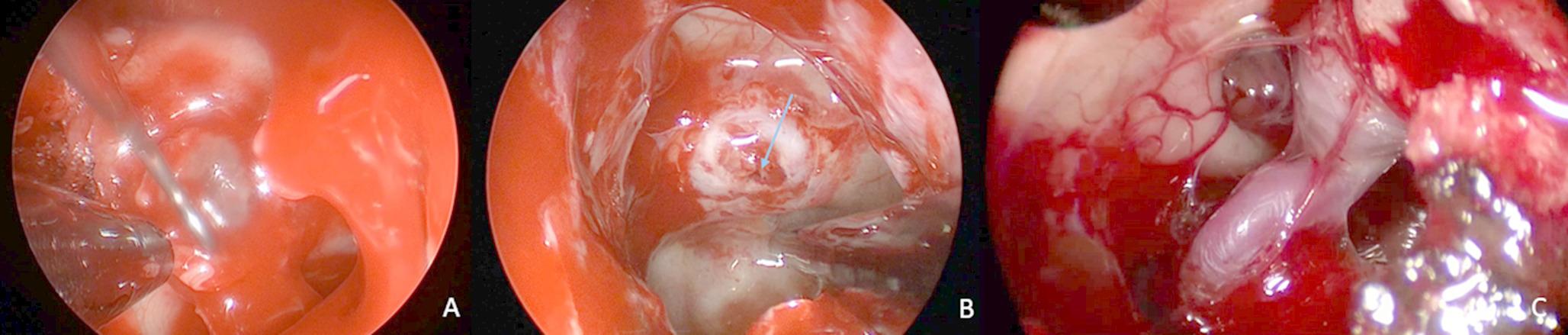
Surgical approaches to the MTR are classified into three groups. Depending on the surface through which the exposure is made, direct exposure is provided by approaches based on the superior, lateral, and posterior aspects (Table 1). Each technique provides direct access to various structures within the MTR, including the hippocampus and amygdala (Table 1). The approach chosen depends on the specific condition being treated as well as the surgeon’s personal preference and level of experience. Although lobectomy operations are done for MTR lesions, there are many studies on selective surgical approaches in the literature. One of these, the transsylvian approach, is a superior approach frequently used for MTR pathologies since 1973 when it was developed by Yasargil [8]. The transsylvian approach is done through the floor of the sylvian fissure close to the anterior inferior portion of the inferior peri-insular sulcus of the insula. It allows for resection of the MTR without neocortical structure resection and temporal lobe retraction [1, 6] (Fig. 1B). Niemeyer introduced the selective amygdalohippocampectomy in 1958; this approach was originally called the transventricular amygdalohippocampectomy. He used a middle temporal cortical incision through the middle temporal gyrus to reach the hippocampus [20], comprising a lateral approach through any sulcus or gyrus of the lateral area of the temporal lobe. If posterior enlargement is necessary, the hippocampus and parahippocampal gyrus can be resected with subpial aspiration; however, homonymous hemianopsia is more prevalent with the transcortical approach than with the transsylvian approach [20, 21]. The sulcus between the superior and middle temporal gyrus is typically utilized for the trans-sulcal approach. The sulci endings nearest the temporal horn of the lateral ventricle require minimal resection of the white matter. Nevertheless, during sulcal dissection, the pia mater and, consequently, the underlying cortex are damaged. Moreover, it may be necessary to coagulate small sulcal veins. This procedure carries the risk of indirectly impairing cortical function [22, 23].
Hori and colleagues developed the subtemporal approach which enables the surgeon to manipulate MTR without damaging the vein of Labb´e. This method can minimize postoperative parenchymal damage [24], but a statistically significant relationship to memory loss was found on long-term follow-up. In addition, long-term follow-up revealed a statistically significant relationship between alcohol consumption and memory loss [25]. Ture and Pamir have recommended the small petrosal approach over the subtemporal approach to increase the chance of preserving the basal vessels and allowing greater surgical exposure or angle [26]. In the presigmoid-retrolabyrinthine approach, variations in venous anatomy can make retracting the basal surface of the temporal lobe difficult or impossible. In the presigmoid- retrolabyrinthine approach, the petrosal bone is drilled to obtain broad exposure of the skull-base structures. It has the benefits of minimizing brain retraction, shortening the working distance, and allowing access to critical neurovascular structures from multiple angles. It also requires less retractile force than alternative posterior techniques. However, the complexity of this technique necessitates a lengthy learning curve [27, 28]. The supratentorial-infraoccipital approach, defined by Smith and Spetzler [27], begins with gentle retraction of the occipital lobe, and the brain retractor is removed after the cerebrospinal fluid has been drained. The vein of Galen and its tributaries remain medially, allowing a surgical corridor. Disadvantages of this approach are dural sinus injury, delayed dural sinus thrombosis, and visual defects. There may also be difficulty reaching the amygdala [27, 29, 30].
The supracerebellar transtentorial approach is an effective, less-invasive avenue. Voigt and Yasargil first used this approach to remove a cavernous angioma in the posterior hippocampus [31]. Subsequently, Yonekawa and colleagues used it to clip the distal segment of a posterior cerebral artery aneurysm [32]. Moftakhar and colleagues also described using this approach for the posterior portion of the MTR [33]. Further, Türe used this approach to reach the entire MTR and has popularized it for selective amygdalohippocampectomy [3]. The risk of injury to the visual cortex and optic radiation is minimal, and it provides a safe corridor for surgical access to the middle and posterior MTR portions. Although there is more than one bridging vein, these are directly visualized in the supracerebellar transtentorial approach. In some cases, access to the MTR may be difficult with this approach, as the venous system may obstruct the superior portion of the posterior portion. In particular, the lateral cerebellar bridging veins, which may be parallel to the MRI sections, cannot be seen on the preoperative MRI.
Presently, these surgical approaches are used frequently and successfully. Nevertheless, selecting an effective surgical approach to the MTR remains difficult. The complex functional anatomical structure and deep location of the MTR have been the focus of many articles regarding the success of selected techniques [3, 21, 23, 25]. Today, technological advances facilitate safer access to localized deep brain lesions similar to those in the MTR. One of these advances, MRI sequences, allow noninvasive preoperative evaluation and are increasingly used for surgical planning and strategy [34, 35]. These MRI images are also useful during surgery, along with neuronavigation devices. Neuronavigation systems function by matching the spatial positions of digital data (MRI or computed tomography) obtained from imaging studies to the surgical field. The images obtained before surgery are matched with the patient’s reference points on the operating table. Therefore, in the image, the intracranial point the surgeon is at during the operation is controlled. This technique significantly decreases morbidity and mortality when combined with neuroanatomical structure-based surgical techniques that have been used for decades. Image-guided neuronavigation systems allow more minimally invasive surgery [9]. In our study, a system to evaluate neuroimages and navigation was developed. First, MRI sequences taken from the EDEN2020 Human Brain MRI Datasets were studied [13, 14]. The BrainSuite tool was used to divide T1-weighted MRI sequences into approximately 139 major regions using an atlas-based segmentation technique. Afterward, the existing images were automatically segmented by selecting them on the 3D Slicer tool (Fig. 2). Free, open-source software was used for registration purposes [17]. This software is used to visualize image-guided procedures and for processing, segmentation, planning, and navigation [14, 16]. Thus, tract systems, arteries, and veins could be labeled with the coordinate system along their routes [10]. The pixel and voxel values of the anatomical point, as well as its anatomical structure, were labeled [15, 36] (Table 2). This method provides the benefit of trading in a cubic system with matrices (Fig. 5). MRI images obtained from the data set were used to validate the system. Subsequently, preoperative images from the Radiology Department were processed using angle and thickness sequences from previous datasets. This step is important as it allows the possibility of a personalized preoperative evaluation of patients. The 294 anatomical areas labeled and defined on 3D MRI sequences were classified as target and non-violated areas (Table 2). The target area was determined as the medial portion of the MTR. The areas detailed in Tables 1 and 2 were taken into consideration as areas not to be violated. The QL algorithm (using the Python programming language) was used to find new pathways.
Fig. 4
With Q learning, approaches are created with more location calculations
Fig. 5
The cubic-coordinate system (x,y,z) for AI calculation. (A) The coordinate system represents a cubic graph as a set of wireframes composed of cubes. To utilize the path-finding algorithms on segmentation images, the images must be converted into graphical representations. Anatomical function can be added as another coordinate. (B) Placing MRI scans into the cubic coordinate system to match the brain parenchyma with this system can reveal the concept of digital anatomy and act as a basis for systems that can be developed and controlled
AI algorithms have evolved on two basic mechanisms classified as unsupervised and supervised. In unsupervised AI applications, the aim is not to establish a cause-and-effect relationship. In this method, the aim is to find similarities and differences in a complex and large data set. In supervised machine training applications, AI has a training and development process. In this training process outputs are given to the computer and the computer software learns the relationship between outputs [10].
Machine learning (ML) is a subfield of AI that focuses on the development of algorithms and statistical models that enable computers to learn from and make predictions based on data. ML algorithms use patterns and insights from data to train models that can be used to make predictions or take actions in real-world applications. ML has a growing presence in the field of neurosurgery, where it is used to improve patient outcomes, speed up diagnosis and treatment, and support surgical planning and decision-making. Some of the applications of ML in neurosurgery include image analysis, predictive modeling, surgical planning, real-time guidance, and clinical decision support. Overall, ML has the potential to significantly improve the efficiency of neurosurgical procedures and patient outcomes [10, 37]. Reinforcement learning (RL) is a type of machine learning in which an agent learns to make decisions by performing actions in an environment to maximize a reward signal, evolved from the main group of supervised learning. The agent interacts with the environment in a sequence of trials and, in each trial, it chooses an action based on its current state, performs the action, and receives a reward signal that tells it how well the action was performed. Over time, the agent learns to map states to actions that lead to high rewards, which forms its policy [10, 18, 38]. Previously generated cubic brain parenchyma was run through the coordinate system in our study. Its target is the MTR, and the locations that should not be touched on the way to this area have been determined. An analysis of approximately 12 trillion points was used to compare the optimal linear pathways. Subsequently, the entrance points to these linear pathways were used in the QL algorithm to find nonlinear pathways similar to the selected standard pathways. Finally, the risk scores and Q-learning scores for these new pathways were listed.
In our image-guided, AI-assisted approach to risk analysis, the Q-learning score of the supracerebellar-transtentorial approach (i.e., the penalty score) was the lowest. According to Q Learning, the MTR can be reached through this method with minimal surgical damage. The trans-sulcal, subtemporal, and supratentorial-infraoccipital approaches followed (Table 3). The transsylvian approach yielded a Q-learning score of 28.30 when the middle cerebral artery and the sylvian veins were omitted, and the system was re-executed without considering them. This value was determined to be third behind the supracerebellar-transtentorial and trans-sulcal values.
In our study, the Q-learning score for the supracerebellar transtentorial approach was the highest among the examined approaches. Of interest, the total risk score of all points with pathways drawn by the reviewers differed from the total risk scores of the pathways formed and followed by QL.
Comments (0)