
Remember me


A 53-year-old woman developed progressive bilateral blurry vision and bilateral eye pain. She had no other signs or symptoms except for a few cervical enlarged lymph nodes, present from two years, and stable. The patient had a diagnosis of CLL stage A (Binet staging) two years earlier based on peripheral blood lymphocyte typing, she was not treated with any specific therapy and underwent regular follow-ups.
One month after symptoms onset, the neuro-ophthalmological examination showed diminished Visual Acuity (0.9 in right eye and 0.8 in left eye), the anterior segment was normal as well as ocular pressure. Fundoscopic examination showed bilateral optic disc edema (Frisen Scale 2) confirmed by Optical coherence tomography that displayed elevated nerve fiber layer. Eye ultrasound confirmed the presence of bilateral papilledema with increased thickness of the optic nerve.
The ocular angiography showed late bilateral papillary leakage in right eye hypofluorescence from upper peripapillary preretinal hemorrhages (Fig. 1 – 2).
Fig. 1
Ocular angiography shows late bilateral papillary leakage (A-B) and in right eye hypofluorescence from upper peripapillary preretinal hemorrhages (A)
Fig. 2
Fundus photography shows bilateral optic disc edema (Frisen Scale 3) A—B and upper peripapillary preretinal hemorrhages in right eye (A)
Then brain MR showed a subtle Fluid-attenuated inversion recovery (FLAIR) hyperintensity in the pre-chiasmatic tract of the left optic nerve. No significant swelling or enhancement of the optic nerve were seen (Fig. 3).
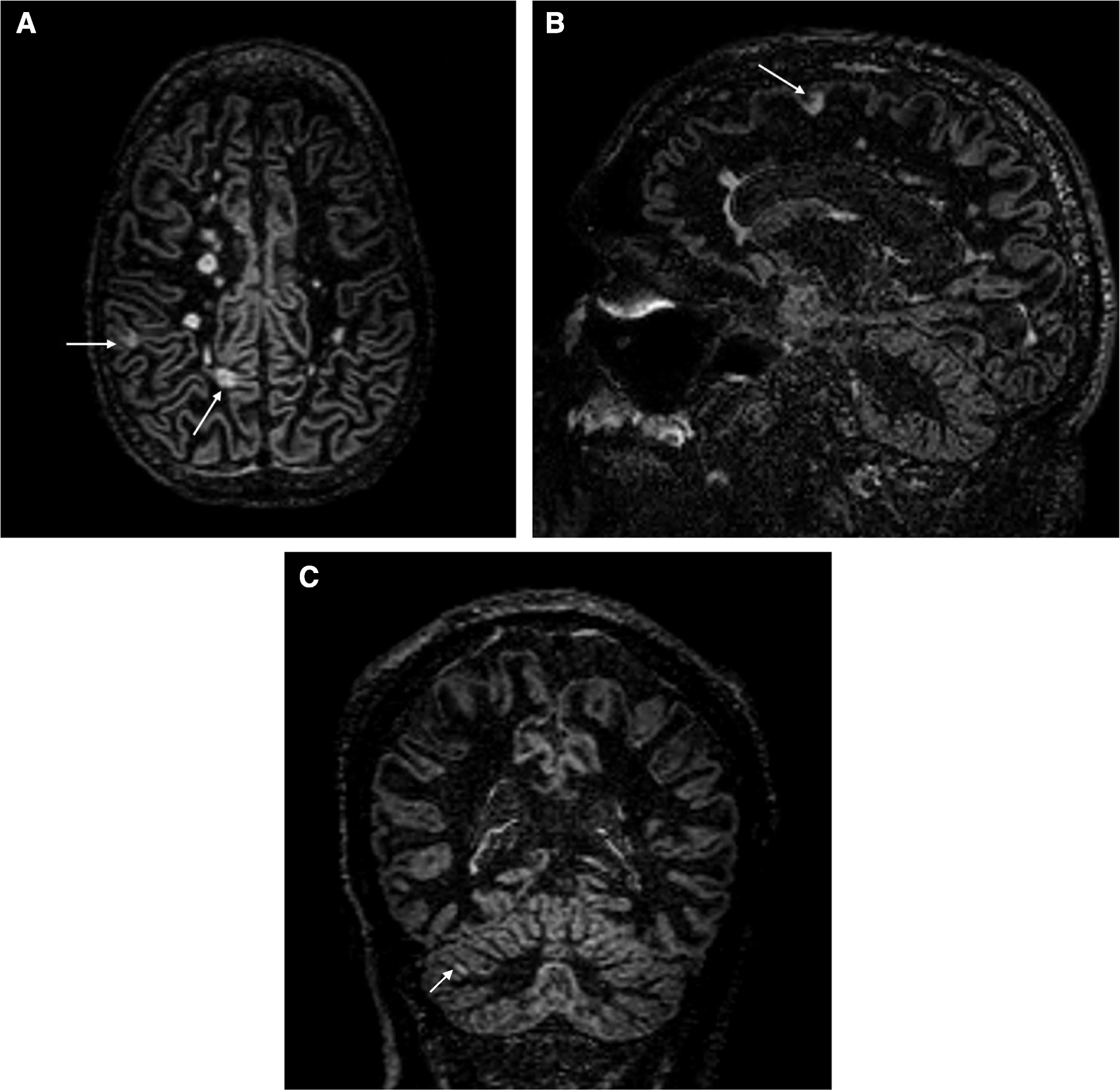
Fig. 3
MR shows subtle hyperintensity along the optic nerve in FLAIR coronal images (arrow in A). No significant swelling of the nerves in T2 axial fat sat images (B) nor clear enhancement in T1 after gadolinium administration (C)
Only 4 months after the onset, she was referred for neurological evaluation (that documented no additional signs except to optic disc edema) and then she was hospitalized for complete diagnostic work-up.
Cerebral spinal fluid (CSF) analysis showed normal glucose, normal protein and the presence of 59 cells/mm3 White Blood Cells (WBC), with 97% of lymphocytes including 79.5% of abnormal CD5 + B-cell population at typing analysis, suggesting the cell B CD5 + lymphoproliferative disease. CSF-Anti EBV antibodies were negative. Complete blood count unremarkable except for elevated revealed WBC count (20,950 cells/mL) (normal: 4,500 − 10,000 WBC/mL) with 58.6% lymphocytes.
Cytometric analysis of peripheral blood showed 81.1% of CD19 + B Lymphocytes with regular shape and the following atypical immunophenotype: CD19 + , heterogenous CD20 + , CD5 + , CD23-/ + (30%, weak), CD79b + (intermediate intensity), CD81 + (weak), CD10-, CD38-/ + , FMC7-, kappa + (weak). Such data are compatible with CD5 + B cells lymphoproliferative disease.
Additional analysis as serological autoimmune profile and Anti-Aquaporin 4 (AQP4) antibodies resulted negative.
Optic nerve biopsy was not performed due to the positivity of CSF markers and the high probability of secondary visual impairment.
The patient started 1 g/day of intravenous methylprednisolone for 5 days, with immediate slight improvement of optic disc edema. No further tapering of steroid was performed.
She then underwent to systemic work-up, including total body Positron Emission Tomography (PET) imaging with 2-[(18)F]fluoro-2-deoxy-D-glucose (FDG) that showed lateral-cervical lymphnodal hypercaptation. A lymph node biopsy was then performed, and showed predominantly B lymphoid proliferation with Ki 67 value of 30%, consistent with CLL. The white cell count was 7200/uL and 1200/L for total blood cells and total lymphocytes respectively.
Based on the absence of histologic transformation in the bone marrow and in lymph nodes and the absence of biological and clinical negative prognostic factors systemic therapy was not started, and the patient continued with periodical stringent follow-up.
She did not complain any blurry vision and pain by the 3-month and 6-month follow up.
The following neuro-ophthalmological evaluation after 3 months of steroid therapy showed a clear improvement: in particular, optical disc edema resolved, associated with a complete disappearance of the preretinal hemorrhages; visual acuity remained stable. A further examination 6 months later confirmed the well-control of neurological alterations, showing visual acuity stability andster mild change, based on reduction of retinal nerve fiber layer and ganglionar cells at OCT analysis.
Further brain MR was stable compared to the exam five months earlier.
The last haematological evaluation did not show systemic disease progression.
Comments (0)