Dementia is a group of neurodegenerative disorders characterized by cognitive impairment, memory loss, and difficulties in daily activities due to the progressive degeneration of brain cells. People living with face numerous challenges, including functional limitations, social isolation, emotional distress, health complications, and financial strain. It is the second leading cause of death in Australia and ranks seventh globally [1]. The social and economic impacts of dementia are profound, as it places immense strain on individuals, families, communities, and healthcare systems. According to the World Health Organization (WHO), over 55 million people currently live with dementia, with cases projected to rise to 78 million by 2030 and 139 million by 2050 [2, 3]. This growing prevalence places a significant burden on healthcare systems and economies, with global dementia-related costs reaching $1.3 trillion USD in 2019, half of which was attributed to informal caregiving [2].
Dementia includes several types, such as Alzheimer’s disease (AD), frontotemporal dementia (FTD), vascular dementia (VaD), and dementia with Lewy bodies (DLB). AD is the most common form, accounting for 60–70% of cases [4]. Mild cognitive impairment (MCI) is often considered a precursor to dementia, with an increased risk of progression, though the rate varies among individuals. This study focuses on two of the most common forms of dementia-AD and FTD, which share overlapping symptoms, making accurate diagnosis and targeted treatment development particularly challenging.
Despite its devastating impact, dementia has no cure or treatment to halt or reverse its progression. However, early detection can help mitigate its effects and improve patients’ quality of life. Traditional diagnostic methods for dementia, such as clinical assessments and neuroimaging techniques (MRI, PET scans), are expensive, time-consuming, and not widely accessible. As a result, researchers are increasingly exploring non-invasive, cost-effective alternatives, such as electroencephalography (EEG). EEG is a promising tool for studying brain activity due to its high temporal resolution, affordability, and ease of use. EEG captures the electrical activity of the brain through electrodes placed on the scalp, allowing researchers to analyze patterns of neural oscillations and connectivity [5]. Dementia-related changes in brain function can be reflected in EEG signals, particularly in different brain lobes responsible for cognition, memory, and sensory processing.
The human brain consists of several lobes-frontal, temporal, parietal, and occipital—each playing a crucial role in cognitive processing. Studies have linked EEG abnormalities in specific lobes to dementia-related neurodegeneration. For example, temporal lobe disruptions are commonly associated with memory and language deficits in AD, while frontal and parietal lobe alterations may indicate cognitive impairment and executive dysfunction [4]. Recent research suggests that identifying lobe-specific EEG biomarkers can enhance dementia detection accuracy.
This study aims to investigate brain lobe biomarkers derived from EEG data to enhance dementia detection. By focusing on lobe-specific EEG alterations, we seek to improve diagnostic accuracy and contribute to the development of non-invasive, accessible, and reliable tools for early dementia detection.
Existing Work/Prior Art
In recent years, numerous studies have focused on detecting various types of dementia using EEG signals [6,7,8,9,10,11,12,13,14,15,16,17,18,19,20,21,22,23,24,25,26,27,28,29]. These studies have employed a range of machine learning (ML) algorithms combined with different feature extraction techniques for EEG-based dementia detection. Feature extraction methods such as time, frequency, and time–frequency analysis [6,7,8,9,10, 19], entropy measures [11, 16], connectivity measures [12, 13], complexity measures [14], and event-related analysis [15] have been used in conjunction with well-established ML models, including support vector machines (SVM) [8, 9, 12, 13, 18, 19], k-nearest neighbors (kNN) [8, 11, 12, 16], logistic regression [15], random forests [16], and neural networks [10].
Traditional machine learning methods, which typically rely on shallow architectures with at most one layer of non-linear feature transformation, have limitations in effectively capturing the complex patterns hidden in EEG signals. As a result, these models may fail to detect the deeper characteristics of AD and related dementias. To address this challenge, deep learning (DL) methodologies have become increasingly popular because of their ability to automatically extract and learn features directly from raw EEG data. This approach eliminates the need for hand-crafted features or prior domain-specific knowledge, making the feature extraction process more efficient and data-driven.
In their 2023 study, Alvi et al. [20] introduced a deep learning framework utilizing a Long Short-Term Memory (LSTM) model. Their approach successfully differentiates individuals with MCI, an early stage of AD, from healthy controls. That same year, Ravikanti and Saravanan [21] proposed an Optimized Transformer-based Attention Long Short-Term Memory (OTA-LSTM) model designed for AD detection using EEG signals. In [22], Chaabene et al. differentiated amnestic MCI (aMCI), non-amnestic MCI (naMCI), and healthy controls (HC) during a verbal fluency task (VFT) using EEG data. A transformer-based MCI detection method is proposed, achieving up to 94.78% accuracy. In 2024, Siuly et al. [5] employed LSTM to discern the most effective EEG rhythms and channels for diagnosing AD. Tawhid et al. [23] introduced a Convolutional Neural Network (CNN)-based framework to pinpoint precise frequency band biomarkers for MCI diagnosis. Hasoon et al. [24] explored EEG functional connectivity to differentiate stable MCI from converting MCI. Patients who progress to dementia show altered EEG connectivity in alpha and beta bands, making these measures valuable for early prediction. Şeker and Özerdem [25] investigated spectral and synchrony neuromarkers from resting-state EEG for MCI detection, identifying peak amplitudes and weighted Phase Lag Index (wPLI) in high-frequency bands as effective biomarkers. Adebisi et al. [26] employed Phase Lag Index (PLI) and Graph Convolution Network (GCN-net) on EEG-based brain functional networks (BFNs), achieving 95.07% accuracy (delta) and 80.62% (theta) in distinguishing MCI, AD, and vascular dementia (VD). Sen et al. [27] introduced an intrinsic time-scale decomposition (ITD)-based EEG method for AD detection, achieving 94% accuracy in Q1, surpassing raw EEG (88.40% in Q2). In 2025, Şeker and Özerdem [28] explored MCI classification using EEG data, transforming raw signals into input images for deep learning. CNNs, transfer learning, hybrid models, and Vision Transformer (ViT) achieve effective classification. Acharya et al. [29] proposed EEGConvNeXt, a lightweight 2D CNN model that converts EEG signals into power spectrogram images for classifying AD, FD, and controls. It features a transformer-based CNN structure with stem, main model, downsampling, and output stages.
Motivation, Problem Description, and Objectives
Detecting dementia using EEG signals is challenging due to the complex and dynamic nature of brain activity, which complicates the extraction of meaningful features to differentiate between healthy individuals and those with dementia. The recent literature reveals a gap in research regarding which brain lobes are most effective as biomarkers for the efficient diagnosis of dementia, such as AD and FTD, using EEG data. Identifying the specific brain regions or lobes that provide critical information for dementia detection is crucial, as these biomarkers are essential for understanding the brain activity patterns associated with the disease. The key aim of this study is to address this gap by developing a deep learning-based model that utilizes spectrogram images and CNN to identify the most effective brain lobes for improved dementia detection.
Our Method
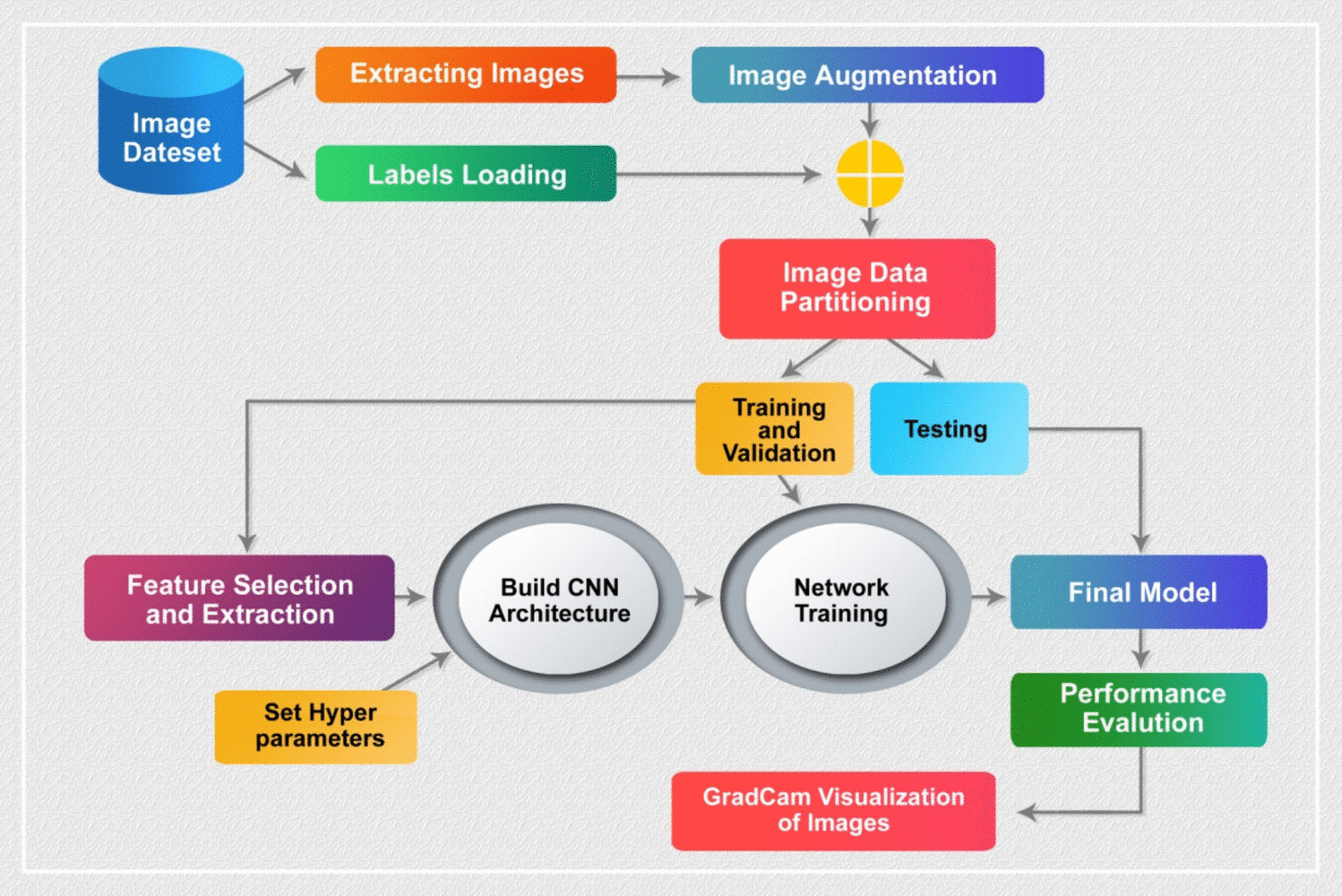
This study proposes a novel framework combining short-time Fourier transform (STFT) and CNN to identify key brain lobes as biomarkers for detecting dementia, specifically AD and FTD, using EEG data. The framework begins by preprocessing the EEG signals, where noise is removed using a butterworth band-pass filter, automatic artifact rejection, and independent component analysis (ICA). The EEG signals are then segmented into smaller time frames. Next, the EEG channels are organized into five brain lobes based on biological principles. Spectrogram images are generated for each brain lobe and the full set of EEG channels using STFT, which provides a time–frequency representation of brain activity. These spectrograms are subsequently fed into a deep learning-based CNN, which is trained to classify dementia. The classification is performed independently on the spectrogram images from each brain lobe as well as from the full channel set.
The combination of STFT and CNN is particularly effective for dementia detection because STFT captures both the time and frequency domains of the brain’s electrical activity, offering insights into cognitive decline. CNNs, known for their ability to detect patterns in 2D data such as spectrograms, are adept at learning complex and subtle features associated with dementia. By leveraging the strengths of both techniques, this approach aims to detect dementia-related abnormalities in brain wave patterns more effectively.
Novelties and Contributions
To our knowledge, no research has explored which brain lobes are most responsive in providing critical information for effective dementia detection. This study offers several significant contributions:
Novel framework development: It introduces a new framework for dementia detection that integrates STFT with CNN algorithms, capable of distinguishing between AD, FTD, and HC using EEG data.
Innovative approach: This study is the first to use STFT-based spectrogram images alongside a CNN model for detecting dementia.
Brain lobe analysis: It investigates which brain lobes are most crucial for extracting representative information for effective dementia detection.
Performance enhancement: The study aims to improve the performance of dementia detection compared to existing methods.
Remainder of the Article
The remainder of this paper is structured as follows: the “Proposed Methodology Framework” section outlines the data analyzed and the proposed methodology. The “Experiments and Results” section presents the experimental setup and results, while the “Discussion” section discusses the experimental results and overall findings. Finally, the “Conclusions and Future Plan” section concludes the paper and discusses future work.


Comments (0)