
Remember me


The Wnt/β-catenin signaling pathway, as an evolutionarily-conserved pathway, employs both proinflammatory and anti-inflammatory functions [12]. Its activation is associated with deficiency of an immune infiltrate in cancer microenvironment [13]. Deregulation of this pathway was detected in liver, osteosarcoma, breast, colorectal, gastric, cervical, and bone cancers [14]. Consist with these findings, several reports corroborate activation of this pathway in ESCC.
Here we manifested that mRNA expression of PYGO2 and IL10, as two important genes involved in Wnt signaling pathway, was upregulated in ESCC and intriguingly, there was a significant correlation between these genes which probably occur through Wnt/β-catenin signaling pathway. Having performed functional study, we demonstrated that enforced expression of PYGO2 changed IL10 expression in ESCC cells. Furthermore, we found that concomitant expression of the genes was strongly correlated with clinical features of ESCC patients and different indices of poor prognosis which may introduce their concomitant expression as a new regulatory axis in ESCC.
PYGO2 is an essential transcriptional coactivator of the Wnt/β-catenin signaling pathway which promotes β-catenin-LEF/TCF transcriptional activation through adapter protein BCL9 [15]. It contributes in expression of highly transcribed RNAs during cell-cycle progression and DNA replication, growth and expansion of cancer cells, as well as histone code interpretation through β-catenin– histone methyltransferase/histone acetyltransferase (HMT/HAT) association [16]. Furthermore, it is applied in a MYC-dependent transcriptional program as an epigenetic accessory protein to regulate cell division [17]. Up-regulated expression of PYGO2 has been illustrated in diverse malignant tumors including ovarian, colon, liver, and cervical cancers. In addition, its oncogenic role is confirmed in hepatic carcinoma, prostate adenocarcinoma, lung and breast cancers as well as renal cell carcinoma and glioma [18,19,20,21]. In line with these reports, PYGO2 overexpression has been reported in ESCC in association with the grade of tumor cell differentiation [22]. Intriguingly, we also observed high level expression of PYGO2 in 31.0% of ESCC samples. It has been revealed that PYGO2 overexpression promotes lymph node metastasis in lung and prostate cancers [23]. Here we found that 7 of 18 PYGO2 overexpressed tumors, were metastasized to the lymph nodes, probably reflecting the importance of PYGO2 in metastasis process. Likewise, PYGO2 activates Wnt target genes such as AXIN1, cyclin D1, and IL10. Surprisingly, we found a significant correlation between PYGO2 and IL10 expression. In tumors with high levels of PYGO2, IL10 expression was significantly higher compared to those with normal PYGO2 expression. In addition, induced expression of PYGO2 in ESCC lines increased expression of IL10 significantly. This finding is noteworthy, suggesting the regulatory role of PYGO2 as a transcriptional activator of IL10 expression probably through Wnt/β-catenin signaling pathway (Fig. 4). Since co-overexpression of PYGO2 and IL10 was significantly correlated with different indices of poor prognosis in ESCC, Wnt/β-catenin signaling may be involved in ESCC tumorigenesis through these genes.
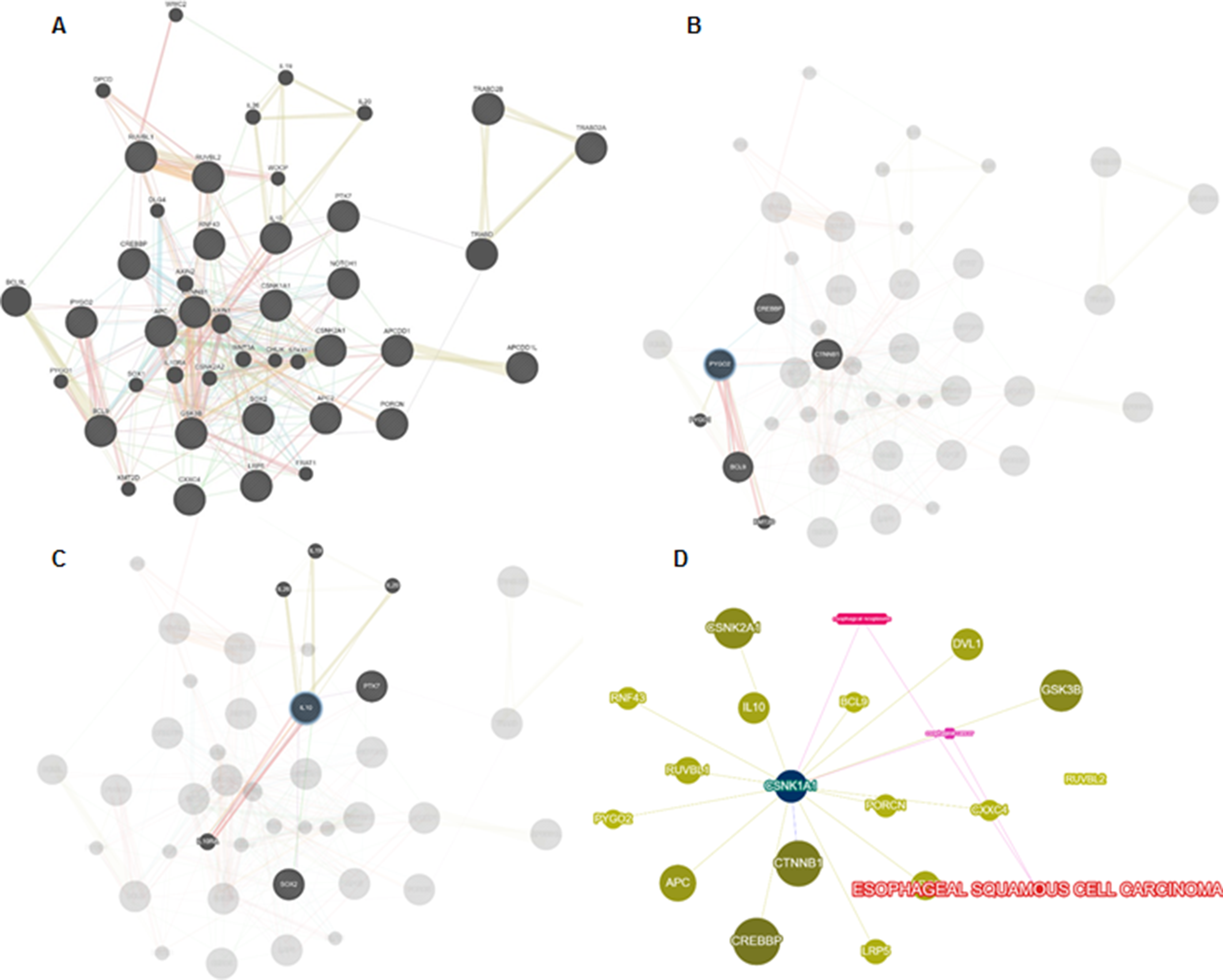
Fig. 4
Potential correlation between PYGO2 and IL10 expression through the Wnt/β-catenin signaling pathway in ESCC progression. Binding of Wnt ligand to FZD and LRP induces inactivation of the AXIN-APC-GSK3β destruction complex via DVL, leading to β-catenin accumulation and transition to the nucleus. Then, PYGO2 as a coactivator of the transcriptional complex (LEF/TCF/β-catenin), may directly activate transcription of target genes such as IL10. Furthermore, IL10 expression manifests direct correlation with FZD, WNT, LRP expression, β-catenin accumulation, and augmentation of EGFR levels in this pathway
Cytokines play critical roles in cancer, inducing tumor metastasis and invasion, as well as apoptosis inhibition [24]. Interleukin-10 is a pleiotropic cytokine with broad anti-inflammatory properties through suppression of both dendritic cell and macrophage function. It suppresses the antitumor immune response, so it is indispensable for tumor development [25]. IL10 has widely been examined in various types of human malignancy. Elevated serum concentrations of IL10 is related with poor prognosis and adverse disease stage in gastric, colon, lung, bone sarcoma, hepatocellular carcinoma, melanoma, and pancreatic malignancies. It is interesting that our findings disclosed IL10 overexpression in 51.7% of ESCC samples which is consisting with previous reports in ESCC [26, 27]. Furthermore, we observed 25 of the 30 IL10 overexpressed ESCCs (83.33%) were invaded to the esophagus adventitia presenting T3 depth of tumor invasion. This data corroborates recent reports in melanoma and cervical cancer [28, 29]. According to IL10 function in tumor development, it may be assumed that IL10 induce tumor invasion and progression by suppressing immune responses and stimulating angiogenesis.
IL10 has been illustrated to contribute in divers signaling pathways. It suppresses starvation induced autophagy in HS-derived fibroblasts (HSFs) through cross talk between the IL10/AKT-mTOR and IL10/IL10R-STAT3 pathways [30]. Moreover, Notch signaling can induce IL10 expression through TH1 cells which involves STAT4-dependent process [31]. This gene is closely linked with various Wnt pathway components, wherein multiple Wnt ligands regulate its responses [32]. In addition, fibroblast-derived Wnt16B induces IL10 secretion in dendritic cells. IL10 expression is associated with the levels of EGFR (as a Wnt target gene) and β-catenin accumulation in lung cancer and melanoma, respectively. Similarly, IL10 level is directly correlated with the level of FZD9 and LRP5/6 in Wnt pathway [33,34,35]. Therefore, all these evidences support the involvement of IL10 in Wnt pathway active state.
The concomitant expression of PYGO2 and IL10 in this study was significantly correlated with depth of tumor invasion. Tumor invasion is a predictor of lymph node metastasis in several cancers such as ESCC. Interestingly, of 25 patients with lymph node metastasis, 6 (24.0%) co-overexpressed of both genes, while in 75.8% of patients (25 of 33) without lymph node metastasis, PYGO2 and IL10 were not co-overexpressed. Having consider these results, the correlation between the genes may be envisaged potentially to activate a cascade leading to malignant invasion and metastasis. In the case of tumor stage, the co-overexpression of the genes was significantly correlated with advanced stage of tumor progression (stage III). It was largely expected, since stage III is associated to lymph node involvement. This result confirms the similar findings in NSCLC, where IL10 overexpression was correlated with high degree of stage and lymph node metastases [36]. Considering this fact that ESCC initiation and progression is a multistep process with poor prognosis, the potential correlation between PYGO2 and IL10 may be considered as an efficient prognostic panel of markers in ESCC patients.
Previous studies have separately considered PYGO2 and IL10 as co-transcription factor and target gene in Wnt pathway, respectively. Subsequently, the involvement of deregulated Wnt/β-catenin pathway has been revealed in ESCC carcinogenesis and progression. Based on these reports and our results, it seems likely that PYGO2 may activate IL10 expression through Wnt/β-catenin signaling pathway in ESCC which may augment ESCC carcinogenesis and aggressiveness.
Comments (0)