Materials
DBP, DEP, DEBP, celecoxib (CCXB), indomethacin (INDO), and NAC were purchased from SigmaAldrich (St. Louis, MO, USA). Phospho-ERK, phospho-JNK, phospho-p38, ERK, JNK, p38, phospho-STAT1, phospho-STAT3, STAT1 and STAT3 antibodies were obtained from Cell Signaling Technology (Danvers, MA, USA). Anti-COX-2 and anti-iNOS antibodies were purchased from Abcam (Cambridge, UK). Anti-β-actin antibody was obtained from Santa Cruz Biotechnology (Dallas, TX, USA).
Cell culture
RAW264.7 macrophages were purchased from the American Type Culture Collection (ATCC, Manassas, VA, USA). Cells were cultured in Dulbecco’s modified Eagle’s medium (DMEM) supplemented with 10% fetal bovine serum (FBS; HyClone, Logan, UT, USA) at 37 °C in a humidified atmosphere (5% CO2 and 95% air). Prior to experiments, cells were plated in 96-well (1.5 × 10⁴ cells per well) or 6-well (5 × 10⁶ cells per well) plates for 24 h and then incubated with an inhibitor or 100 and 300 µM of phthalates for 24–48 h. In each experiment, treatments were performed in triplicate.
Phthalates
The phthalate mixture was dissolved in dimethyl sulfoxide (DMSO) and diluted to achieve the desired phthalate concentrations of 100 and 300 µM. The concentrations used in this study were based on previous reports; measurements of phthalate monoester levels in human plasma indicate that environmental exposure can reach 0.4 µM and occupational and medical procedure exposures can reach 40 µM [17, 18]. Although above the human exposure range, previous in vitro studies used 400 µM as the highest concentration [19,20,21]. Therefore, DEP concentrations of 100 and 300 µM were used in the present study.
Inhibitor treatment
To investigate the potential inhibitory effects of COX inhibitors and antioxidants on phthalate-induced oxidative stress, RAW264.7 cells were pretreated with or without CCXB (10 µM), INDO (10 µM), or NAC (10 mM) for 2 h before incubation with phthalates. Thereafter, proteins were harvested from the cells via trypsin digestion and centrifugation.
NO measurement
Culture media were collected and centrifuged at 1,500 rpm and 4 °C for 10 min. The Griess Reagent System (Promega) determined NO levels in the supernatant. Briefly, 50 µL of sulfanilamide reagent and 50 µL of 0.1% naphthalene ethylenediamine reagent (NED) were added to 50 µL of supernatant from each well, followed by incubation in the dark for 15 min at room temperature to measure NO levels. The absorbance was measured at 550 nm using a microplate reader. Sodium nitrite was used to prepare a standard curve.
Measurement of ROS production
To determine ROS levels, cells were seeded in plates and treated with NAC and DEP. After washing with serum-free medium, 10 µM of DCFDA-Cell ROS Assay Buffer (Abcam, Cambridge, UK) was added to each well, and the plate was incubated at 37 °C for 30 min. The fluorescence intensity was measured using a microplate reader at excitation and emission wavelengths of 485 and 535 nm, respectively. The images were obtained using a fluorescence microscope (200x magnification).
GSH/GSSG determination
Cells were seeded onto clear 96-well microtiter plates at a density of 1 × 10⁵ cells/well. Cell lysates were analyzed for glutathione using a GSH/GSSG assay kit (Promega Corporation, Madison, WI, USA). The GSH/GSSG ratio was calculated according to the manufacturer’s instructions. Luminescence was measured with a microplate reader, and the GSH/GSSG ratio was calculated as [(net treated total glutathione RLU − net treated GSSG RLU)/(net treated GSSG RLU)] × 2, where RLU is relative light units.
Reverse transcription–quantitative polymerase chain reaction
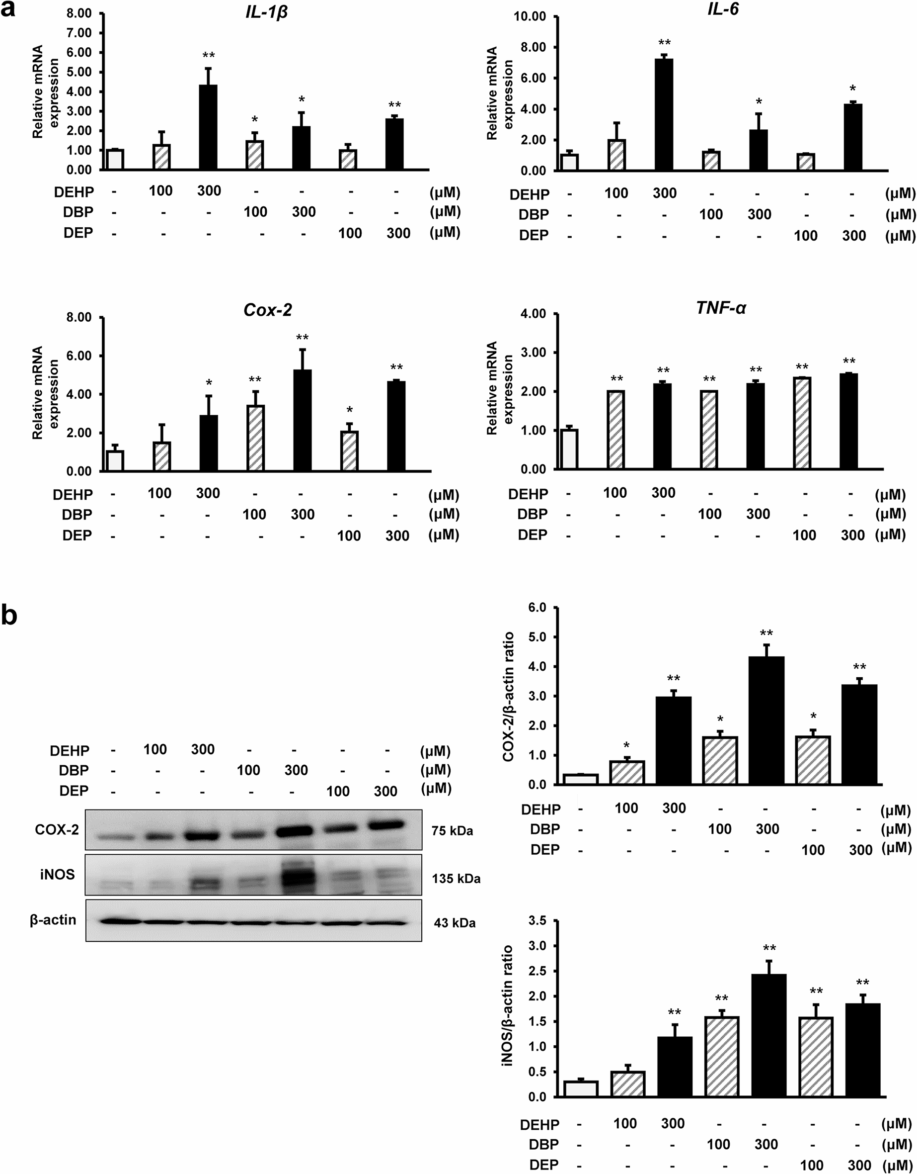
Total RNA was extracted from cells using an RNeasy Mini kit (Qiagen, Valencia, CA, USA) and reverse-transcribed to generate cDNA using CycleScript RT PreMix (Bioneer, Daejeon, South Korea). Quantitative real-time RT–PCR was performed using SYBR Green PCR Master Mix (Applied Biosystems, Foster City, CA, USA) and specific primers (Table 1). COX-2, iNOS, IL-1β, and IL-6 mRNA levels were normalized to that of GAPDH.
Table 1 Primers used for qPCRWestern blot analysis
Cells were lysed in RIPA buffer (Cell Signaling Technology) containing a protease inhibitor cocktail (cOmplete™ Mini Protease Inhibitor Tablet; Roche Diagnostics GmbH, Mannheim, Germany) to extract the proteins. Protein concentrations were determined using a Pierce BCA Protein Assay kit (Thermo Fisher Scientific, Waltham, MA, USA). Proteins (30 µg) were separated on 8–16% and 6% SDS–PAGE gels (Bio-rad) and transferred to nitrocellulose membranes (Thermo Fisher Scientific). After blocking with 5% (w/v) skim milk (Bio-rad) for 1 h at room temperature, the membranes were incubated overnight at 4 °C with primary antibodies against COX-2 (1:1,000), iNOS (1:1,000), p-Akt (1:1,000), p-JNK (1:1,000), and β-actin (1:5,000). Thereafter, the membranes were washed with Tris-buffered saline containing 0.1% Tween 20 and incubated with secondary horseradish peroxidase-conjugated anti-rabbit/mouse IgG antibodies (1:5000; Thermo Fisher Scientific) for 2 h at room temperature. Protein bands were visualized using a Pierce-enhanced chemiluminescence substrate (Thermo Fisher Scientific) and quantified using ImageJ software. Protein expression was normalized to that of β-actin.
Enzyme-linked immunosorbent assay
The levels of IL-1β and PGE2 in the culture supernatant were assessed using a commercially available enzyme-linked immunosorbent assay (ELISA) kit (Abcam and R&D Systems, Minneapolis, MN, USA), according to the manufacturer’s instructions.
Statistical analysis
All statistical analyses were performed using SPSS Windows software (version 22.0; SPSS Inc., Chicago, IL, USA). Quantitative data are presented as the mean ± standard error of the mean from three experimental replicates. Significant differences between groups were determined using two-tailed Student’s t-test (two groups) or one-way analysis of variance with Tukey’s post hoc comparisons (multiple groups). Statistical significance was set at p < 0.05 (*p < 0.05 and **p < 0.01).


Comments (0)