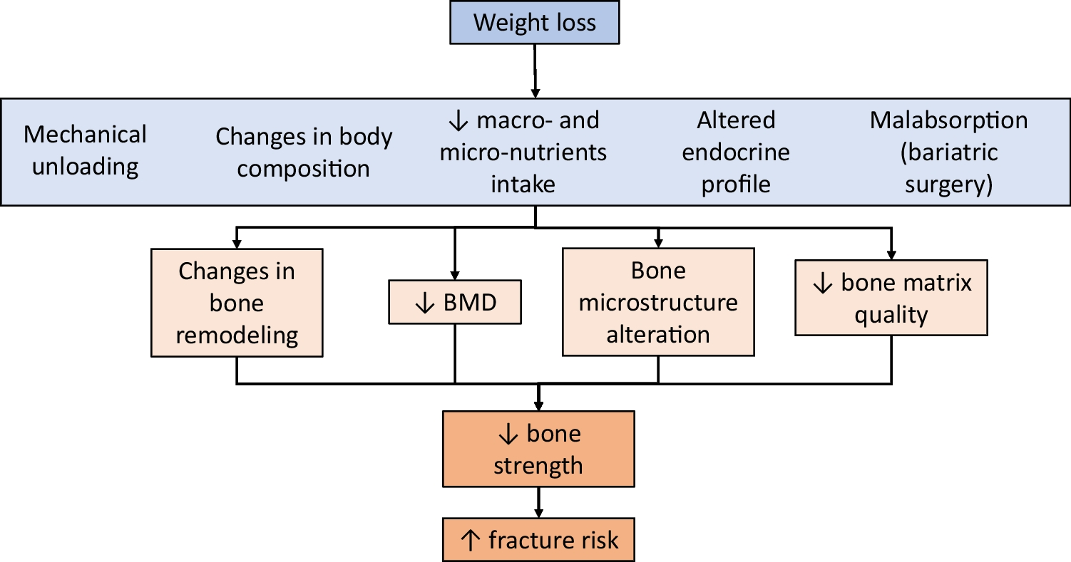
Osteoporosis is a systemic skeletal pathology distinguished by diminished bone mineral density (BMD) and compromised bone microarchitecture, consequently resulting in heightened susceptibility to bone fragility and fracture. In 2022, the global prevalence of osteoporosis was estimated to be 19.7% [1], with postmenopausal osteoporosis constituting the majority of cases linked to physiological changes resulting from menopause and the aging process. Owing to its asymptomatic characteristic prior to the occurrence of a fracture, osteoporosis is frequently characterized as a "silent" epidemic of the twenty-first century. Common medications for osteoporosis may be antiresorptive, anabolic, or have a dual mode of action [2, 3]. Examples of antiresorptive medications such as bisphosphonates, which inhibit the activity of osteoclasts that break down bone, while anabolic agents like teriparatide stimulate new bone growth to enhance overall skeletal strength. In addition to these, other medications such as denosumab and selective estrogen receptor modulators (SERMs) are also utilized in the management of osteoporosis, each offering unique mechanisms to help maintain bone health and prevent fractures. While medication for osteoporosis can be beneficial, many of the commonly prescribed medications, such as bisphosphonates and anabolic agents, come with potential side effects that can sometimes outweigh their benefits. For instance, long-term use of bisphosphonates has been associated with rare but serious complications like osteonecrosis of the jaw and atypical femur fractures [4]. Additionally, anabolic agents like teriparatide (PTH1-34) are often expensive and may not be accessible for all patients. While pharmaceutical medications can have a role in osteoporosis management, natural non-pharmaceutical alternatives have gained attention for their potential to support bone health without the associated risks. Kefir is one of them that has been extensively studied in various pathological backgrounds and recently been reported with promising effects on osteoporosis.
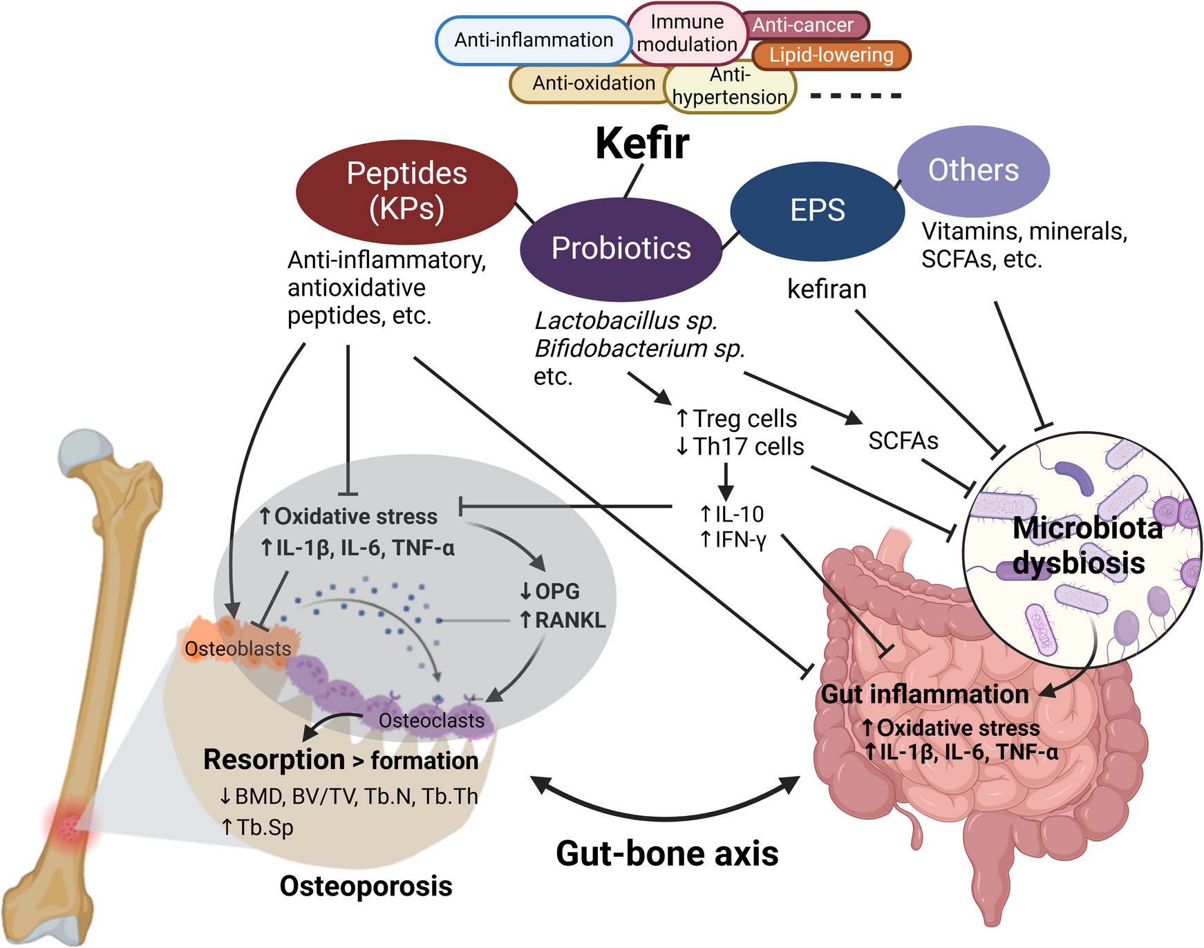
Kefir is an alcoholic and acidic beverage made by the milk fermentation with kefir grains, which comprise a diverse assemblage of bacteria and yeast that contribute to the probiotic characteristics of kefir with anti-inflammatory, antioxidant, anti-hypertensive, antimicrobial, anticancer, lipid-lowering, and immune modulation activities, alongside the advantageous effects on brain, liver, gastrointestinal, and skeletal health in both animal and human subjects [5, 6]. These health-promoting characteristics are attributed to the abundance of probiotics in kefir grains and the wide range of bioactive substances produced during fermentation, such as bioactive peptides, exopolysaccharides, organic acids, vitamins, and minerals that enhance the overall nutritional profile of the beverage. The unique combination of these components not only supports gut health but also plays a significant role in regulating metabolism and fosters a balanced immune response, thereby reducing the risk of various chronic diseases and rendering kefir a valuable nutritious diet. The type of kefir can vary significantly depending on the source of kefir grains and the fermentation process used, leading to different flavors, textures, and probiotic profiles that can cater to diverse dietary preferences and health needs [7].
Over the past decade, our laboratory has concentrated on using animal models and clinical trials to demonstrate the value of kefir in preventing and treating osteoporosis. In this article, we aim to review the related studies and discuss the advantages of milk protein-derived peptides, probiotics, and exopolysaccharides present in kefir or kefir grains on the bone health.
Kefir Peptides for Osteoporosis Prevention and Treatment
Chen et al. [8] were the pioneering investigators to clarify the efficacy of kefir supplementation in the prevention of osteoporosis in ovariectomized (OVX) rats. OVX models are extensively employed to elucidate the pathophysiologic mechanism underlying menopausal osteoporosis and to assess the efficacy of various natural substances and pharmacological agents for its prophylaxis and therapeutic intervention [9]. Without the implementation of suitable therapeutic measures, OVX rats were shown to exhibit marked osteopenia in the proximal tibial and distal femoral metaphysis within 14 days [10, 11], and further demonstrated enhanced bone resorption in the femoral neck and lumbar vertebrae after 30 and 60 days, respectively [12, 13]. Chen et al. [8]. provided evidence that a 12-week regimen of kefir administered at varying dosages (164, 328, and 656 mg per kilogram of body weight per day (mg/kg BW/day)) caused a significant reduction in the systemic bone resorption biomarker C-terminal telopeptides of type I collagen (CTX) and a notable enhancement in femoral microarchitecture by increasing the parameters of BMD, bone volume (BV/TV), trabecular number (Tb.N), and trabecular thickness (Tb.Th) while decreasing trabecular separation (Tb.Sp). It also enhanced the mechanical properties of cortical bone, with effects comparable to alendronate, a first-line antiresorptive bisphosphonate agent for osteoporosis therapy, at the highest dose [8]. While the underlying mechanism was not fully explored, findings from the Caco-2 cell model suggested that kefir may promote intestinal calcium absorption.
A follow-up clinical assessment conducted by Tu et al. [14] delineated the short-term effects of kefir consumption in those diagnosed with osteoporosis. Tu’s study revealed that, over a 6-month period, the administration of kefir (1,600 mg/day) in conjunction with CaCO3 (1,500 mg/day) significantly improved BMD compared to the administration of CaCO3 alone. Furthermore, the concomitant treatment of kefir and CaCO3 resulted in a reduction of serum β-CTX in patients with T-scores > −1 after 3 months, as well as an elevation of serum osteocalcin (OC) after a 6-month duration, alongside an increase in serum parathyroid hormone (PTH). This pilot study illustrated the advantages of kefir intake in reducing bone resorption and increasing bone formation, resulting in improved hip BMD in patients with osteoporosis. Based on previous findings in Caco-2 cells [8], it is reasonable to suggest that the positive impact of kefir on bone is partly connected to better intestinal calcium uptake. It is essential to note that the principal active constituents in the aforementioned two studies are primarily small peptides, referred to as "kefir peptides (KPs)" (KEFPEP®), which are generated through the hydrolysis of milk protein during the fermentation process, wherein the probiotic elements derived from kefir grains are eliminated subsequent to fermentation.
Subsequently, the identical research group assessed the effectiveness of KPs across a diverse array of murine models of osteoporosis [15,16,17,18]. These studies were executed through the daily administration of KPs via the oral route at a dosage of 100 mg/kg BW [15] or at concentrations varying from 164 to 654 mg/kg BW [16,17,18] over a minimum duration of 8 weeks. In OVX mice, CaCO3 alone conferred limited protection against bone loss; however, KPs exhibited CaCO3-independent osteoprotective effects, as well as altering gut microbiota by enriching beneficial bacteria and reducing potential pathogens, thus enhancing bone health [15]. In the context of the aldo–keto reductase family 1 member A1 (AKR1A1) knockout (KO) murine model, the condition of osteoporosis was instigated by a deficiency in vitamin C [19, 20], and KPs proficiently ameliorated this condition [16]. The osteoprotective effects of KPs in AKR1A1-KO mice have been ascribed to the attenuation of systemic inflammation and oxidative stress, alongside the restoration of bone remodeling markers, particularly through the modulation of the ratio of receptor activator of nuclear factor kappa-B ligand (RANKL) and osteoprotegerin (OPG), which serves as a critical indicator of bone mass and structural integrity [16]. Moreover, KPs inhibited RANKL-induced osteoclastogenesis in bone marrow macrophages (BMMs) by downregulating NFATc1, c-Fos, c-Src, and cathepsin K expression, as well as attenuating signal transduction via p38, NF-κB, Akt, PLCγ2, and CREB-1 phosphorylation [16]. Conversely, KPs facilitated osteoblastic differentiation and mineralization in bone marrow mesenchymal stem cells (BMMSCs) through the upregulation of osteogenic factors, including RUNX2, BMP-2, NFATc1, collagen I, HO-1, and β-catenin, while simultaneously downregulating the negative regulator p53 [16]. These findings illuminated the fundamental mechanisms that contribute to the antiresorptive and anabolic properties of KPs.
Hemophilia is also a notable risk factor for the development of osteoporosis, as a substantial number of individuals afflicted with diverse forms of hemophilia are concurrently diagnosed with osteoporosis [21, 22]. A murine model demonstrating significant deficits in coagulation factor VIII (F8) is routinely used to explore impaired bone remodeling in hemophilic subjects [23]. Utilizing this murine model, Yen et al. [17] identified that F8-KO mice exhibited significant bone loss by the age of 20 weeks; however, following an 8-week regimen, KPs facilitated the restoration of the trabecular architecture within the femurs and lumbar vertebrae, concurrently enhancing the mechanical strength of the femoral cortical bones in F8-KO mice. Furthermore, KPs treatments led to a reduction in the serum RANKL/OPG ratio and the concentration of the proinflammatory cytokine IL-6, in addition to an elevation in the bone formation biomarker alkaline phosphatase (ALP) and a decrement in the bone resorption biomarker CTX. These results illustrate the potential of KPs for the management of hemophilia-induced osteoporosis.
Additionally, cardiovascular disease patients with vascular calcification symptoms often have concomitant osteoporosis, a condition referred to as the "bone-vascular axis" [24, 25]. Chang et al. [18] found that apolipoprotein E (ApoE)-KO mice fed an atherogenic diet for 13 weeks developed aortic calcification and severe bone loss in the femur. Intervention with KPs attenuated systemic and vessel-specific inflammation and oxidative stress, reducing lipid, collagen, and calcium mineral deposition in the vessels and mitigating bone loss in the femur.
Beyond osteoporosis prevention, KPs have demonstrated significant benefits in fracture repair. This was evidenced in our previous rat model of femoral fracture with KP intervention [26]. Unlike untreated rats, which showed incomplete bone healing, rats administered 400 mg/kg of KPs daily for 4 weeks exhibited complete bone healing, with bone gaps reunited and initial fracture lines entirely vanished. The expression of osteogenic factors such as Col1a1, Alp, Spp1, Vegfa, and Cox2 was upregulated in these KP-treated rats. Given that osteoporosis is a major cause of clinical fractures, the findings by Lai et al. [26] suggest the potential of KPs as a nutritional supplement to accelerate bone healing in clinical fracture cases.
Table 1 summarizes the relevant experimental outcomes using KPs in the aforementioned studies, highlighting their potential as a nutritional supplement for osteoporosis prevention, treatment, and fracture healing acceleration.
Table 1 Studies using KPs for osteoporosis prevention and treatmentIndividual Bioactive Peptides With Osteoprotective Effects
Although bioactive peptides with osteoprotective effects have not yet been extensively identified from kefir-fermented products, earlier studies on milk protein-derived peptides offer valuable insights. Caseinophosphopeptides (CPPs) are the most thoroughly studied milk casein (CN)-derived bioactive peptides, known for their calcium-binding activity [27,28,29,30]. The benefits of CPPs for bone health are based on the premise that these peptides can promote intestinal calcium absorption, thereby enhancing bone calcification. This premise is supported by findings that dietary calcium-bound CPPs prevented bone loss in OVX rats [31]. Subsequently, Liu et al. [32, 33] isolated a calcium-binding peptide, β-CN (16–40), from a commercially available CPP mixture and found that this peptide enhanced calcium transport in Caco-2 cells via the transcellular route by upregulating the expression of the TRPV6 calcium channel protein. Additionally, this peptide improved bone formation and prevented bone resorption in rats.
Ebner et al. [34] identified a peptide profile of bovine milk protein released from a commercial kefir starter culture, which included many casein phosphopeptides with sequences overlapping β-CN (16–40) and other known CPPs, such as αS1-CN (59–79) [27, 29] and β-CN (1–25) [30, 31]. However, most of these peptides have not yet been functionally characterized. The efficacy of CPPs in promoting calcium absorption remains a controversial issue, possibly due to differences in methods used for assessing calcium bioavailability [35].
To date, ACE-inhibitory tripeptides (IPP and VPP) [36,37,38], casein-derived antioxidative peptides (EDVPSER, NAVPITPTL, VLPVPQK, and HPHPHLSF) [39,40,41,42,43], and whey-derived ACE-inhibitory (YLLF and WLAHK) and antioxidative (YVEEL) peptides [44] have been characterized for their ability to stimulate in vitro osteoblast proliferation and differentiation. Among these peptides, VLPVPQK, YVELL, and YLLF have been further demonstrated for their osteoprotective potential in OVX rats [45]. Oxidative stress plays a significant role in the development of chronic inflammatory bone disease, as excessive generation of reactive oxygen species (ROS) negatively impacts bone remodeling by promoting osteoclastogenesis and inhibiting osteogenesis [46]. This explains why antioxidative peptides often have osteoprotective effects.
The mechanism of ACE-inhibitory peptides on osteoblasts may involve their activity in inhibiting ACE, thereby preventing the conversion of angiotensin I to angiotensin II, which has been shown to induce osteoblast mitochondrial dysfunction and apoptosis, thus decreasing osteoblast differentiation [47]. Notably, all the mentioned casein-derived peptides can be found in previous peptide profiles of bovine milk-based kefir with complete sequence coverage, suggesting their osteoprotective effects [34, 48]. Undoubtedly, high-throughput screening of osteoprotective peptides from kefir deserves attention in future research.
Earlier, we isolated a bioactive peptide named KFP-1 from the < 3-kDa fraction of KPs [49]. The identity of KFP-1 covers the sequence of κ-CN (138–154) and has been characterized for its functions in binding calcium, promoting calcium influx in Caco-2 cells through TRPV6 channels, and enhancing intestinal calcium absorption in mice. Furthermore, KFP-1 was found to inhibit osteoclastogenesis and promote osteoblastogenesis in vitro, as well as exert osteoprotective effects in the AKR1A1-KO mouse model of osteoporosis. Techniques such as LC/MS/MS and high-throughput analysis of antioxidative and anti-inflammatory peptides using Raw264.7 cell models may aid in the screening and characterization of more milk protein-derived bioactive peptides with osteoprotective effects. These experiments are currently being undertaken in our laboratory.
The known milk protein-derived bioactive peptides with osteoprotective effects are summarized in Table 2.
Table 2 Milk protein-derived bioactive peptides with osteoprotective effectsProbiotics with Osteoprotective Benefits
The utilization of molecular biology methodologies in microbiome analysis, including 16S rRNA sequencing and metagenomic next-generation sequencing, has enhanced the investigation of intricate microbial ecosystems in kefir grains and associated kefir products. A recent metagenomic analysis conducted by Zeng et al. [50]. revealed the microflora structure of Tibetan kefir grains obtained from three distinct sources, in which species under the genera Lactobacillus comprise the predominant microbial consortium in kefir grains. Many species identified in the study of Zeng et al. align with prior studies and literature [51, 52]. A lot of species identified in kefir grains or milk kefir beverages have been shown to exert osteoprotective effects in animals and humans, including Lactoacillus acidophilus [53,54,55,56,57,58], Lactoacillus casei [56,57,58], Lactoacillus fermentum [59], Lactoacillus helveticus [60, 61], Lactoacillus plantarum [62,


Comments (0)