Ceftobiprole was obtained from Basilea Pharmaceutica International Ltd. (Basel, Switzerland). Commercial ceftobiprole medocaril powder for injection (Zevtera®, Advanz Pharma, Spain) was kindly supplied by Advanz Pharma (Madrid, Spain). Sodium chloride 0.9% and dextrose 5% were purchased from Baxter (Madrid, Spain). Portable elastomeric infusion devices (Accufuser C0100L 10 mL/h 300 mL) were purchased from Grifols (Barcelona, Spain). According to the technical data sheet, the balloon is composed of medical-grade silicone, and the infusion line is PVC-TOTM (DEHP-free), with a flow rate accuracy within ± 10% under controlled conditions. Purified water was obtained using an Elix 3, Millipore purified water system (Merck, Massachusetts, USA). All other chemicals and solvents were at least of ACS reagent grade and used without further purification.
This article does not contain any new studies with human participants or animals performed by any of the authors. Ethical committee approval was not required for this work, as it involved only in vitro experiments with pharmaceutical products.
Ceftobiprole Quantification by Liquid Chromatography Triple Quadrupole Mass Spectrometry (LC-QQQ-MS)
Ceftobiprole and ceftobiprole medocaril were quantified using a previously validated LC-QQQ-MS method (see the appendix in the electronic supplementary material). The analytic column was a Phenomenex Gemini 5 μm C18 110 Å 150 × 2 mm. Analysis was performed using an LCMS-8030 Shimadzu (Kyoto, Japan) in a gradient mode. Mobile phase A was composed of H2O + 0.1% formic acid and phase B was composed of acetonitrile + 0.1% formic acid. For the detection of ceftobiprole medocaril, the gradient mode consisted of 0–5 min at 3–95% phase B, followed by 5–7 min at 95% phase B, 7–8.5 min at 95–3% phase B, and 8.5–10 min at 3% phase B with a flow rate of 0.4 mL/min and an injection volume of 40 μL. For the detection of ceftobiprole, the gradient mode consisted of 0–4 min at 3–40% phase B, followed by 4–6 min at 40–90% phase B, 6–7.5 min at 90% phase B, 7.5–9 min at 90–3% phase B, and 9–10 min at 3% phase B with a flow rate of 0.3 mL/min and an injection volume of 20 μL.
The multiple reaction monitoring (MRM) mode was employed to detect the following quantifier ion transitions: 712.50 → 22.90 (ceftobiprole medocaril) and 534.90 → 69.20 (ceftobiprole) and the following qualifier ion transitions: 712.50 → 398.00 and 534.90 → 203.20.
For the preparation of the standards, methanol (0.1% formic acid) was used as a solvent for ceftobiprole medocaril detection, and methanol (0.05% formic acid) for ceftobiprole quantification.
Chemical Stability
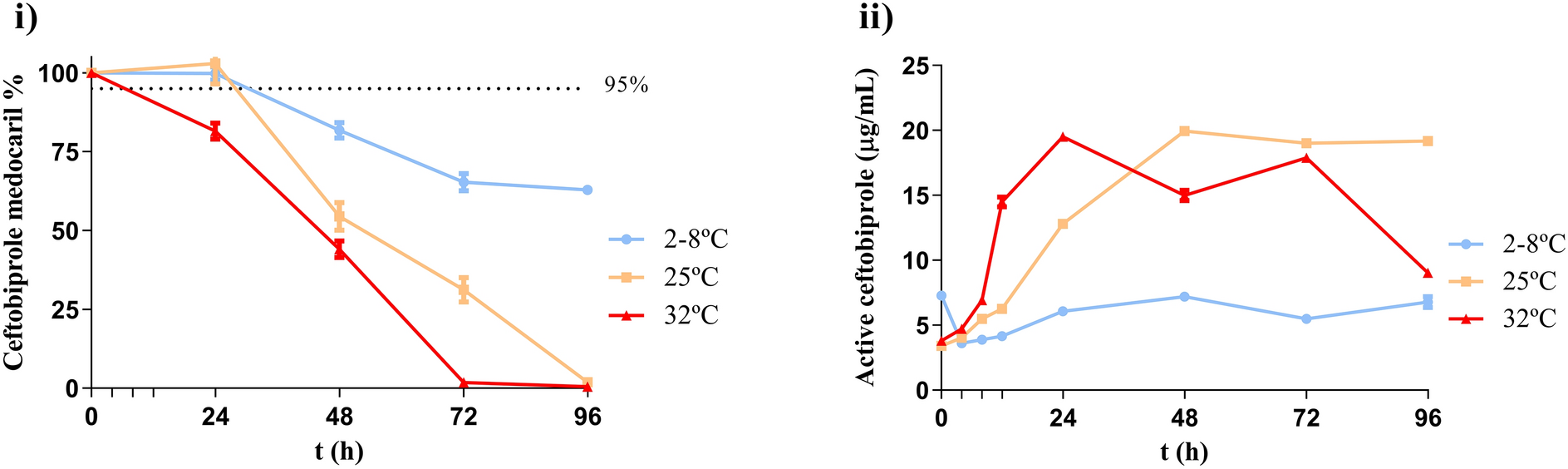
Ceftobiprole medocaril (Zevtera®) was reconstituted in 10 mL of water for injection. Portable elastomeric infusion devices (Accufuser C0100L 10 mL/h 300 mL) were filled with solutions diluted in either sodium chloride 0.9% or dextrose 5% to a final concentration of 6.25 mg/mL and a total volume of 240 mL. This reflects a 24-h continuous infusion regimen delivering 1500 mg daily (equivalent to 500 mg every 8 h). Note: 500 mg of active ceftobiprole equals 667 mg of ceftobiprole medocaril. Three portable elastomeric infusion devices were filled with each diluent and were stored at 2–8 °C, 25 °C, and 32 °C, respectively, in the refrigerator and temperature-controlled ovens. Samples (5 mL) were taken at 0, 4, 8, 12, 24, 48, 72, and 96 h. Each was diluted in duplicate with deionized water (1/100) and analyzed directly to reflect the actual conditions within the elastomeric infusion devices. Chemical stability was assessed by calculating the percentage of ceftobiprole medocaril remaining at each time. Stability was considered acceptable if drug recovery was ≥ 95%. Ceftobiprole concentrations were also determined to evaluate prodrug conversion.
Modelling of Chemical Stability
Mathematical calculations were based on the Arrhenius equation:
where K is the chemical reaction rate, A is a constant referred to as the preexponential factor, Ea is the activation energy of the reaction, typically measured in kJ mol−1 or kcal mol−1, that describes the “temperature sensitivity” of the drug, R is the universal gas constant whose value is 1.987 cal K−1 mol−1, and T is the absolute temperature expressed in Kelvin.
Drug concentrations were fitted to zero-order, first-order, second-order, Avrami, and diffusion models [9]. Model suitability was assessed by the regression coefficient (R2). The Arrhenius equation was used to estimate activation energy and the temperature effect on degradation.
Physical Stability
Physical stability (visible/subvisible particles and pH) was evaluated according to the YCD at the same time points as chemical stability. Visible particles and color changes were assessed visually. Subvisible particles were measured using a Zetasizer (Malvern, UK). A zero was performed initially with each of the diluent solutions, either dextrose 5% or sodium chloride 0.9%. Analysis was performed following United States Pharmacopoeia (USP) <787>, <788> and European Pharmacopoeia (EP) 2.9.19. pH was measured using a Mettler Toledo MP230 GLP Research pH meter (Madrid, Spain).
Antimicrobial Activity Quantification
Antimicrobial activity was assessed at 0, 24, 48, 72, and 96 h via agar diffusion test on Mueller Hinton agar (MH) following EUCAST guidelines. The quality control strain reference strain was methicillin-susceptible S. aureus CECT 438, cultured for 72 h at 35 °C (± 1 °C) to ensure viability and absence of contamination. The inoculum was prepared by suspending a single colony in 3 mL of sterile sodium chloride 0.9%. The suspension was vortexed, and its turbidity was adjusted to match a 0.5 McFarland standard using a spectrophotometer (600 nm), resulting in a bacteria stock suspension of 1 × 10⁶ cells/mL. The bacterial suspension was mixed with 250 mL of MH agar and poured into sterile Petri dishes. After solidification, 6 mm paper disks were impregnated with 8 µL of the test solutions. The in vitro activity of the test solutions was compared to control disks impregnated with 5 μg of ceftobiprole. Plates were incubated at 35 °C (± 1 °C) within 15 min of disk placement and inhibition zones were measured after 24 h using a caliper.
Adsorption Study
At the end of the experiment, each device was fully emptied. Residual drug was removed by rinsing twice with 100 mL deionized water. Once the devices were emptied, they were filled with 50 mL of H2O/MeOH (50:50; v/v) and sonicated for 20 min. A 1-mL aliquot was analyzed in duplicate by LC-QQQ-MS to determine the percentage of drug adsorbed on the device surface.
Translation of Results into Clinical Practice
A multidisciplinary team of three pharmacists and two infectious disease specialists developed proposed administration schedules based on the study results.
Data Analysis
Data were analyzed using GraphPad Prism version 5.0 (GraphPad Software, California, USA). Statistical significance (p < 0.05) was assessed by analysis of variance (ANOVA) and Newman–Keuls multiple-comparison test.


Comments (0)